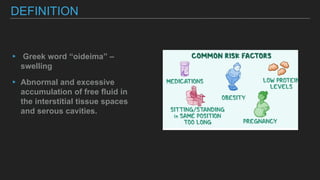
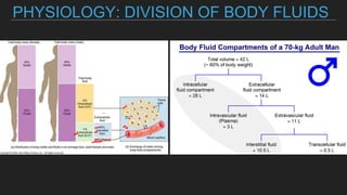
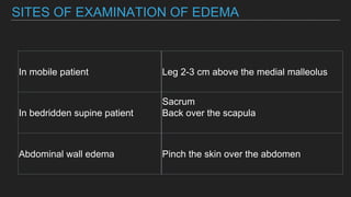
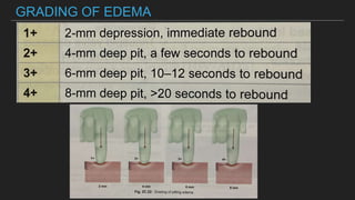
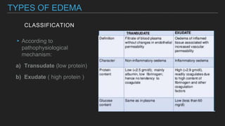
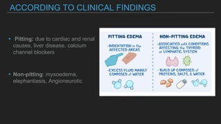
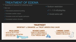
KRISHNA PATEL
DEFINITION
▸ Greek word “oideima” –
swelling
▸ Abnormal and excessive
accumulation of free fluid in
the interstitial tissue spaces
and serous cavities.
PHYSIOLOGY: DIVISION OF BODY FLUIDS
_
PATHOPHYSIOLOGY OF
EDEMA
▸ Fluid movement across capillaries
1. Hydrostatic pressure
2. Oncotic pressure
3. Capillary permeability
‣ Lymphatic flow
A perturbation of these factors singly or in
combination results in Edema
CAUSES
A. INCREASE IN hydrostatic pressure
Normal serum proteins
‣ Congestive heart failure
‣ Renal failure
‣ Pericarditis
‣ Cirrhosis of liver
‣ Pregnancy
B. DECREASED ONCOTIC
PRESSURE
Low serum protein
▸ Increased protein loss
Burns
Nephrotic syndrome
Bowel disease
▸ Decreased intake / synthesis
Kwashiorkor
Malabsorption
Liver disease
C. LYMPHATIC
OBSTRUCTION
LYMPHOEDEMA
Malignancy
Radiation induced
Filariasis
Myxoedema- hypothyroidism
Pretibial myxedema- Graves’ disease
UNILATERAL EDEMA
Cellulitis
Thrombophlebitis
DVT
Acute trauma
FACIAL EDEMA
▸ Hypothyroidism
▸Allergies
▸Nephrotic syndrome
▸Angio-oedema
▸Trichinosis
CONGENITAL EDEMA
▸ Milroy’s disease
▸ Meigs disease
▸ Lymphoedema Praecox
▸ Lymphoedema Tarda
DRUG INDUCED
EDEMA
▸ Nifedipine
▸Corticosteroids
▸Oestrogen
▸NSAIDs
▸Insulin
TECHNIQUE
Inspect the legs for any swelling.
Look at the skin between the medial malleolus and Achilles
tendon for the presence or absence of convexity - flat or
convex means edema
Apply pressure 1 inch above the medial malleolus using
the pulp of the thumb for a minimum of 30seconds on both
lower limbs simultaneously.
Inspect and palpate the area for any dimple or pitting.
When there is edema repeat the test in the rest of the limb
from below up to note the extent of edema
Check for oedema in thighs, genitalia & rest of the body .
Bed ridden patients- Turn the patient to a side and uncover
the sacral area. Apply pressure over the sacrum with right
thumb for 30seconds and palpate and look and for dimple.
SITES OF EXAMINATION OF EDEMA
In mobile patient Leg 2-3 cm above the medial malleolus
In bedridden supine patient
Sacrum
Back over the scapula
Abdominal wall edema Pinch the skin over the abdomen
GRADING OF EDEMA
CLASSIFICATION
TYPES OF EDEMA
▸According to
pathophysiological
mechanism:
a) Transudate (low protein)
b) Exudate ( high protein )
ACCORDING TO LOCATION
▸ Localised: Venous edema, lymphatic
Edema, allergy/angio-
oedema/inflammation
▸Generalised- cardiac Edema, hepatic
Edema, renal Edema
ACCORDING TO CLINICAL FINDINGS
▸ Pitting: due to cardiac and renal
causes, liver disease, calcium
channel blockers
▸Non-pitting: myxoedema,
elephantiasis, Angioneurotic
ORGAN SPECIFIC EDEMA
▸ Brain – cerebral Edema
▸Lung -(intra alveolar) pulmonary Edema
▸(Intra pleural) pleural effusion
▸Peritoneum-ascites
GENERAL INVESTIGATIONS
▸ Urine dipstick and microscopy
Proteinuria
Hematuria
Casts
▸Renal function test
Serum urea
Serum creatinine
▸Thyroid function tests.
▸Serum electrolytes
▸CBC and PBF
▸Liver function test
Transaminases eg ALT,AST
Serum albumin
Serum bilirubin
▸Chest x-ray
Cardiomegaly
Left ventricular hypertrophy
Perihilar vascular margins
▸ECG
ST elevation with T wave inversion
(pericarditis)
TREATMENT OF EDEMA
▸ Supportive & treatment of underlying cause.
▸Bed rest
✓ Decreased peripheral pooling
✓ Increase cardiac output
✓ Increase renal and hepatic perfusion
✓ Increased sodium diuresis
▸Sodium restriction
✓ 1 - 1.5 mEq/kg/day
✓ Avoid extra salt
FOLLOW-UP OF PATIENT
WITH EDEMA
▸ Weight
▸ Abdominal girth
▸ Input/output chart
▸ Edema status
THANK YOU

Edema

  • 1.
  • 2.
    DEFINITION ▸ Greek word“oideima” – swelling ▸ Abnormal and excessive accumulation of free fluid in the interstitial tissue spaces and serous cavities.
  • 3.
  • 4.
    PATHOPHYSIOLOGY OF EDEMA ▸ Fluidmovement across capillaries 1. Hydrostatic pressure 2. Oncotic pressure 3. Capillary permeability ‣ Lymphatic flow A perturbation of these factors singly or in combination results in Edema
  • 5.
    CAUSES A. INCREASE INhydrostatic pressure Normal serum proteins ‣ Congestive heart failure ‣ Renal failure ‣ Pericarditis ‣ Cirrhosis of liver ‣ Pregnancy B. DECREASED ONCOTIC PRESSURE Low serum protein ▸ Increased protein loss Burns Nephrotic syndrome Bowel disease ▸ Decreased intake / synthesis Kwashiorkor Malabsorption Liver disease
  • 6.
  • 7.
    UNILATERAL EDEMA Cellulitis Thrombophlebitis DVT Acute trauma FACIALEDEMA ▸ Hypothyroidism ▸Allergies ▸Nephrotic syndrome ▸Angio-oedema ▸Trichinosis
  • 8.
    CONGENITAL EDEMA ▸ Milroy’sdisease ▸ Meigs disease ▸ Lymphoedema Praecox ▸ Lymphoedema Tarda DRUG INDUCED EDEMA ▸ Nifedipine ▸Corticosteroids ▸Oestrogen ▸NSAIDs ▸Insulin
  • 9.
    TECHNIQUE Inspect the legsfor any swelling. Look at the skin between the medial malleolus and Achilles tendon for the presence or absence of convexity - flat or convex means edema Apply pressure 1 inch above the medial malleolus using the pulp of the thumb for a minimum of 30seconds on both lower limbs simultaneously. Inspect and palpate the area for any dimple or pitting. When there is edema repeat the test in the rest of the limb from below up to note the extent of edema Check for oedema in thighs, genitalia & rest of the body . Bed ridden patients- Turn the patient to a side and uncover the sacral area. Apply pressure over the sacrum with right thumb for 30seconds and palpate and look and for dimple.
  • 10.
    SITES OF EXAMINATIONOF EDEMA In mobile patient Leg 2-3 cm above the medial malleolus In bedridden supine patient Sacrum Back over the scapula Abdominal wall edema Pinch the skin over the abdomen
  • 11.
  • 12.
    CLASSIFICATION TYPES OF EDEMA ▸Accordingto pathophysiological mechanism: a) Transudate (low protein) b) Exudate ( high protein )
  • 13.
    ACCORDING TO LOCATION ▸Localised: Venous edema, lymphatic Edema, allergy/angio- oedema/inflammation ▸Generalised- cardiac Edema, hepatic Edema, renal Edema
  • 14.
    ACCORDING TO CLINICALFINDINGS ▸ Pitting: due to cardiac and renal causes, liver disease, calcium channel blockers ▸Non-pitting: myxoedema, elephantiasis, Angioneurotic
  • 15.
    ORGAN SPECIFIC EDEMA ▸Brain – cerebral Edema ▸Lung -(intra alveolar) pulmonary Edema ▸(Intra pleural) pleural effusion ▸Peritoneum-ascites
  • 19.
    GENERAL INVESTIGATIONS ▸ Urinedipstick and microscopy Proteinuria Hematuria Casts ▸Renal function test Serum urea Serum creatinine ▸Thyroid function tests. ▸Serum electrolytes ▸CBC and PBF ▸Liver function test Transaminases eg ALT,AST Serum albumin Serum bilirubin ▸Chest x-ray Cardiomegaly Left ventricular hypertrophy Perihilar vascular margins ▸ECG ST elevation with T wave inversion (pericarditis)
  • 20.
    TREATMENT OF EDEMA ▸Supportive & treatment of underlying cause. ▸Bed rest ✓ Decreased peripheral pooling ✓ Increase cardiac output ✓ Increase renal and hepatic perfusion ✓ Increased sodium diuresis ▸Sodium restriction ✓ 1 - 1.5 mEq/kg/day ✓ Avoid extra salt
  • 21.
    FOLLOW-UP OF PATIENT WITHEDEMA ▸ Weight ▸ Abdominal girth ▸ Input/output chart ▸ Edema status
  • 22.