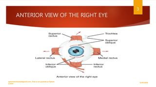
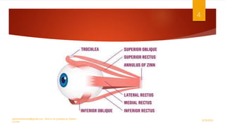
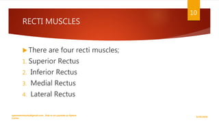
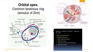
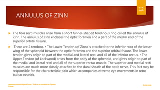
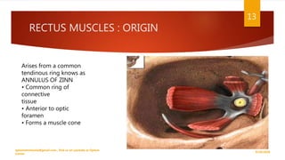
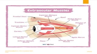
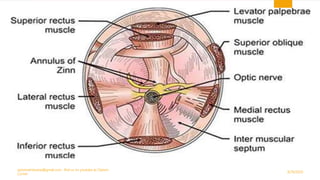
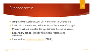
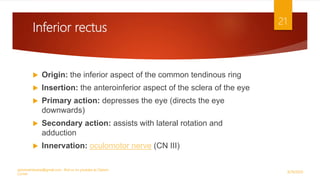
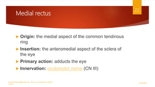
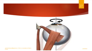
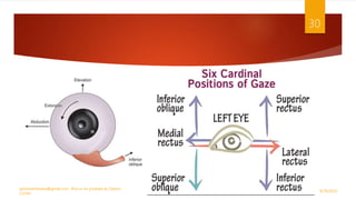
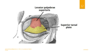
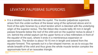
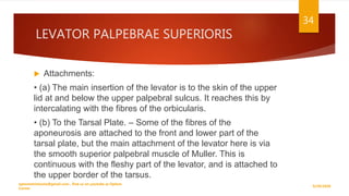
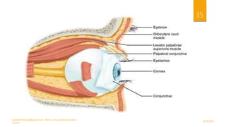
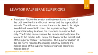
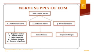
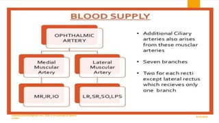
EXTRA OCULAR MUSCLES
LECTURE BY
DR. SANIA ASLAM OD
KEMU LAHORE
9/29/2020
optometristsania@gmail.com , find us on youtube as Optom
Corner
2
ANTERIOR VIEW OF THE RIGHT EYE
9/29/2020
optometristsania@gmail.com , find us on youtube as Optom
Corner
3
9/29/2020
optometristsania@gmail.com , find us on youtube as Optom
Corner
4
 The extraocular muscles (EOM) are responsible
for controlling the movements of the eyeball and
upper eyelid. These muscles are also known as the
extrinsic eye muscles, distinguishing them from
intrinsic eye muscles which are responsible for
controlling the movement of the iris. In this lecture
we will discuss the anatomy of the EOM including
their relevant attachments, innervation and actions.
9/29/2020
optometristsania@gmail.com , find us on youtube as Optom
Corner
5
 It is useful to classify the extraocular muscles into two sub-groups; muscles
that move the eye and muscles that move the upper eyelid. We discuss both
groups below.
9/29/2020
optometristsania@gmail.com , find us on youtube as Optom
Corner
6
TYPES
Functionally, they can be divided into two groups:
1. Responsible for eye movement:
 Recti Muscles
 Oblique Muscles.
2. Responsible for superior eyelid Elevation:
 Levator Palpebrae Superioris
9/29/2020
optometristsania@gmail.com , find us on youtube as Optom
Corner
7
Muscles responsible for the movement of
the eye
 This group contains six muscles;
four muscles that run almost a straight course
from origin to insertion and hence are
called recti (Latin for straight),
and two muscles that run a diagonal course,
the oblique muscles.
9/29/2020
optometristsania@gmail.com , find us on youtube as Optom
Corner
8
RECTI MUSCLES
 The name recti is derived from the latin for
‘straight’ – this represents the fact that the
recti muscles have direct path from origin to
attachment. This is in contrast with the
oblique eye muscles, which have an angular
approach to the eyeball.
9/29/2020
optometristsania@gmail.com , find us on youtube as Optom
Corner
9
RECTI MUSCLES
 There are four recti muscles;
1. Superior Rectus
2. Inferior Rectus
3. Medial Rectus
4. Lateral Rectus
9/29/2020
optometristsania@gmail.com , find us on youtube as Optom
Corner
10
9/29/2020
optometristsania@gmail.com , find us on youtube as Optom
Corner
11
ANNULUS OF ZINN
 The four recti muscles arise from a short funnel-shaped tendinous ring called the annulus of
Zinn. The annulus of Zinn encloses the optic foramen and a part of the medial end of the
superior orbital fissure.
 There are 2 tendons. • The Lower Tendon (of Zinn) is attached to the inferior root of the lesser
wing of the sphenoid between the optic foramen and the superior orbital fissure. The lower
tendon gives origin to part of the medial and lateral recti and all of the inferior rectus. • The
Upper Tendon (of Lockwood) arises from the body of the sphenoid, and gives origin to part of
the medial and lateral recti and all of the superior rectus muscle. The superior and medial recti
muscles are much more closely attached to the dural sheath of the optic nerve. This fact may be
responsible for the characteristic pain which accompanies extreme eye movements in retro-
bulbar neuritis.
9/29/2020
optometristsania@gmail.com , find us on youtube as Optom
Corner
12
RECTUS MUSCLES : ORIGIN
9/29/2020
optometristsania@gmail.com , find us on youtube as Optom
Corner
13
Arises from a common
tendinous ring knows as
ANNULUS OF ZINN
• Common ring of
connective
tissue
• Anterior to optic
foramen
• Forms a muscle cone
9/29/2020
optometristsania@gmail.com , find us on youtube as Optom
Corner
14
9/29/2020
optometristsania@gmail.com , find us on youtube as Optom
Corner
15
SPIRAL OF TILLAUX
 This is the “spiral” that describes the insertion
of the rectus muscles in relation to the
limbus. The average values are
medial=5.5mm, inferior=6.5, lateral=6.9mm,
and superior=7.7mm. In the OR, however, I
just remember “5,6,7,8.
9/29/2020
optometristsania@gmail.com , find us on youtube as Optom
Corner
16
9/29/2020
optometristsania@gmail.com , find us on youtube as Optom
Corner
17
Optom Corner
RECTI MUSCLES THICKNESS
9/29/2020
optometristsania@gmail.com , find us on youtube as Optom
Corner
18
EXTRAOCULAR MUSCLES MOTOR UNITS
 The motor units are small, with only from 5 to 18
 muscle fibers contact by each motor nerve
9/29/2020
optometristsania@gmail.com , find us on youtube as Optom
Corner
19
Superior rectus
 Origin: the superior aspect of the common tendinous ring
 Insertion: the antero-superior aspect of the sclera of the eye
 Primary action: elevates the eye (directs the eye upwards)
 Secondary action: assists with medial rotation and
adduction
 Innervation: oculomotor nerve (CN III)
9/29/2020
optometristsania@gmail.com , find us on youtube as Optom
Corner
20
Inferior rectus
 Origin: the inferior aspect of the common tendinous ring
 Insertion: the anteroinferior aspect of the sclera of the eye
 Primary action: depresses the eye (directs the eye
downwards)
 Secondary action: assists with lateral rotation and
adduction
 Innervation: oculomotor nerve (CN III)
9/29/2020
optometristsania@gmail.com , find us on youtube as Optom
Corner
21
Medial rectus
 Origin: the medial aspect of the common tendinous
ring
 Insertion: the anteromedial aspect of the sclera of
the eye
 Primary action: adducts the eye
 Innervation: oculomotor nerve (CN III)
9/29/2020
optometristsania@gmail.com , find us on youtube as Optom
Corner
22
Lateral rectus
 Origin: the lateral aspect of the common tendinous
ring
 Insertion: the anterolateral aspect of the sclera of
the eye
 Primary action: abducts the eye
 Innervation: abducens nerve (CN VI)
9/29/2020
optometristsania@gmail.com , find us on youtube as Optom
Corner
23
OBLIQUE MUSCLES
 There are two oblique muscles:
1. Superior Oblique
2. Inferior Oblique
• Unlike the recti group of muscles, they do not originate from
the common tendinous ring. • From their origin, the oblique
muscles take an angular approach to the eyeball (in contrast to
the straight approach of the recti muscles). They attach to the
posterior surface of the sclera.
9/29/2020
optometristsania@gmail.com , find us on youtube as Optom
Corner
24
SUPERIOR OBLIQUE
 It is longest and thinnest eye muscle.
 Origin – arises above and medial to the optic foramen by a narrow tendon which
partially overlaps the origin of the levator.
 Insertion – inserted to trochlea at orbital rim, on the medial wall of the antero-
superior-medial orbit on the frontal bone. The muscle stops just before the
trochlea and then proceeds as tendon under superior rectus posterior to insert on
the temporal aspect of the eye behind the equator.
 Blood supply – the superior muscular branch of ophthalmic artery supply blood
 Size – 40 mm long; tendon is 20 mm long and 10.8 mm wide.
9/29/2020
optometristsania@gmail.com , find us on youtube as Optom
Corner
25
SUPERIOR OBLIQUE
 Trochlea- The trochlea consists of a U-shaped piece of fibrocartilage.
The cartilage merges imperceptibly above with fibrous tissue, and is
attached to the fovea or spina trochlearis on the frontal bone a few
millimeters behind the orbital margin on the medial wall of the orbit.
Immediately before entering the pulley striated muscle joins the tendon,
which is enclosed in a synovial sheath, beyond which a strong fibrous
sheath accompanies the tendon to the eye.
 Innervation – The superior oblique is supplied by the 4th or trochlear
nerve which, having divided into three or four branches, enters the
muscle on the upper-surface near its lateral border; the most anterior
branch at the junction of the posterior and middle thirds, the most
posterior about 8 mm. from its origin.
9/29/2020
optometristsania@gmail.com , find us on youtube as Optom
Corner
26
9/29/2020
optometristsania@gmail.com , find us on youtube as Optom
Corner
27
INFERIOR OBLIQUE
 Origin – The inferior oblique is the only extrinsic muscle to
take origin from the front of the orbit; arises from a rounded
tendon in a depression on orbital floor near orbital rim
(maxilla), just behind the orbital margin and lateral to orifice
of the naso-lacrimal duct. Some of its fibres may arise from
the fascia covering the lacrimal sac. • Insertion – inserted
posterior inferior temporal quadrant at level of macula •
Blood supply – the inferior branch of ophthalmic artery and
infraorbital artery • Size – 37 mm long; the shortest tendon of
insertion (essentially no tendon) and it is 9.6 mm wide at
insertion.
9/29/2020
optometristsania@gmail.com , find us on youtube as Optom
Corner
28
INFERIOR OBLIQUE
 Relations – Near its origin the lower surface of the muscle contacts the
periosteum of the orbital floor, laterally it is separated from the floor by
fat. Just before the insertion of the muscle, this surface which now faces
laterally is covered by the lateral rectus and Tenon's capsule. The upper
aspect contacts fat, then the inferior rectus, then finally spreading out
and becoming concave it moulds itself on the eye. • Innervation– the
inferior division of the oculomotor nerve, crosses above the posterior
border to enter the muscle on its upper-surface at about the middle of
the muscle. • Blood-supply -comes from the infraorbital artery and the
inferior muscular branch of the ophthalmic artery.
9/29/2020
optometristsania@gmail.com , find us on youtube as Optom
Corner
29
9/29/2020
optometristsania@gmail.com , find us on youtube as Optom
Corner
30
LEVATOR PALPEBRAE SUPERIORIS
 The levator palpebrae superioris (LPS) is the only
muscle involved in raising the superior eyelid. A
small portion of this muscle contains a collection of
smooth muscle fibres – known as the superior tarsal
muscle. In contrast to the LPS, the superior tarsal
muscle is innervated by the sympathetic nervous
system.
9/29/2020
optometristsania@gmail.com , find us on youtube as Optom
Corner
31
9/29/2020
optometristsania@gmail.com , find us on youtube as Optom
Corner
32
LEVATOR PALPEBRAE SUPERIORIS
 It is a striated muscle to elevate the eyelid. The levator palpebrae superioris
arises from the under-surface of the lesser wing of the sphenoid above and in
front of the optic foramen by a short tendon which is blended with the underlying
origin of the superior rectus. The flat ribbon-like muscle belly 40 mm in length
passes forwards below the roof of the orbit and on the superior rectus to about 1
cm. behind the orbital septum (at the upper fornix or a few millimeters in front of
the equator of the eye), where it ends in a membranous expansion or
aponeurosis. The tendon is about 10-15 mm in length and extend from the
equator forward. This spreads out in a fan-shaped manner, so as to occupy the
whole breadth of the orbit and thus gives the whole muscle tendon complex the
approximate form of an isosceles triangle.
9/29/2020
optometristsania@gmail.com , find us on youtube as Optom
Corner
33
LEVATOR PALPEBRAE SUPERIORIS
 Attachments:
• (a) The main insertion of the levator is to the skin of the upper
lid at and below the upper palpebral sulcus. It reaches this by
intercalating with the fibres of the orbicularis.
• (b) To the Tarsal Plate. – Some of the fibres of the
aponeurosis are attached to the front and lower part of the
tarsal plate, but the main attachment of the levator here is via
the smooth superior palpebral muscle of Muller. This is
continuous with the fleshy part of the levator, and is attached to
the upper border of the tarsus.
9/29/2020
optometristsania@gmail.com , find us on youtube as Optom
Corner
34
9/29/2020
optometristsania@gmail.com , find us on youtube as Optom
Corner
35
LEVATOR PALPEBRAE SUPERIORIS
 Relations– Above the levator and between it and the roof of
the orbit are the 4th and frontal nerves and the supraorbital
vessels. The 4th nerve crosses the muscle close to its origin
from lateral to medial to reach the superior oblique. The
supraorbital artery is above the muscle in its anterior half
only. The frontal nerve crosses the muscle obliquely from the
lateral to the medial side. Below the levator is the medial part
of the superior rectus. • Innervation– The superior division of
the 3rd nerve reaches the muscle either by piercing the
medial edge of the superior rectus or curving around its
medial border.
9/29/2020
optometristsania@gmail.com , find us on youtube as Optom
Corner
36
NERVE SUPPLY OF EOM
9/29/2020
optometristsania@gmail.com , find us on youtube as Optom
Corner
37
Blood Supply
9/29/2020
optometristsania@gmail.com , find us on youtube as Optom
Corner
38
Ophthalmic Artery
Medial Muscular Branch:
• Inferior Rectus
• Inferior Oblique
• Medial Rectus
Lateral Muscular Branch:
• Superior Rectus
• Superior Oblique
• Lateral Rectus
• Levator Palpebrae Superioris
9/29/2020
optometristsania@gmail.com , find us on youtube as Optom
Corner
39
Mnemonic
 A useful mnemonic for remembering the nerve supply to
the extraocular muscles is:
 LR6SO4AO3
 LR = Lateral rectus: innervated by the sixth nerve
 SO = Superior oblique: innervated by the fourth nerve
 AO = All other extraocular muscles: innervated by the third
nerve
9/29/2020
optometristsania@gmail.com , find us on youtube as Optom
Corner
40
9/29/2020
optometristsania@gmail.com , find us on youtube as Optom
Corner
41
Schematic diagram of the actions of the extraocular muscles and their innervation.
Clinical relevance: cranial nerve palsy
 Damage to any of the three cranial nerves innervating the extraocular muscles can result in
paralysis of the corresponding muscles.
 Oculomotor nerve palsy (CN III)
 The oculomotor nerve supplies all extraocular muscles except the superior oblique(CNIV)
and the lateral rectus (CNVI). Oculomotor palsy (a.k.a. ‘third nerve palsy’), therefore, results in
the unopposed action of both the lateral rectus and superior oblique muscles, which pull the
eye inferolaterally. As a result, patients typically present with a ‘down and out’ appearance of
the affected eye.
 Oculomotor nerve palsy can also cause ptosis (due to a lack of innervation to levator palpebrae
superioris) as well as miosis due to the loss of parasympathetic fibres responsible for
innervating to the sphincter pupillae muscle.
9/29/2020
optometristsania@gmail.com , find us on youtube as Optom
Corner
42
Trochlear nerve palsy (CN IV)
 The only muscle the trochlear nerve innervates is the superior oblique
muscle. As a result, trochlear nerve palsy (‘fourth nerve palsy’)
typically results in vertical diplopia when looking inferiorly, due to loss
of the superior oblique’s action of pulling the eye downwards.
 Patients often try to compensate for this by tilting their head forwards
and tucking their chin in, which minimises vertical diplopia. Trochlear
nerve palsy also causes torsional diplopia (as the superior oblique
muscle assists with intorsion of the eye when the head tilts). To
compensate for this, patients with trochlear nerve palsy tilt their head
to the opposite side, in order to fuse the two images.
9/29/2020
optometristsania@gmail.com , find us on youtube as Optom
Corner
43
Abducens nerve palsy (CN VI)
 The abducens nerve (CN VI) innervates
the lateral rectus muscle. Abducens nerve
palsy (‘sixth nerve palsy’) results in unopposed
adduction of the eye (by the medial rectus
muscle), resulting in a convergent squint.
Patients typically present with horizontal diplopia
which is worsened when they attempt to look
towards the affected side.
9/29/2020
optometristsania@gmail.com , find us on youtube as Optom
Corner
44
9/29/2020
optometristsania@gmail.com , find us on youtube as Optom
Corner
45

Extra ocular muscles

  • 1.
    EXTRA OCULAR MUSCLES LECTUREBY DR. SANIA ASLAM OD KEMU LAHORE
  • 2.
    9/29/2020 optometristsania@gmail.com , findus on youtube as Optom Corner 2
  • 3.
    ANTERIOR VIEW OFTHE RIGHT EYE 9/29/2020 optometristsania@gmail.com , find us on youtube as Optom Corner 3
  • 4.
    9/29/2020 optometristsania@gmail.com , findus on youtube as Optom Corner 4
  • 5.
     The extraocularmuscles (EOM) are responsible for controlling the movements of the eyeball and upper eyelid. These muscles are also known as the extrinsic eye muscles, distinguishing them from intrinsic eye muscles which are responsible for controlling the movement of the iris. In this lecture we will discuss the anatomy of the EOM including their relevant attachments, innervation and actions. 9/29/2020 optometristsania@gmail.com , find us on youtube as Optom Corner 5
  • 6.
     It isuseful to classify the extraocular muscles into two sub-groups; muscles that move the eye and muscles that move the upper eyelid. We discuss both groups below. 9/29/2020 optometristsania@gmail.com , find us on youtube as Optom Corner 6
  • 7.
    TYPES Functionally, they canbe divided into two groups: 1. Responsible for eye movement:  Recti Muscles  Oblique Muscles. 2. Responsible for superior eyelid Elevation:  Levator Palpebrae Superioris 9/29/2020 optometristsania@gmail.com , find us on youtube as Optom Corner 7
  • 8.
    Muscles responsible forthe movement of the eye  This group contains six muscles; four muscles that run almost a straight course from origin to insertion and hence are called recti (Latin for straight), and two muscles that run a diagonal course, the oblique muscles. 9/29/2020 optometristsania@gmail.com , find us on youtube as Optom Corner 8
  • 9.
    RECTI MUSCLES  Thename recti is derived from the latin for ‘straight’ – this represents the fact that the recti muscles have direct path from origin to attachment. This is in contrast with the oblique eye muscles, which have an angular approach to the eyeball. 9/29/2020 optometristsania@gmail.com , find us on youtube as Optom Corner 9
  • 10.
    RECTI MUSCLES  Thereare four recti muscles; 1. Superior Rectus 2. Inferior Rectus 3. Medial Rectus 4. Lateral Rectus 9/29/2020 optometristsania@gmail.com , find us on youtube as Optom Corner 10
  • 11.
    9/29/2020 optometristsania@gmail.com , findus on youtube as Optom Corner 11
  • 12.
    ANNULUS OF ZINN The four recti muscles arise from a short funnel-shaped tendinous ring called the annulus of Zinn. The annulus of Zinn encloses the optic foramen and a part of the medial end of the superior orbital fissure.  There are 2 tendons. • The Lower Tendon (of Zinn) is attached to the inferior root of the lesser wing of the sphenoid between the optic foramen and the superior orbital fissure. The lower tendon gives origin to part of the medial and lateral recti and all of the inferior rectus. • The Upper Tendon (of Lockwood) arises from the body of the sphenoid, and gives origin to part of the medial and lateral recti and all of the superior rectus muscle. The superior and medial recti muscles are much more closely attached to the dural sheath of the optic nerve. This fact may be responsible for the characteristic pain which accompanies extreme eye movements in retro- bulbar neuritis. 9/29/2020 optometristsania@gmail.com , find us on youtube as Optom Corner 12
  • 13.
    RECTUS MUSCLES :ORIGIN 9/29/2020 optometristsania@gmail.com , find us on youtube as Optom Corner 13 Arises from a common tendinous ring knows as ANNULUS OF ZINN • Common ring of connective tissue • Anterior to optic foramen • Forms a muscle cone
  • 14.
    9/29/2020 optometristsania@gmail.com , findus on youtube as Optom Corner 14
  • 15.
    9/29/2020 optometristsania@gmail.com , findus on youtube as Optom Corner 15
  • 16.
    SPIRAL OF TILLAUX This is the “spiral” that describes the insertion of the rectus muscles in relation to the limbus. The average values are medial=5.5mm, inferior=6.5, lateral=6.9mm, and superior=7.7mm. In the OR, however, I just remember “5,6,7,8. 9/29/2020 optometristsania@gmail.com , find us on youtube as Optom Corner 16
  • 17.
    9/29/2020 optometristsania@gmail.com , findus on youtube as Optom Corner 17 Optom Corner
  • 18.
    RECTI MUSCLES THICKNESS 9/29/2020 optometristsania@gmail.com, find us on youtube as Optom Corner 18
  • 19.
    EXTRAOCULAR MUSCLES MOTORUNITS  The motor units are small, with only from 5 to 18  muscle fibers contact by each motor nerve 9/29/2020 optometristsania@gmail.com , find us on youtube as Optom Corner 19
  • 20.
    Superior rectus  Origin:the superior aspect of the common tendinous ring  Insertion: the antero-superior aspect of the sclera of the eye  Primary action: elevates the eye (directs the eye upwards)  Secondary action: assists with medial rotation and adduction  Innervation: oculomotor nerve (CN III) 9/29/2020 optometristsania@gmail.com , find us on youtube as Optom Corner 20
  • 21.
    Inferior rectus  Origin:the inferior aspect of the common tendinous ring  Insertion: the anteroinferior aspect of the sclera of the eye  Primary action: depresses the eye (directs the eye downwards)  Secondary action: assists with lateral rotation and adduction  Innervation: oculomotor nerve (CN III) 9/29/2020 optometristsania@gmail.com , find us on youtube as Optom Corner 21
  • 22.
    Medial rectus  Origin:the medial aspect of the common tendinous ring  Insertion: the anteromedial aspect of the sclera of the eye  Primary action: adducts the eye  Innervation: oculomotor nerve (CN III) 9/29/2020 optometristsania@gmail.com , find us on youtube as Optom Corner 22
  • 23.
    Lateral rectus  Origin:the lateral aspect of the common tendinous ring  Insertion: the anterolateral aspect of the sclera of the eye  Primary action: abducts the eye  Innervation: abducens nerve (CN VI) 9/29/2020 optometristsania@gmail.com , find us on youtube as Optom Corner 23
  • 24.
    OBLIQUE MUSCLES  Thereare two oblique muscles: 1. Superior Oblique 2. Inferior Oblique • Unlike the recti group of muscles, they do not originate from the common tendinous ring. • From their origin, the oblique muscles take an angular approach to the eyeball (in contrast to the straight approach of the recti muscles). They attach to the posterior surface of the sclera. 9/29/2020 optometristsania@gmail.com , find us on youtube as Optom Corner 24
  • 25.
    SUPERIOR OBLIQUE  Itis longest and thinnest eye muscle.  Origin – arises above and medial to the optic foramen by a narrow tendon which partially overlaps the origin of the levator.  Insertion – inserted to trochlea at orbital rim, on the medial wall of the antero- superior-medial orbit on the frontal bone. The muscle stops just before the trochlea and then proceeds as tendon under superior rectus posterior to insert on the temporal aspect of the eye behind the equator.  Blood supply – the superior muscular branch of ophthalmic artery supply blood  Size – 40 mm long; tendon is 20 mm long and 10.8 mm wide. 9/29/2020 optometristsania@gmail.com , find us on youtube as Optom Corner 25
  • 26.
    SUPERIOR OBLIQUE  Trochlea-The trochlea consists of a U-shaped piece of fibrocartilage. The cartilage merges imperceptibly above with fibrous tissue, and is attached to the fovea or spina trochlearis on the frontal bone a few millimeters behind the orbital margin on the medial wall of the orbit. Immediately before entering the pulley striated muscle joins the tendon, which is enclosed in a synovial sheath, beyond which a strong fibrous sheath accompanies the tendon to the eye.  Innervation – The superior oblique is supplied by the 4th or trochlear nerve which, having divided into three or four branches, enters the muscle on the upper-surface near its lateral border; the most anterior branch at the junction of the posterior and middle thirds, the most posterior about 8 mm. from its origin. 9/29/2020 optometristsania@gmail.com , find us on youtube as Optom Corner 26
  • 27.
    9/29/2020 optometristsania@gmail.com , findus on youtube as Optom Corner 27
  • 28.
    INFERIOR OBLIQUE  Origin– The inferior oblique is the only extrinsic muscle to take origin from the front of the orbit; arises from a rounded tendon in a depression on orbital floor near orbital rim (maxilla), just behind the orbital margin and lateral to orifice of the naso-lacrimal duct. Some of its fibres may arise from the fascia covering the lacrimal sac. • Insertion – inserted posterior inferior temporal quadrant at level of macula • Blood supply – the inferior branch of ophthalmic artery and infraorbital artery • Size – 37 mm long; the shortest tendon of insertion (essentially no tendon) and it is 9.6 mm wide at insertion. 9/29/2020 optometristsania@gmail.com , find us on youtube as Optom Corner 28
  • 29.
    INFERIOR OBLIQUE  Relations– Near its origin the lower surface of the muscle contacts the periosteum of the orbital floor, laterally it is separated from the floor by fat. Just before the insertion of the muscle, this surface which now faces laterally is covered by the lateral rectus and Tenon's capsule. The upper aspect contacts fat, then the inferior rectus, then finally spreading out and becoming concave it moulds itself on the eye. • Innervation– the inferior division of the oculomotor nerve, crosses above the posterior border to enter the muscle on its upper-surface at about the middle of the muscle. • Blood-supply -comes from the infraorbital artery and the inferior muscular branch of the ophthalmic artery. 9/29/2020 optometristsania@gmail.com , find us on youtube as Optom Corner 29
  • 30.
    9/29/2020 optometristsania@gmail.com , findus on youtube as Optom Corner 30
  • 31.
    LEVATOR PALPEBRAE SUPERIORIS The levator palpebrae superioris (LPS) is the only muscle involved in raising the superior eyelid. A small portion of this muscle contains a collection of smooth muscle fibres – known as the superior tarsal muscle. In contrast to the LPS, the superior tarsal muscle is innervated by the sympathetic nervous system. 9/29/2020 optometristsania@gmail.com , find us on youtube as Optom Corner 31
  • 32.
    9/29/2020 optometristsania@gmail.com , findus on youtube as Optom Corner 32
  • 33.
    LEVATOR PALPEBRAE SUPERIORIS It is a striated muscle to elevate the eyelid. The levator palpebrae superioris arises from the under-surface of the lesser wing of the sphenoid above and in front of the optic foramen by a short tendon which is blended with the underlying origin of the superior rectus. The flat ribbon-like muscle belly 40 mm in length passes forwards below the roof of the orbit and on the superior rectus to about 1 cm. behind the orbital septum (at the upper fornix or a few millimeters in front of the equator of the eye), where it ends in a membranous expansion or aponeurosis. The tendon is about 10-15 mm in length and extend from the equator forward. This spreads out in a fan-shaped manner, so as to occupy the whole breadth of the orbit and thus gives the whole muscle tendon complex the approximate form of an isosceles triangle. 9/29/2020 optometristsania@gmail.com , find us on youtube as Optom Corner 33
  • 34.
    LEVATOR PALPEBRAE SUPERIORIS Attachments: • (a) The main insertion of the levator is to the skin of the upper lid at and below the upper palpebral sulcus. It reaches this by intercalating with the fibres of the orbicularis. • (b) To the Tarsal Plate. – Some of the fibres of the aponeurosis are attached to the front and lower part of the tarsal plate, but the main attachment of the levator here is via the smooth superior palpebral muscle of Muller. This is continuous with the fleshy part of the levator, and is attached to the upper border of the tarsus. 9/29/2020 optometristsania@gmail.com , find us on youtube as Optom Corner 34
  • 35.
    9/29/2020 optometristsania@gmail.com , findus on youtube as Optom Corner 35
  • 36.
    LEVATOR PALPEBRAE SUPERIORIS Relations– Above the levator and between it and the roof of the orbit are the 4th and frontal nerves and the supraorbital vessels. The 4th nerve crosses the muscle close to its origin from lateral to medial to reach the superior oblique. The supraorbital artery is above the muscle in its anterior half only. The frontal nerve crosses the muscle obliquely from the lateral to the medial side. Below the levator is the medial part of the superior rectus. • Innervation– The superior division of the 3rd nerve reaches the muscle either by piercing the medial edge of the superior rectus or curving around its medial border. 9/29/2020 optometristsania@gmail.com , find us on youtube as Optom Corner 36
  • 37.
    NERVE SUPPLY OFEOM 9/29/2020 optometristsania@gmail.com , find us on youtube as Optom Corner 37
  • 38.
    Blood Supply 9/29/2020 optometristsania@gmail.com ,find us on youtube as Optom Corner 38
  • 39.
    Ophthalmic Artery Medial MuscularBranch: • Inferior Rectus • Inferior Oblique • Medial Rectus Lateral Muscular Branch: • Superior Rectus • Superior Oblique • Lateral Rectus • Levator Palpebrae Superioris 9/29/2020 optometristsania@gmail.com , find us on youtube as Optom Corner 39
  • 40.
    Mnemonic  A usefulmnemonic for remembering the nerve supply to the extraocular muscles is:  LR6SO4AO3  LR = Lateral rectus: innervated by the sixth nerve  SO = Superior oblique: innervated by the fourth nerve  AO = All other extraocular muscles: innervated by the third nerve 9/29/2020 optometristsania@gmail.com , find us on youtube as Optom Corner 40
  • 41.
    9/29/2020 optometristsania@gmail.com , findus on youtube as Optom Corner 41 Schematic diagram of the actions of the extraocular muscles and their innervation.
  • 42.
    Clinical relevance: cranialnerve palsy  Damage to any of the three cranial nerves innervating the extraocular muscles can result in paralysis of the corresponding muscles.  Oculomotor nerve palsy (CN III)  The oculomotor nerve supplies all extraocular muscles except the superior oblique(CNIV) and the lateral rectus (CNVI). Oculomotor palsy (a.k.a. ‘third nerve palsy’), therefore, results in the unopposed action of both the lateral rectus and superior oblique muscles, which pull the eye inferolaterally. As a result, patients typically present with a ‘down and out’ appearance of the affected eye.  Oculomotor nerve palsy can also cause ptosis (due to a lack of innervation to levator palpebrae superioris) as well as miosis due to the loss of parasympathetic fibres responsible for innervating to the sphincter pupillae muscle. 9/29/2020 optometristsania@gmail.com , find us on youtube as Optom Corner 42
  • 43.
    Trochlear nerve palsy(CN IV)  The only muscle the trochlear nerve innervates is the superior oblique muscle. As a result, trochlear nerve palsy (‘fourth nerve palsy’) typically results in vertical diplopia when looking inferiorly, due to loss of the superior oblique’s action of pulling the eye downwards.  Patients often try to compensate for this by tilting their head forwards and tucking their chin in, which minimises vertical diplopia. Trochlear nerve palsy also causes torsional diplopia (as the superior oblique muscle assists with intorsion of the eye when the head tilts). To compensate for this, patients with trochlear nerve palsy tilt their head to the opposite side, in order to fuse the two images. 9/29/2020 optometristsania@gmail.com , find us on youtube as Optom Corner 43
  • 44.
    Abducens nerve palsy(CN VI)  The abducens nerve (CN VI) innervates the lateral rectus muscle. Abducens nerve palsy (‘sixth nerve palsy’) results in unopposed adduction of the eye (by the medial rectus muscle), resulting in a convergent squint. Patients typically present with horizontal diplopia which is worsened when they attempt to look towards the affected side. 9/29/2020 optometristsania@gmail.com , find us on youtube as Optom Corner 44
  • 45.
    9/29/2020 optometristsania@gmail.com , findus on youtube as Optom Corner 45