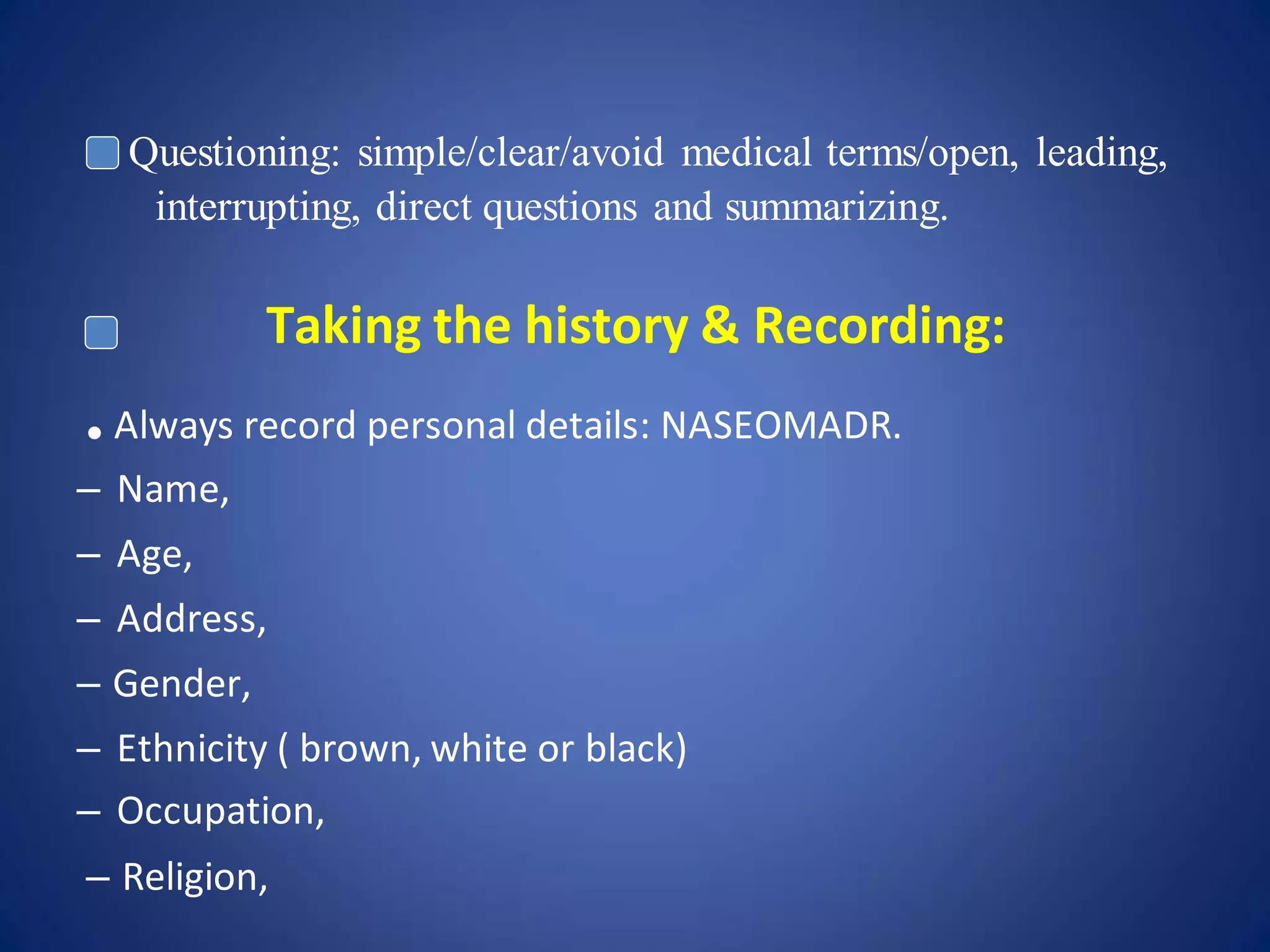
The document discusses the importance and process of taking a thorough patient history in optometry. It notes that 80% of diagnoses are based on history alone and outlines key principles like establishing rapport, maintaining privacy, and documenting clearly. It then details the specific components of a history, including demographic data, presenting complaint assessed using LOFTSEA, past ocular/medical/drug/family histories, and reviewing other body systems. The importance of summarizing, addressing patient concerns, and being available to answer questions is also covered.
This document provides guidance on how to properly conduct a history taking for patients in ophthalmology. It emphasizes the importance of establishing rapport, focusing on the key problem, and understanding how the problem impacts the patient's life. The document outlines the key areas to cover in a history, including chief complaints, history of present illness, past medical/ocular history, family history, medications, and social/allergies. It provides guidance on documenting the history thoroughly and organized using templates like SOAP notes. The goal is to obtain all relevant information to understand the problem and guide appropriate examination and management.
This document contains a sample case assessment from an uveitis outpatient department. It provides demographic information, chief complaints, ocular and medical history, examination findings, and assessment for a 51-year-old female patient presenting with recurrent redness and eye pain in the right eye for 15 days. On examination, the right eye shows conjunctival congestion, stromal edema with deep vessels in the cornea, cells in the anterior chamber, and posterior synechiae of the iris. The left eye also demonstrates similar findings but to a lesser degree.
The optometric examination involves 4 main parts: obtaining a patient history, performing a preliminary examination, conducting a refractive examination, and assessing binocular vision. The patient history is the most important first step, as it guides the exam and allows the optometrist to make a tentative diagnosis. A thorough history involves learning about the chief complaint, history of present illness, review of systems, ocular history, medical history, family history, social history, and medications.
Keratometry is a technique used to measure the curvature of the anterior surface of the cornea. It works by reflecting light off the cornea's convex surface and measuring the size of the reflected image to calculate the radius of curvature. The cornea acts as a convex mirror. Keratometry is important for assessing corneal astigmatism, estimating refractive error, monitoring conditions like keratoconus, and calculating intraocular lens power. Factors like improper calibration, positioning, focusing, or corneal irregularities can introduce errors in keratometry measurements.
Contrast sensitivity refers to the ability to see objects that have low contrasts or do not stand out clearly from their backgrounds. It is measured using charts with different spatial frequencies and contrast levels to determine the minimum contrast needed to see a target. Contrast sensitivity is affected by many eye diseases and conditions more subtly than visual acuity and can provide early detection of problems. It is tested using various charts like Pelli-Robson, Cambridge Low Contrast Gratings, and Functional Acuity Contrast Testing (FACT) that evaluate contrast sensitivity levels at different spatial frequencies.
Keratometry is a technique used to measure the radius of curvature of the anterior corneal surface using an instrument called a keratometer. The keratometer utilizes the reflective properties of the cornea to measure the size of an image formed by reflection of an object of known size and position, allowing the radius of curvature to be calculated. Commonly, keratometers either use a fixed doubling system with variable mires or a variable doubling system with fixed mires. Measurements from keratometry are used to fit contact lenses and monitor corneal changes from contact lens wear.
This document discusses pediatric refraction and various techniques used for refracting children. Pediatric refraction is different from adult refraction due to active accommodation in children. Cycloplegic refraction is preferable to paralyze accommodation. Different techniques are used based on the age of the child, including near retinoscopy, dynamic retinoscopy, and book retinoscopy. Cycloplegics help obtain an accurate refraction by paralyzing accommodation.
Real pediatric visual acuity assessmentBipin Koirala
This document discusses various methods for assessing visual acuity in pediatric patients from infants to school-aged children. It begins by outlining visual milestones in infant development and different techniques used for infants, including optokinetic nystagmus testing, preferential looking tests, Cardiff acuity testing, and visually evoked potentials. Methods for toddlers are then reviewed, such as dot visual acuity tests, coin tests, miniature toy tests, Sheridan's ball test, and Boek's candy test. The document concludes by emphasizing the importance of early visual acuity assessment and addressing challenges in pediatric assessment.
- Aphakia is the absence of the crystalline lens from the eye. It can be congenital or caused by surgery or trauma.
- In aphakia, the eye becomes highly hyperopic, the anterior focal point moves forward, and the retinal image is magnified. This decreases visual acuity and field of view.
- Aphakia is treated with spectacles, contact lenses, or intraocular lenses. Spectacles cause issues like increased image size, ring scotomas, and reduced field of view. Contact lenses and IOLs provide better image quality but have risks of complications.
Accommodation/ Accommodation of Eye, Measurement of Accommodation of Eye (hea...Bikash Sapkota
CLICK HERE TO DOWNLOAD FULL PPT ❤❤ https://healthkura.com/measurement-of-accommodation-of-eye/ ❤❤
Dear viewers Check Out my other piece of works at ❤❤❤ https://healthkura.com ❤❤❤
Measurement of Accommodation of eye:
Amplitude, Facility,
Relative Accommodation, Fatigue, Lag,
Dynamic Retinoscopy
Presentation Layout:
-Introduction to accommodation of eye
-Mechanism
-Components
-Measurement of accommodation of eye
- Amplitude
- Facility
- Relative accommodation
- Lag
-Dynamic Retinoscopy
Accommodation
-dioptric adjustment of the crystalline lens of the eye
- to obtain clear vision for a given target of regard
-process by which the refractive power of eye is altered
- to ensure a clear retinal image
For further reading
-Clinical Procedures in Optometry by J.D. Bartlett, J.B. Eskridge, J.F. Amos
-Primary Care Optometry by Theodere Grosvenor
-Borish’s Clinical Refraction by W.J. Benjamin
-Clinical Procedures for Ocular examination by Carlson et al
-American Academy of Ophthalmology
-Optometric Clinical Practice Guideline by American Optometric Association
-Internet
Follow me to get in touch with optometric and ophthalmic updates
This document discusses objective refraction techniques, primarily retinoscopy. It begins by explaining the principles of retinoscopy, including far point concept and how different ametropias affect the far point. It then describes the components and optics of the retinoscope, how it works, and retinoscopy techniques. Key aspects covered include neutralization, prerequisites for retinoscopy, and problems that can occur. Autorefractometry is also briefly discussed. In under 3 sentences:
Retinoscopy is the primary objective refraction technique discussed, which uses a retinoscope to illuminate the retina and observe the movement of the red reflex to determine the refractive error, neutralizing with trial lenses. The document covers the optics
This document contains information about conducting a low vision assessment, including sections on collecting demographic data, chief complaints, medical and ocular history, visual functioning, goals, and potential low vision devices. It also includes 4 case studies: an 89-year-old with macular degeneration who needs help reading small print, a graduate student who needs magnification for lab work, an aphakic patient with distance and near vision difficulties, and a teacher with retinitis pigmentosa. The case studies demonstrate evaluating patients' needs, calculating required optical powers, testing devices, and selecting appropriate low vision aids.
The keratometer is an instrument used to measure the curvature of the cornea, which provides information such as the radii of curvature, astigmatism level and direction, and presence of distortions. Keratometry is important for contact lens fitting, monitoring keratoconus, and determining intraocular lens power for cataract surgery. There are two main types - single-position keratometers measure two meridians simultaneously while double-position keratometers measure one meridian at a time. The procedure involves aligning and focusing the instrument before taking radius and astigmatism measurements from the scales.
The document summarizes the Amsler grid, a diagnostic tool used since 1945 to screen for and monitor macular diseases. It consists of a grid with a central dot that patients look at to detect any distortions, gaps, or blurred areas in their central vision. Various versions are available, including ones with different colors, patterns of lines, or dot sizes to test specific parts of the visual field and detect different types of visual abnormalities that could indicate conditions like macular degeneration or glaucoma. The procedure involves having patients view the grid with each eye separately at 16 inches and report any anomalies in the lines of the grid.
The LogMAR chart is designed to provide a more accurate measurement of visual acuity compared to other charts like the Snellen chart. Each line of the LogMAR chart contains the same number of letters and the letter sizes decrease logarithmically between lines, making it easy to use at different distances. The LogMAR chart is now commonly used in clinical settings and recommended for research due to its improved accuracy over other charts, especially for testing children's vision. Visual acuity is scored on the LogMAR chart by referring to the logarithm of the minimum angle of resolution, with more positive values indicating poorer vision.
This document provides information about fundus fluorescein angiography (FFA). It begins with basic principles of FFA and the dyes used, including sodium fluorescein and indocyanine green. The purpose, indications, contraindications, technique, phases, and interpretation of FFA are described. Abnormal fluorescence patterns like hyperfluorescence and hypofluorescence are discussed. Recent advances in wide-field imaging and indocyanine green angiography are also summarized.
Ultrasonography uses ultrasound to image tissues within the body. A-scan ultrasonography provides a one-dimensional view of the eye by measuring the echoes of ultrasound waves. It can be used to detect and measure tumors, assess eye structures for IOL calculation, and interpret pathology. The ultrasound is reflected at interfaces between tissues, appearing as spikes on the display. Immersion techniques provide more accurate measurements than contact techniques by avoiding compression artifacts. Limitations include artifacts, small lesions, missed foreign bodies, and misalignment issues.
The cover test is used to qualitatively measure strabismus. It involves covering each eye separately while having the patient fixate on a target. This allows the examiner to observe any movement in the uncovered eye, indicating the presence or absence of a manifest deviation. There are three main types of cover tests: direct cover test to detect manifest squint, cover-uncover test to detect heterophoria, and alternate cover test to differentiate between unilateral and alternating squint and determine if the deviation is concomitant or paralytic. The results of the cover test help diagnose the type of strabismus present.
The document provides information about the Jackson Crossed-Cylinder (JCC) technique for determining astigmatism during eye exams. It discusses the optics and proper use of the JCC. It describes the historical origins of the JCC, how it works, and the step-by-step procedure for using it to refine the axis and power of astigmatic corrections. Common sources of error are also outlined. The JCC is presented as an important tool for optometrists to accurately measure and correct astigmatism in clinical practice.
The document describes the components and uses of a trial box, which is a set of lenses, frames, and accessories used to test vision. It contains trial frames that hold spherical, cylindrical, and prismatic lenses in various diopters for refraction testing. Accessories include occluders, filters, charts, and tools like Maddox rods and cross cylinders. The trial box is used for objective and subjective refraction, diagnosing conditions like squint, and assessing binocular vision.
Frame measurements are essential for ordering prescription glasses correctly. The boxing system uses geometric center, lens size (eye size A), depth (B), and width (C) in millimeters. Distance between lenses (DBL) and geometric center distance (GCD) are also in millimeters. Temple length is overall length from center barrel to end. Frames are marked with eye size, DBL, temple length, manufacturer, and country of origin. Safety frames are marked with "Z87". Metal frames indicate gold content in karats.
what is Duochrome Test, Why do we take Red and Green color only,
What is the Principal of Duochrome Test, Why Hyperopic Pt sees green better than red and vice versa
Spherical and cylindrical lenses are the two main types of lenses. Spherical lenses have a constant curvature across all meridians, while cylindrical lenses have varying curvatures between meridians. Common spherical lens forms include plano-concave, plano-convex, and bi-convex. Tilting a lens can induce astigmatism, with the cylinder power equal to the sphere power and axis along the tilt meridian. The spherical equivalent represents the average power of a lens and is determined by combining half the cylinder power with the sphere power.
Soft Contact Lenses: Material, Fitting, and EvaluationZahra Heidari
Soft contact lenses are made from various materials like silicone and hydrogels, with advantages like comfort and easier fitting but disadvantages like potential for complications. The document discusses the history and evolution of contact lens materials, characteristics of different lens types, factors to consider for patient fitting like base curve and power selection, and how to evaluate fit and make modifications if needed. Proper patient selection and evaluation is important for successful fitting of soft contact lenses.
Soft contact lens complications can include ocular discomfort, inflammation, infection, and other issues. Ocular discomfort is the most common complication and has many potential causes, both physical and physiological. Inflammation like bulbar redness, corneal infiltrates, and contact lens peripheral ulcers can occur due to factors like tight lenses, deposits, hypoxia, or infection. Proper lens care and frequent replacement are important to prevent complications, along with addressing any underlying causes like dryness or infection. Complications generally require discontinuing lens wear until signs and symptoms resolve.
Cycloplegic refraction involves temporarily paralyzing the ciliary muscle with eye drops in order to determine a person's full refractive error. This is important for children who accommodate too much. Common cycloplegic agents include atropine, homatropine, and cyclopentolate. Cyclopentolate is often the drug of choice due to its faster onset and shorter duration. A cycloplegic refraction allows an accurate assessment of refractive error, especially in children and other patients where accommodation can affect results.
This document discusses various binocular refraction techniques including binocular balancing and binocular best sphere. It describes several methods for achieving binocular balancing such as Humphiss fogging, alternate occlusion testing, duochrome testing with fogging, prism dissociation, and Turville's infinity balance test. The goal of binocular balancing is to achieve equal accommodation between the two eyes rather than just matching visual acuity. Proper binocular balancing is important to reduce asthenopia from an imbalanced refraction.
The document provides guidance on taking an effective ophthalmic patient history. It emphasizes the importance of obtaining an accurate history, which can often provide a diagnosis. The history should include introducing oneself, chief complaint, history of present illness, past medical history, drug history, family history, and social history. Key details and tips are provided on questioning patients and documenting each component of the history.
History Taking for Health Professionals, Nurses Pooja Koirala
This document provides guidelines for taking a patient's medical history. It outlines the key components of a history, including biographical information, chief complaints, history of present illness, past medical history, family history, and review of systems. The guidelines describe how to systematically collect information on symptoms, onset, severity, treatments received, and associated factors. Proper techniques for history taking are also covered, such as establishing rapport, active listening, maintaining privacy, and using a structured format to document the patient's history in a clear and organized manner.
History taking (History of Physical Examination)pankaj rana
A History of Physical Examination Texts and the Conception of Bedside Diagnosis. ... Throughout this paper we construct a difference between a “bedside diagnosis,” made when the physician and patient are in each other's presence, and a “remote diagnosis,” made when the patient and physician are separated.
This document provides guidance on taking a patient's medical history. It discusses obtaining information from sources other than the patient if they are unable to provide their own history. It also outlines the components of a thorough history, including chief complaint, history of present illness, past medical history, allergies, medications, social history, and review of systems. The goal is to understand the nature and progression of the patient's condition.
This document provides guidance on performing a neurological history and physical examination. It emphasizes that history taking is one of the most important skills, as it can help identify and localize neurological pathology. The key aspects of history taking outlined are introducing oneself to the patient, obtaining consent, listening to the patient, and documenting the assessment clearly. The document then describes the components of a complete history, including chief complaint, history of present illness, past medical history, medications, and systems review. It also provides details on performing a neurological examination and using tools like the Mini-Mental State Examination to evaluate cognition.
This document provides guidance on performing a thorough patient history. It outlines the key components of a patient history, including chief complaint, history of present illness, past medical history, drug history, family history, and social history. The importance of obtaining an accurate history is emphasized as it allows the healthcare provider to determine the etiology of the patient's problem. Guidelines are provided on how to conduct each part of the history respectfully and obtain relevant information through active listening and open-ended questioning.
The document outlines the process and importance of history taking in medical diagnosis and care. It explains that obtaining an accurate history from the patient is the critical first step, and that a physician can make a diagnosis in 70% of cases based on the history alone. The document then describes the components of a thorough history, including the patient's profile, chief complaint, history of present illness, past medical history, family history, social history, and a systems review. An effective history takes practice and focuses on active listening, building rapport, and involving the patient to understand their experience fully.
The document provides information on the diagnosis of diseases. It discusses diagnosis methods in conventional medicine including lab investigations and radiological investigations. It defines medical diagnosis as the identification of a disease based on objective and subjective symptoms. The document also outlines different types of medical diagnoses such as clinical, laboratory, radiology diagnoses as well as differential, pre-natal, and self-diagnoses. It describes the process of taking a patient's history, including chief complaints, present illness, past medical history, and social history, which are important for making an accurate diagnosis.
This document discusses electronic medical records (EMRs) and electronic health records (EHRs). EMRs contain patient medical data entered by doctors, while EHRs also include additional information like demographics and test results. EMRs are part of EHRs and are used for registration, billing, screenings, and scheduling. EHRs provide benefits like comprehensive patient histories and improved care, but also risks like security issues, hacking vulnerabilities, and data loss. Taking an accurate patient history is important for determining the cause of illness, and involves listening carefully and asking common sense questions. History taking is both an art and a science, requiring social and medical skills to build patient confidence and direct objective principles for maximum benefit.
History taking is a critical process for physicians to obtain useful information from patients to formulate diagnoses and provide medical care. It involves asking specific questions to gain information about a patient's chief complaint, history of present illness, past medical history, family history, and systems review. An accurate history obtained through good communication skills is important, as the diagnosis can often be determined from the history alone in about 70% of cases. The history should be taken in a structured manner, with open-ended questions to allow the patient to provide their full account before asking focused questions.
The document provides guidance on taking a patient's medical history. It discusses the importance of obtaining an accurate history and outlines the general approach and structure for conducting a history, including introducing oneself, ensuring confidentiality, listening to the patient, and asking open-ended questions. It then covers how to record specific components of the history, such as the chief complaint, history of present illness, past medical history, drug history, family history, and social history.
This document outlines the process and components of taking a patient's medical history. It discusses introducing oneself to the patient, obtaining their chief complaint, history of present illness, past medical history, family history, drug history, and social history. It emphasizes listening to the patient, asking open-ended questions, avoiding medical terminology, and recording all information in the patient's own words. The goal is to accurately determine the etiology of the patient's illness based on their history.
Cardiovascular history taking is an important skill that is often assessed in bedside teaching . It’s important to have a systematic approach to ensure you don’t miss any key information. The guide below provides a framework to take a thorough cardiovascular history.
The document provides guidance on how to conduct a thorough patient history, including how to structure the history taking session and how to approach each component of the history. It discusses taking the chief complaint, history of present illness, past medical history, drug history, family history, and social history. For each component, it provides tips on what information to obtain and how to record it in the patient's own words. It also describes doing a review of all body systems to check for any associated symptoms.
Key points of obstetrics and gynaecological historyNaila Memon
This document contains a template for taking a thorough medical history. It includes sections for collecting the patient's biodata, chief complaints, history of present illness, obstetric history, gynecologic history, past medical history, family history, medications, allergies, social history, and systems review. The template provides guidance on the key information to collect under each section to fully understand the patient's history and current medical concerns.
Here are the key points to ascertain the genuine nature of a complaint:
- Verify details like duration, frequency, severity, associated symptoms
- Ask others who interact with the patient like family members
- Look for objective signs that correlate with the complaint
- Record and monitor vital parameters over time
- Consider potential confounding factors like efforts for secondary gain
This document provides guidance on taking a thorough medical history. It outlines the key components of a medical history, including identifying data, chief complaint, present illness, past medical history, family history, social history, and review of systems. The present illness section should provide a chronological account of the patient's symptoms and issues that prompted them to seek care. Gathering detailed information about symptoms, such as location, quality, timing and exacerbating/relieving factors is important for diagnosis. A comprehensive history helps health workers understand the patient's perspective and identify pertinent medical factors.
This document outlines the components and structure of taking a medical history. It discusses introducing yourself to the patient, obtaining their personal details, and asking open-ended questions about their presenting complaint. It also covers exploring the patient's medical, surgical, drug, and family histories. The document emphasizes active listening skills and tailoring your history based on the individual patient. The goal is to accurately understand the patient's condition in order to inform their management.
HISTORY AND EXAMINATION LECTURE PART 4 (1).pptxDanaiChiwara
1. The document outlines the components of a patient history, including demographics, presenting complaint, history of presenting complaint, review of systems, past medical history, drug and family histories, and physical examination.
2. Key aspects of the history of presenting complaint are to obtain a chronological account using a timeline and details of symptoms.
3. The review of systems thoroughly examines each body system for current and past issues.
4. Other important sections are past medical, surgical, social and family histories to identify relevant risk factors.
Similar to History taking in optometry or ophthalmology (20)
The document discusses the extraocular muscles (EOM) that control eye and eyelid movement. It describes the six muscles responsible for eye movement - the four rectus muscles (superior, inferior, medial, lateral) and two oblique muscles (superior, inferior). It also discusses the levator palpebrae superioris muscle responsible for raising the upper eyelid. The origins, insertions, innervations and actions of each muscle are defined.
The document discusses image formation using convex mirrors. It notes that convex mirrors always produce virtual, upright images that are located behind the mirror and are smaller than the object. As the object distance decreases, both the image distance and size increase. So as an object approaches a convex mirror, its virtual image also approaches the mirror and becomes larger. Additionally, the relationship between object and image distances is such that the distance between the image and mirror is always less than between the object and mirror for convex mirrors.
The document discusses the characteristics of images formed by a concave mirror based on the location of the object. It divides object locations into 5 cases: (1) beyond the center of curvature, (2) at the center of curvature, (3) between the center of curvature and focal point, (4) at the focal point, (5) in front of the focal point. For each case, it describes whether the image is real or virtual, upright or inverted, magnified or reduced compared to the object. Key factors that determine the image characteristics are the object's position relative to the focal point and center of curvature.
Optics is the study of light, including its interactions with matter. There are three main subfields: geometrical optics studies light as rays, physical optics studies light as waves, and quantum optics studies light as particles. Mirrors form images through the reflection of light rays according to specific rules. Plane mirrors form virtual upright images that are laterally inverted. Spherical mirrors can be concave or convex and form images using rules for tracing the path of light rays. Constructing ray diagrams involves using rays that pass through the center of curvature, focal point, or pole to locate the image point.
The document discusses various types of ocular injuries including open globe injuries, closed globe injuries, and periocular injuries. Open globe injuries involve a full thickness break of the eye wall, while closed globe injuries do not penetrate the full thickness. Periocular injuries involve structures surrounding the eye such as the eyelids. Specific injuries like corneal abrasions, hyphema, and lid lacerations are described along with their typical presentations, evaluations, and treatments. Ocular trauma requires careful examination and many cases need referral to an ophthalmologist.
The nervous system is composed of the central nervous system (CNS), which includes the brain and spinal cord, and the peripheral nervous system (PNS), which includes nerves and ganglia. The CNS is protected by meninges and cerebrospinal fluid. The brain and spinal cord contain neurons and neuroglia cells. Neurons have dendrites, a cell body, and an axon and transmit nerve impulses. Neuroglia cells provide support and protection for neurons. The PNS transmits signals between the CNS and other body systems.
The document summarizes the gross anatomy of the head and neck. It describes the bones that make up the neurocranium (skull vault) and viscerocranium (facial skeleton). Key bones include the frontal, parietal, occipital, temporal, maxilla, mandible, zygomatic and nasal bones. It notes differences in a newborn skull, such as fontanelles between unfused bone plates that close during infancy. Clinical implications of skull fractures and suture obliteration with age are also discussed.
The document provides an introduction to the muscular system. It discusses that the muscular system is composed of muscle fibers which are specialized cells that are contractile. Muscles are responsible for movement when they are attached to bones or internal organs. There are three main types of muscle - skeletal, smooth, and cardiac muscle. The document then goes on to describe the structure of skeletal muscle and provides examples of muscle groups in the head, neck, trunk, upper extremities and more.
Histology is the study of tissues and organs under the microscope. It involves using light or electron microscopes to examine the microscopic anatomy of cells, tissues, and organs. The document provides an introduction to histology, describing the parts of light and electron microscopes. It also discusses the basic tissues - epithelial, connective, muscular and nervous tissue - and explains how histology can be classified into general and systemic histology. Histological techniques like staining and the structures of cells and their organelles are also outlined.
Epidemiological trends and health care implicationsania aslam
Epidemiology is the study of patterns, causes, and effects of health and disease conditions in defined populations. It informs public health policy and evidence-based medicine by identifying risk factors and targets for prevention. Epidemiologists conduct descriptive and analytical studies to understand disease distribution and determinants. They help with study design, data analysis, and dissemination of results. The bureau of epidemiology conducts epidemiological studies to protect public health from infectious and environmental diseases. It investigates outbreaks, conducts disease surveillance, and develops prevention strategies.
How to Fix the Import Error in the Odoo 17Celine George
An import error occurs when a program fails to import a module or library, disrupting its execution. In languages like Python, this issue arises when the specified module cannot be found or accessed, hindering the program's functionality. Resolving import errors is crucial for maintaining smooth software operation and uninterrupted development processes.
This presentation was provided by Steph Pollock of The American Psychological Association’s Journals Program, and Damita Snow, of The American Society of Civil Engineers (ASCE), for the initial session of NISO's 2024 Training Series "DEIA in the Scholarly Landscape." Session One: 'Setting Expectations: a DEIA Primer,' was held June 6, 2024.
Main Java[All of the Base Concepts}.docxadhitya5119
This is part 1 of my Java Learning Journey. This Contains Custom methods, classes, constructors, packages, multithreading , try- catch block, finally block and more.
LAND USE LAND COVER AND NDVI OF MIRZAPUR DISTRICT, UPRAHUL
This Dissertation explores the particular circumstances of Mirzapur, a region located in the
core of India. Mirzapur, with its varied terrains and abundant biodiversity, offers an optimal
environment for investigating the changes in vegetation cover dynamics. Our study utilizes
advanced technologies such as GIS (Geographic Information Systems) and Remote sensing to
analyze the transformations that have taken place over the course of a decade.
The complex relationship between human activities and the environment has been the focus
of extensive research and worry. As the global community grapples with swift urbanization,
population expansion, and economic progress, the effects on natural ecosystems are becoming
more evident. A crucial element of this impact is the alteration of vegetation cover, which plays a
significant role in maintaining the ecological equilibrium of our planet.Land serves as the foundation for all human activities and provides the necessary materials for
these activities. As the most crucial natural resource, its utilization by humans results in different
'Land uses,' which are determined by both human activities and the physical characteristics of the
land.
The utilization of land is impacted by human needs and environmental factors. In countries
like India, rapid population growth and the emphasis on extensive resource exploitation can lead
to significant land degradation, adversely affecting the region's land cover.
Therefore, human intervention has significantly influenced land use patterns over many
centuries, evolving its structure over time and space. In the present era, these changes have
accelerated due to factors such as agriculture and urbanization. Information regarding land use and
cover is essential for various planning and management tasks related to the Earth's surface,
providing crucial environmental data for scientific, resource management, policy purposes, and
diverse human activities.
Accurate understanding of land use and cover is imperative for the development planning
of any area. Consequently, a wide range of professionals, including earth system scientists, land
and water managers, and urban planners, are interested in obtaining data on land use and cover
changes, conversion trends, and other related patterns. The spatial dimensions of land use and
cover support policymakers and scientists in making well-informed decisions, as alterations in
these patterns indicate shifts in economic and social conditions. Monitoring such changes with the
help of Advanced technologies like Remote Sensing and Geographic Information Systems is
crucial for coordinated efforts across different administrative levels. Advanced technologies like
Remote Sensing and Geographic Information Systems
9
Changes in vegetation cover refer to variations in the distribution, composition, and overall
structure of plant communities across different temporal and spatial scales. These changes can
occur natural.
The simplified electron and muon model, Oscillating Spacetime: The Foundation...RitikBhardwaj56
Discover the Simplified Electron and Muon Model: A New Wave-Based Approach to Understanding Particles delves into a groundbreaking theory that presents electrons and muons as rotating soliton waves within oscillating spacetime. Geared towards students, researchers, and science buffs, this book breaks down complex ideas into simple explanations. It covers topics such as electron waves, temporal dynamics, and the implications of this model on particle physics. With clear illustrations and easy-to-follow explanations, readers will gain a new outlook on the universe's fundamental nature.
How to Manage Your Lost Opportunities in Odoo 17 CRMCeline George
Odoo 17 CRM allows us to track why we lose sales opportunities with "Lost Reasons." This helps analyze our sales process and identify areas for improvement. Here's how to configure lost reasons in Odoo 17 CRM
This slide is special for master students (MIBS & MIFB) in UUM. Also useful for readers who are interested in the topic of contemporary Islamic banking.
Strategies for Effective Upskilling is a presentation by Chinwendu Peace in a Your Skill Boost Masterclass organisation by the Excellence Foundation for South Sudan on 08th and 09th June 2024 from 1 PM to 3 PM on each day.
2. • Introduction and Describing Aim &Objectives
• Chief complaint
• History of present illness
• Past medical history
• Systemic enquiry
• Family history
• Drug history
• Social history
3. Importance of History Taking
• Obtaining an accurate history is the critical first step in determining
the aetiology of a patient's problem.
• A large percentage of the time 70%, you will actually be able to
make a diagnosis based on the history alone.
4. How to take a history?
• The sense of what constitutes important data will grow exponentially
in future as you learn about the pathophysiology of disease.
• You are already in possession of the tools that will enable you to
obtain a good history.
• An ability to listen &ask common-sense questions that help define
the nature of a particular problem.
• A vast & sophisticated fund of knowledge not needed to successfully
interview a patient.
5. General Approach
Introduce yourself.
• Note – never forget patient names
• Create patient interaction appropriately in a friendly relaxed
way.
•Confidentiality and respect patient privacy.
Try to see things from patient`s point of view. Understand
patient underneath mental status, anxiety, irritation or
depression.
Always exhibit neutral position.
Listening
6. Questioning: simple/clear/avoid medical terms/open, leading,
interrupting, direct questions and summarizing.
Taking the history & Recording:
.Always record personal details: NASEOMADR.
– Name,
– Age,
– Address,
– Gender,
– Ethnicity ( brown, white or black)
– Occupation,
– Religion,
7. – Marital status.
– Date of examination
Complete History Taking
• Chief complaint
• History of present illness
• Past medical /surgical history
• Systemic review
• Family history
• Drug Allergy history
• Social history
• Present medical history.
8. CHIEF COMPLAINT
Chief Complaint
• The main reason push the pt. to seek for visiting an ophthalmic
consultation.
• Usually a single symptoms, occasionally more than one complaints
e.g. blurred vision, swelling, pain, trauma, inflammation etc.
• The patient describes the problem in their own words.
• It should be recorded in his/her own words.
9. • What brings you here? How can I help you? What seems to be the
problem?
Chief Complaint
Chief Complaint (CC):
• Short/specific in one clear sentence communicating present/major
problem/issue.
• Timing
• Recurrent
• Any major disease important e.g. DM, asthma, HT, pregnancy.
• Note: CC should be put in patient language.
10. Duration: tips
• Exact duration.
• For how long you are suffering.
• When you were completely normal.
• Is this complain for the first time or you have other episodes.
11. History of Present Illness
Details & progression, regression of the CC:
History of Present Illness - Tips
• Elaborate on the chief complaint in detail
• Ask relevant associated symptoms
• Have differential diagnosis in mind
• Lead the conversation & thoughts
• Decide & weight the importance of minor complaints
12. History of Presenting Complaint (HPC)
In details of present problem with- time of onset/ mode of
evolution/ any investigation, treatment & outcome/any associated
+’ve or -’ve symptoms.
Sequential presentation
•Always relay story in days before admission e.g. 1 week
before the admission, the patient fell while gardening&
causes ocular trauma
•Narrate in details – By that evening, the eye became swollen
and patient was unable to see. Next day patient attended
hospital and they gave him some oral and topical antibiotics.
He doesn’t know the name. There is no effect on his
13. condition and two days prior to admission, the eye continued
to swell and started to discharge and pain.
History of Presenting Complaint (HPC)
In details of symptomatic presentation
•If patient has more than one symptoms, like pain, foreign body
sensation and discharge, take each symptom individually and follow it
through fully mentioning significant negatives as well.
14. History of Present Illness - Tips
• Avoid medical terminology & make use of a descriptive language that
is familiar to them
• Ask OPQRSTA for each symptoms
15. Pain OPQRST
Onset of disease
Position/site
Quality, nature, character – burning sharp, stabbing, and crushing; also
explain depth of pain – superficial or deep.
Relationship to anything or other bodily function/position.
Radiation: where moved to
Relieving or aggravating factors – any activities or position
Severity – how it affects daily work/physical activities. Wakes him
up at night, cannot sleep/do any work.
16. Timing – mode of onset (abrupt or gradual), progression (continuous
or intermittent – if intermittent ask frequency/ nature.)
Treatment received or/and outcome.
Are there any associated symptoms?
Past Medical Illness Past Medical
/Surgical History
• Start by asking the patient if they have any medical problems
• IHD(ischemic heart disease), / Heart
Attack/DM/Asthma/HT/RHD(rheumaticheart disease),
TB/Jaundice/Fits :E.g. if diabetic- mention time of diagnosis/current
medication/clinic check up
• Past surgical/operation history
17. • E.g. time/place/ what type of operation.
• Note any blood transfusion / blood grouping.
• H/O dental extractions/circumcision & any excessive bleeding during
these procedures.
• History of trauma/accidents
• E.g. time/place/ and what type of accident
• Any minor operations or procedures including endoscopies, dental
interventions, biopsies.
Drug History
• Drug History (DH)
18. • Always use generic name or put trade name in brackets with dosage,
timing &how long.
• Example: Ranitidine 150 mg BD PO
• Note: do not forget to mention: OCT/Vitamins/Traditional /Herbal
medicine & alternative medicine as cupping or cattery or
acupuncture.
• Blood transfusion.
Drug History
• Bd (Bis die) - Twice daily (usually morning and night)
• Tds (ter die sumendus)/Tid (ter in die) = Three times a day mainly 8
hourly
19. • Qds (quarter die sumendus)/Qid (quarter in die) = four times daily
mainly 6 hourly
• Mane/(om – omni mane) = morning
• Nocte/(on – omni nocte) = night
• Ac (ante cibum) = before food
• Pc (post cibum) = after food
• Po (per orum/os) = by mouth
• Stat – statim = immediately as initial dose
• Rx (recipe) = treat with
20. Family History
• Any familial disease/running in families e.g. breast cancer, IHD
(ischemic heart disease), DM, schizophrenia, Developmental delay,
asthma, and albinism.
• Infections running in families as TB, Leprosy.
• typhoid in case of epidemics.
21. Social History
• Smoking history - amount, duration & type.
• A strong risk factor for IHD
• Alcohol history - amount, duration & type.
• Occupation, social & educational background, ADL (activity of daily
living ) , family social support& financial situation.
• Social class.
• Home conditions as:
• Water supply.
• Sanitation status in his home & surrounding.
• Animals / birds in his/her house.
22. Social History: Smoking
• The most important cause of preventable diseases.
• Smoking history - amount, duration & type.
• Amount: pack”year calculations.
• Duration: continuous or interrupted.
• Any trials of quitting & how many.
• Deep inhalation or superficial.
• Active or passive smoker.
• Type: packs, self-made, Cigars, Shesha , chewing etc.
23. Social History: Smoking
• Ask the smoker whether he is willing to quit or not.
• Do not forget to encourage the smoker to quit whenever contacting
a smoker as it is proved to increase quitting rate.
• If he is willing to quit, but cannot, help him by NRT, bupropion.
• Nicotine replacement therapy
24. Social History: Alcohol.
• Whether drinking alcohol or not.
• If drinking know whether it is healthy or not.
• Healthy alcohol use:
• Men: 14 units/week, not > 4 units/session.
• Women: 7 units/week, not > 2 units/session.
• Don’t forget that healthy alcohol use is associated with less IHD &
Ischemic CVA. Cerebrovascular accident or stroke.
• Unhealthy alcohol use is associated with cardiomyopathy, CVA,
Myopathies, liver cirrhosis & CPNS dysfunction.
25. Social History: Alcohol.
• Note:
Do not advice patients or individuals , to drink for health, because of:
• Religious & cultural reasons.
• Possibility of addiction with its known health problems.
26. Other Relevant History
• Immunization if small child
• Note: Look for the child health card.
• Travel / other history if suspected STDs or infectious disease
• Note:
• If small child, obtain the history from the care giver. Make sure; talk
to right care giver.
• If someone does not talk to your language, get an interpreter
(neutral not family friend or member also familiar with both
language). Ask simple & straight question but do not go for yes or no
answer.
28. SOAP
Subjective: how patient feels/thinks about him. How does he look.
Includes PC (post sebum-after meals) and general
appearance/condition of patient
Objective relevant points of patient complaints/vital sings, physical
examination/daily weight, fluid balance, diet/laboratory investigation
and interpretation
Assessment: address each active problem after making a problem
list. Make differential diagnosis.