SlideShare a Scribd company logo
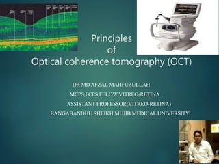
Principles
of
Optical coherence tomography (OCT)
DR MD AFZAL MAHFUZULLAH
MCPS,FCPS,FELOW VITREO-RETINA
ASSISTANT PROFESSOR(VITREO-RETINA)
BANGABANDHU SHEIKH MUJIB MEDICAL UNIVERSITY
Introduction
 OCT is noncontact noninvasive technique for imaging
biological tissues.
 In 1990, Fercher presented cross-sectional topographic
image of the retinal pigment epithelium (RPE) of a
human eye.
 The first commercial instrument, OCT 1,was launched
in 1996.
Basic Principles of Optical Coherence Tomography.
It accomplishes this by directing –
A beam of near-infrared light from a broadband coherent light source
at target tissue.
Capturing light that is back-scattered from that tissue.
Scattering is a fundamental property of a
heterogeneous medium, and occurs because of
variations in the refractive index within tissue.
4
Principle of OCT
 Interferometry is the technique
of superimposing (interfering )
two or more waves, to detect
differences between them.
 Interferometry works because
two waves with the same
frequency that have the same
phase will add each other while
two waves that have opposite
phase will subtract.
 Light from a source is
directed onto a partially
reflecting mirror and is split
into a reference and a
measurement beam.
 The measurement beam
reflected from the specimen
with different time delays
according to its internal
microstructure.
 The light in the reference beam is
reflected from a reference mirror
at a variable distance which
produces a variable time delay.
 The light from the specimen,
consisting of multiple echoes,
and the light from the reference
mirror, consisting of a single
echo at a known delay are
combined and detected.
Formation of OCT image
A-SCAN, B-SCAN, 3D-SCAN
A-scan
B-scan
Time Domain OCT
 The Michelson interferometer splits the light from the broadband
source into two paths, the reference and sample arms.
 The interference signal between the reflected reference wave and
the backscattered sample wave is then recorded.
 The axial optical sectioning ability of the technique is inversely
proportional to its optical bandwidth.
Time Domain OCT
 Transverse scanning of the sample is achieved via rotation of a
sample arm galvonometer mirror.
 In order to measure the time delays of light echoes coming
from different structures within the eye, the position of the
reference mirror is changed so that the time delay of the
reference light pulse is adjusted accordingly
Fourier domain OCT
 In FD-OCT ,the detector arm of the Michelson interferometer
uses a spectrometer instead a single detector.
 The spectrometer measures spectral modulations produced by
interference between the sample and reference reflections.
 No physical scanning of the reference mirror is required; thus,
FD-OCT can be much faster than TDOCT.
 The simultaneous detection of reflections from a broad range
of depths is much more efficient than TD-OCT, in which
signals from various depths are scanned sequentially.
 FD-OCT is also fast enough for sequential image frames to
track the pulsation of blood vessels during the cardiac cycle.
 Time domain Optical Coherence
tomography:
 Fourier domain Optical Coherence
tomography:
Time vs Fourier domain OCT
Time domain OCT
 A scan generated
sequentially, one pixel at a
time of 1.6 seconds
 Moving reference mirror
 400 scans/sec
 Resolution – 10 micron
 Slower than eye
movement
Fourier domain OCT
 Entire A scan is generated
at once based on Fourier
transformation of
spectrometer analysis
 Stationary reference
mirror
 26,000 scans/sec
 Resolution – 5 micron
 Faster than eye movement
Spectral OCT
 Limitation of OCT technology was difficulty in
accurately localizing the cross-sectional images and
correlating them with a conventional en face view of the
fundus.
 To localize and visually interpret the images, integrating
a scanning laser ophthalmoscopy (SLO) into the OCT
was needed.
 This rationale was used by OTI technologies (Toronto,
Canada) to develop the Spectral OCT/SLO.
 The Spectral OCT/SLO is a computerized optical scanner
device providing high-resolution, high-definition images
of the fundus anatomy.
 It integrats SLO’s confocal imaging principles with OCT’s
high resolution tomographic images.
 The system simultaneously produces SLO and OCT
images that are created through the same optical path,
and therefore correspond pixel to pixel.
 It produces a new image format called as C scan
 The technique has already become established as a standard
imaging modality for imaging of the eye.
 The application of OCT imaging to other biomedical areas such
as endoscopic imaging of gastro-intestinal and cardiovascular
systems is currently an active field of research.
Applications:
Oct(optical cohorence tomography)

More Related Content

What's hot

Femtosecond laser assistedcataractsurgery
Femtosecond laser assistedcataractsurgeryFemtosecond laser assistedcataractsurgery
Femtosecond laser assistedcataractsurgery
solinskyeyecare
 
Optical coherence tomography in glaucoma - Dr Shylesh Dabke
Optical coherence tomography in glaucoma - Dr Shylesh DabkeOptical coherence tomography in glaucoma - Dr Shylesh Dabke
Optical coherence tomography in glaucoma - Dr Shylesh Dabke
Shylesh Dabke
 
Refractive changes after posterior segment surgeries (Dr. Nesma, TA ophthalam...
Refractive changes after posterior segment surgeries (Dr. Nesma, TA ophthalam...Refractive changes after posterior segment surgeries (Dr. Nesma, TA ophthalam...
Refractive changes after posterior segment surgeries (Dr. Nesma, TA ophthalam...
Hind Safwat
 
Optical coherence tomography
Optical coherence tomographyOptical coherence tomography
Optical coherence tomography
Samuel Ponraj
 
Transpupillary Thermotherapy (TTT)
Transpupillary Thermotherapy (TTT)Transpupillary Thermotherapy (TTT)
Transpupillary Thermotherapy (TTT)
Pushkar Dhir
 

What's hot (20)

Femtosecond laser assistedcataractsurgery
Femtosecond laser assistedcataractsurgeryFemtosecond laser assistedcataractsurgery
Femtosecond laser assistedcataractsurgery
 
Perimetry
PerimetryPerimetry
Perimetry
 
OCT Angiography
OCT AngiographyOCT Angiography
OCT Angiography
 
IMAGING TECHNIQUES IN GLAUCOMA
IMAGING TECHNIQUES IN GLAUCOMAIMAGING TECHNIQUES IN GLAUCOMA
IMAGING TECHNIQUES IN GLAUCOMA
 
IOL power calculation special situations
IOL power calculation special situations IOL power calculation special situations
IOL power calculation special situations
 
OPTICAL COHERENCE TOMOGRAPHY ANGIOGRAPHY
OPTICAL COHERENCE TOMOGRAPHY  ANGIOGRAPHY  OPTICAL COHERENCE TOMOGRAPHY  ANGIOGRAPHY
OPTICAL COHERENCE TOMOGRAPHY ANGIOGRAPHY
 
Specular microscopy
Specular microscopySpecular microscopy
Specular microscopy
 
Optical coherence tomography in glaucoma - Dr Shylesh Dabke
Optical coherence tomography in glaucoma - Dr Shylesh DabkeOptical coherence tomography in glaucoma - Dr Shylesh Dabke
Optical coherence tomography in glaucoma - Dr Shylesh Dabke
 
HRT and GDx VCC
HRT and GDx VCCHRT and GDx VCC
HRT and GDx VCC
 
Toric iol
Toric iolToric iol
Toric iol
 
Rosek2 part1
Rosek2 part1Rosek2 part1
Rosek2 part1
 
Corneal Topography
Corneal TopographyCorneal Topography
Corneal Topography
 
Refractive changes after posterior segment surgeries (Dr. Nesma, TA ophthalam...
Refractive changes after posterior segment surgeries (Dr. Nesma, TA ophthalam...Refractive changes after posterior segment surgeries (Dr. Nesma, TA ophthalam...
Refractive changes after posterior segment surgeries (Dr. Nesma, TA ophthalam...
 
Optical coherence tomography
Optical coherence tomographyOptical coherence tomography
Optical coherence tomography
 
Transpupillary Thermotherapy (TTT)
Transpupillary Thermotherapy (TTT)Transpupillary Thermotherapy (TTT)
Transpupillary Thermotherapy (TTT)
 
Monovision
MonovisionMonovision
Monovision
 
Eccentric Fixation
Eccentric FixationEccentric Fixation
Eccentric Fixation
 
Scheimpflug imaging in ophthalmology
Scheimpflug imaging in ophthalmologyScheimpflug imaging in ophthalmology
Scheimpflug imaging in ophthalmology
 
IOL Master
IOL MasterIOL Master
IOL Master
 
Diabetic retinopathy Trials
Diabetic retinopathy TrialsDiabetic retinopathy Trials
Diabetic retinopathy Trials
 

Similar to Oct(optical cohorence tomography)

Lasers in ophthalmology
Lasers in ophthalmologyLasers in ophthalmology
Lasers in ophthalmology
Ajay Gulati
 
Guillet mottin 2005 spie_ 5964
Guillet mottin 2005 spie_ 5964Guillet mottin 2005 spie_ 5964
Guillet mottin 2005 spie_ 5964
Stéphane MOTTIN
 

Similar to Oct(optical cohorence tomography) (20)

Principles of optical coherence tomography
Principles of optical coherence tomographyPrinciples of optical coherence tomography
Principles of optical coherence tomography
 
OCT Machines
OCT Machines OCT Machines
OCT Machines
 
Oct introduction
Oct  introductionOct  introduction
Oct introduction
 
Optical coherence tomography
Optical coherence tomographyOptical coherence tomography
Optical coherence tomography
 
11
1111
11
 
Role of oct in ophthalmology
Role of oct in ophthalmologyRole of oct in ophthalmology
Role of oct in ophthalmology
 
OCT MACULA INTERPRETATION.
OCT MACULA  INTERPRETATION.OCT MACULA  INTERPRETATION.
OCT MACULA INTERPRETATION.
 
OPTICAL COHERENCE DEMYSTIFIED
OPTICAL COHERENCE DEMYSTIFIED OPTICAL COHERENCE DEMYSTIFIED
OPTICAL COHERENCE DEMYSTIFIED
 
Final oct
Final octFinal oct
Final oct
 
Lasers in ophthalmology
Lasers in ophthalmologyLasers in ophthalmology
Lasers in ophthalmology
 
Report on EYE-OCT.(OPTICAL COHERANCE TOMOGRAPHY)
Report on EYE-OCT.(OPTICAL COHERANCE TOMOGRAPHY)Report on EYE-OCT.(OPTICAL COHERANCE TOMOGRAPHY)
Report on EYE-OCT.(OPTICAL COHERANCE TOMOGRAPHY)
 
Optical Coherence Tomography(OCT) in posterior segment diseases
Optical Coherence Tomography(OCT) in posterior segment diseasesOptical Coherence Tomography(OCT) in posterior segment diseases
Optical Coherence Tomography(OCT) in posterior segment diseases
 
ASOCT NEW SLIDES.pptx
ASOCT NEW SLIDES.pptxASOCT NEW SLIDES.pptx
ASOCT NEW SLIDES.pptx
 
OCT
OCTOCT
OCT
 
Confocal microscopy by prabhat
Confocal microscopy by prabhatConfocal microscopy by prabhat
Confocal microscopy by prabhat
 
Guillet mottin 2005 spie_ 5964
Guillet mottin 2005 spie_ 5964Guillet mottin 2005 spie_ 5964
Guillet mottin 2005 spie_ 5964
 
Lec 3 Medical Equipment Endoscopes
Lec 3 Medical Equipment EndoscopesLec 3 Medical Equipment Endoscopes
Lec 3 Medical Equipment Endoscopes
 
Sd oct
Sd octSd oct
Sd oct
 
SKIN OCT
SKIN OCTSKIN OCT
SKIN OCT
 
Biomedical Optical Imaging
Biomedical Optical ImagingBiomedical Optical Imaging
Biomedical Optical Imaging
 

More from Md Afzal Mahfuzullah (12)

Hypertensive retinopthy
Hypertensive retinopthyHypertensive retinopthy
Hypertensive retinopthy
 
Orbital blood supply
Orbital blood supplyOrbital blood supply
Orbital blood supply
 
Rop Retinopathy of prematurity
Rop Retinopathy of prematurityRop Retinopathy of prematurity
Rop Retinopathy of prematurity
 
Conjunctival anatomy & and blood supply
Conjunctival anatomy & and blood supplyConjunctival anatomy & and blood supply
Conjunctival anatomy & and blood supply
 
Oct angiogram
Oct angiogramOct angiogram
Oct angiogram
 
Conjunctival anatomy
Conjunctival anatomyConjunctival anatomy
Conjunctival anatomy
 
Diabeticretinopathy30 3-2011-121109075116-phpapp01
Diabeticretinopathy30 3-2011-121109075116-phpapp01Diabeticretinopathy30 3-2011-121109075116-phpapp01
Diabeticretinopathy30 3-2011-121109075116-phpapp01
 
Anti vegf
Anti vegf Anti vegf
Anti vegf
 
FFA Dr Md Afzal Mahfuzullah
FFA Dr Md Afzal MahfuzullahFFA Dr Md Afzal Mahfuzullah
FFA Dr Md Afzal Mahfuzullah
 
Smart phone in ophthalmology
Smart phone in ophthalmologySmart phone in ophthalmology
Smart phone in ophthalmology
 
Smart phone in ophthalmology
Smart phone in ophthalmologySmart phone in ophthalmology
Smart phone in ophthalmology
 
Rubinstein taybi syndrome
Rubinstein taybi syndromeRubinstein taybi syndrome
Rubinstein taybi syndrome
 

Recently uploaded

Recently uploaded (20)

Ocular injury ppt Upendra pal optometrist upums saifai etawah
Ocular injury  ppt  Upendra pal  optometrist upums saifai etawahOcular injury  ppt  Upendra pal  optometrist upums saifai etawah
Ocular injury ppt Upendra pal optometrist upums saifai etawah
 
US E-cigarette Summit: Taming the nicotine industrial complex
US E-cigarette Summit: Taming the nicotine industrial complexUS E-cigarette Summit: Taming the nicotine industrial complex
US E-cigarette Summit: Taming the nicotine industrial complex
 
TEST BANK For Timby's Introductory Medical-Surgical Nursing, 13th American Ed...
TEST BANK For Timby's Introductory Medical-Surgical Nursing, 13th American Ed...TEST BANK For Timby's Introductory Medical-Surgical Nursing, 13th American Ed...
TEST BANK For Timby's Introductory Medical-Surgical Nursing, 13th American Ed...
 
Prix Galien International 2024 Forum Program
Prix Galien International 2024 Forum ProgramPrix Galien International 2024 Forum Program
Prix Galien International 2024 Forum Program
 
The History of Diagnostic Medical imaging
The History of Diagnostic Medical imagingThe History of Diagnostic Medical imaging
The History of Diagnostic Medical imaging
 
ANATOMY OF THE LOWER URINARY TRACT AND MALE [Autosaved] [Autosaved].pptx
ANATOMY OF THE LOWER URINARY TRACT AND MALE [Autosaved] [Autosaved].pptxANATOMY OF THE LOWER URINARY TRACT AND MALE [Autosaved] [Autosaved].pptx
ANATOMY OF THE LOWER URINARY TRACT AND MALE [Autosaved] [Autosaved].pptx
 
Compare home pulse pressure components collected directly from home
Compare home pulse pressure components collected directly from homeCompare home pulse pressure components collected directly from home
Compare home pulse pressure components collected directly from home
 
Is preeclampsia and spontaneous preterm delivery associate with vascular and ...
Is preeclampsia and spontaneous preterm delivery associate with vascular and ...Is preeclampsia and spontaneous preterm delivery associate with vascular and ...
Is preeclampsia and spontaneous preterm delivery associate with vascular and ...
 
"Central Hypertension"‚ in China: Towards the nation-wide use of SphygmoCor t...
"Central Hypertension"‚ in China: Towards the nation-wide use of SphygmoCor t..."Central Hypertension"‚ in China: Towards the nation-wide use of SphygmoCor t...
"Central Hypertension"‚ in China: Towards the nation-wide use of SphygmoCor t...
 
TEST BANK For Williams' Essentials of Nutrition and Diet Therapy, 13th Editio...
TEST BANK For Williams' Essentials of Nutrition and Diet Therapy, 13th Editio...TEST BANK For Williams' Essentials of Nutrition and Diet Therapy, 13th Editio...
TEST BANK For Williams' Essentials of Nutrition and Diet Therapy, 13th Editio...
 
TEST BANK For Wong’s Essentials of Pediatric Nursing, 11th Edition by Marilyn...
TEST BANK For Wong’s Essentials of Pediatric Nursing, 11th Edition by Marilyn...TEST BANK For Wong’s Essentials of Pediatric Nursing, 11th Edition by Marilyn...
TEST BANK For Wong’s Essentials of Pediatric Nursing, 11th Edition by Marilyn...
 
Report Back from SGO 2024: What’s the Latest in Cervical Cancer?
Report Back from SGO 2024: What’s the Latest in Cervical Cancer?Report Back from SGO 2024: What’s the Latest in Cervical Cancer?
Report Back from SGO 2024: What’s the Latest in Cervical Cancer?
 
The Normal Electrocardiogram - Part I of II
The Normal Electrocardiogram - Part I of IIThe Normal Electrocardiogram - Part I of II
The Normal Electrocardiogram - Part I of II
 
Relationship between vascular system disfunction, neurofluid flow and Alzheim...
Relationship between vascular system disfunction, neurofluid flow and Alzheim...Relationship between vascular system disfunction, neurofluid flow and Alzheim...
Relationship between vascular system disfunction, neurofluid flow and Alzheim...
 
hypertensive-disorders-of-pregnancy.pptx
hypertensive-disorders-of-pregnancy.pptxhypertensive-disorders-of-pregnancy.pptx
hypertensive-disorders-of-pregnancy.pptx
 
Why invest into infodemic management in health emergencies
Why invest into infodemic management in health emergenciesWhy invest into infodemic management in health emergencies
Why invest into infodemic management in health emergencies
 
DECIPHERING COMMON ECG FINDINGS IN ED.pptx
DECIPHERING COMMON ECG FINDINGS IN ED.pptxDECIPHERING COMMON ECG FINDINGS IN ED.pptx
DECIPHERING COMMON ECG FINDINGS IN ED.pptx
 
The hemodynamic and autonomic determinants of elevated blood pressure in obes...
The hemodynamic and autonomic determinants of elevated blood pressure in obes...The hemodynamic and autonomic determinants of elevated blood pressure in obes...
The hemodynamic and autonomic determinants of elevated blood pressure in obes...
 
Ophthalmology Clinical Tests for OSCE exam
Ophthalmology Clinical Tests for OSCE examOphthalmology Clinical Tests for OSCE exam
Ophthalmology Clinical Tests for OSCE exam
 
Anuman- An inference for helpful in diagnosis and treatment
Anuman- An inference for helpful in diagnosis and treatmentAnuman- An inference for helpful in diagnosis and treatment
Anuman- An inference for helpful in diagnosis and treatment
 

Oct(optical cohorence tomography)

  • 1. Principles of Optical coherence tomography (OCT) DR MD AFZAL MAHFUZULLAH MCPS,FCPS,FELOW VITREO-RETINA ASSISTANT PROFESSOR(VITREO-RETINA) BANGABANDHU SHEIKH MUJIB MEDICAL UNIVERSITY
  • 2. Introduction  OCT is noncontact noninvasive technique for imaging biological tissues.  In 1990, Fercher presented cross-sectional topographic image of the retinal pigment epithelium (RPE) of a human eye.  The first commercial instrument, OCT 1,was launched in 1996.
  • 3. Basic Principles of Optical Coherence Tomography. It accomplishes this by directing – A beam of near-infrared light from a broadband coherent light source at target tissue. Capturing light that is back-scattered from that tissue.
  • 4. Scattering is a fundamental property of a heterogeneous medium, and occurs because of variations in the refractive index within tissue. 4
  • 5.
  • 6.
  • 7. Principle of OCT  Interferometry is the technique of superimposing (interfering ) two or more waves, to detect differences between them.  Interferometry works because two waves with the same frequency that have the same phase will add each other while two waves that have opposite phase will subtract.
  • 8.  Light from a source is directed onto a partially reflecting mirror and is split into a reference and a measurement beam.  The measurement beam reflected from the specimen with different time delays according to its internal microstructure.
  • 9.  The light in the reference beam is reflected from a reference mirror at a variable distance which produces a variable time delay.  The light from the specimen, consisting of multiple echoes, and the light from the reference mirror, consisting of a single echo at a known delay are combined and detected.
  • 12.
  • 13. Time Domain OCT  The Michelson interferometer splits the light from the broadband source into two paths, the reference and sample arms.  The interference signal between the reflected reference wave and the backscattered sample wave is then recorded.  The axial optical sectioning ability of the technique is inversely proportional to its optical bandwidth.
  • 14. Time Domain OCT  Transverse scanning of the sample is achieved via rotation of a sample arm galvonometer mirror.  In order to measure the time delays of light echoes coming from different structures within the eye, the position of the reference mirror is changed so that the time delay of the reference light pulse is adjusted accordingly
  • 15. Fourier domain OCT  In FD-OCT ,the detector arm of the Michelson interferometer uses a spectrometer instead a single detector.  The spectrometer measures spectral modulations produced by interference between the sample and reference reflections.
  • 16.  No physical scanning of the reference mirror is required; thus, FD-OCT can be much faster than TDOCT.  The simultaneous detection of reflections from a broad range of depths is much more efficient than TD-OCT, in which signals from various depths are scanned sequentially.  FD-OCT is also fast enough for sequential image frames to track the pulsation of blood vessels during the cardiac cycle.
  • 17.  Time domain Optical Coherence tomography:  Fourier domain Optical Coherence tomography:
  • 18. Time vs Fourier domain OCT Time domain OCT  A scan generated sequentially, one pixel at a time of 1.6 seconds  Moving reference mirror  400 scans/sec  Resolution – 10 micron  Slower than eye movement Fourier domain OCT  Entire A scan is generated at once based on Fourier transformation of spectrometer analysis  Stationary reference mirror  26,000 scans/sec  Resolution – 5 micron  Faster than eye movement
  • 19. Spectral OCT  Limitation of OCT technology was difficulty in accurately localizing the cross-sectional images and correlating them with a conventional en face view of the fundus.  To localize and visually interpret the images, integrating a scanning laser ophthalmoscopy (SLO) into the OCT was needed.  This rationale was used by OTI technologies (Toronto, Canada) to develop the Spectral OCT/SLO.
  • 20.  The Spectral OCT/SLO is a computerized optical scanner device providing high-resolution, high-definition images of the fundus anatomy.  It integrats SLO’s confocal imaging principles with OCT’s high resolution tomographic images.  The system simultaneously produces SLO and OCT images that are created through the same optical path, and therefore correspond pixel to pixel.  It produces a new image format called as C scan
  • 21.
  • 22.  The technique has already become established as a standard imaging modality for imaging of the eye.  The application of OCT imaging to other biomedical areas such as endoscopic imaging of gastro-intestinal and cardiovascular systems is currently an active field of research. Applications:

Editor's Notes

  1. In a first approach towards tomographic imaging a cross-sectional topographic image of the retinal pigment epithelium (RPE) of a human eye obtained in vivo by the dual beam LCI technique was presented at the ICO-15 SAT conference by Fercher (1990) and published by Hitzenberger (1991). OCT using fibre optic Michelson LCI was pioneered by Fujimoto and co-workers (Huang et al 1991). First in vivo tomograms of the human retina were published by Fercher et al (1993a) and Swanson et al (1993). Later Chinn et al (1997) used wavelength tuning interferometry (WTI) to synthesize OCT images, whereas H¨ausler and Lindner (1998) generated OCT images using spectral interferometry. For a review of early work in LCI and OCT see the selection of key papers published by Masters (2001).
  2. Based on the principle of low-coherence interferometry where distance information concerning various ocular structures is extracted from time delays of reflected signals Light incident onto a scattering or turbid medium such as tissue is either transmitted, absorbed, or scattered. Absorbed light is converted into heat in the tissue and is effectively removed from the incident beam.
  3. Optical coherence tomography (OCT) is an imaging technique which works similar to ultrasound, simply using light waves instead of sound waves. By using the time information contained in the light waves which have been reflected from different depths inside a sample, an OCT system can reconstruct a depth-profile of the sample structure. Three-dimensional images can then be created by scanning the light beam laterally across the sample surface. Whilst the lateral resolution is determined by the spot size of the light beam, the depth (or axial) resolution depends primarily on the optical bandwidth of the light source.