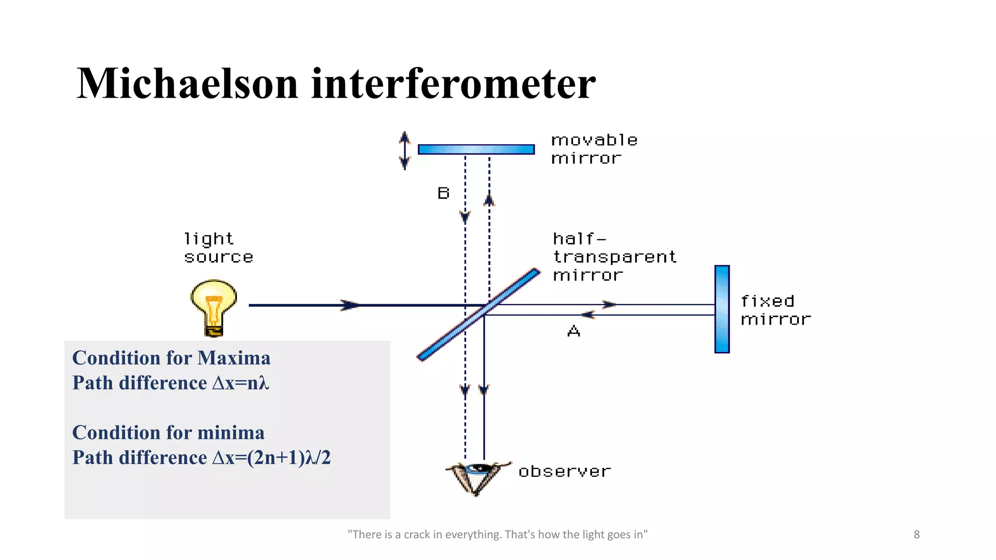
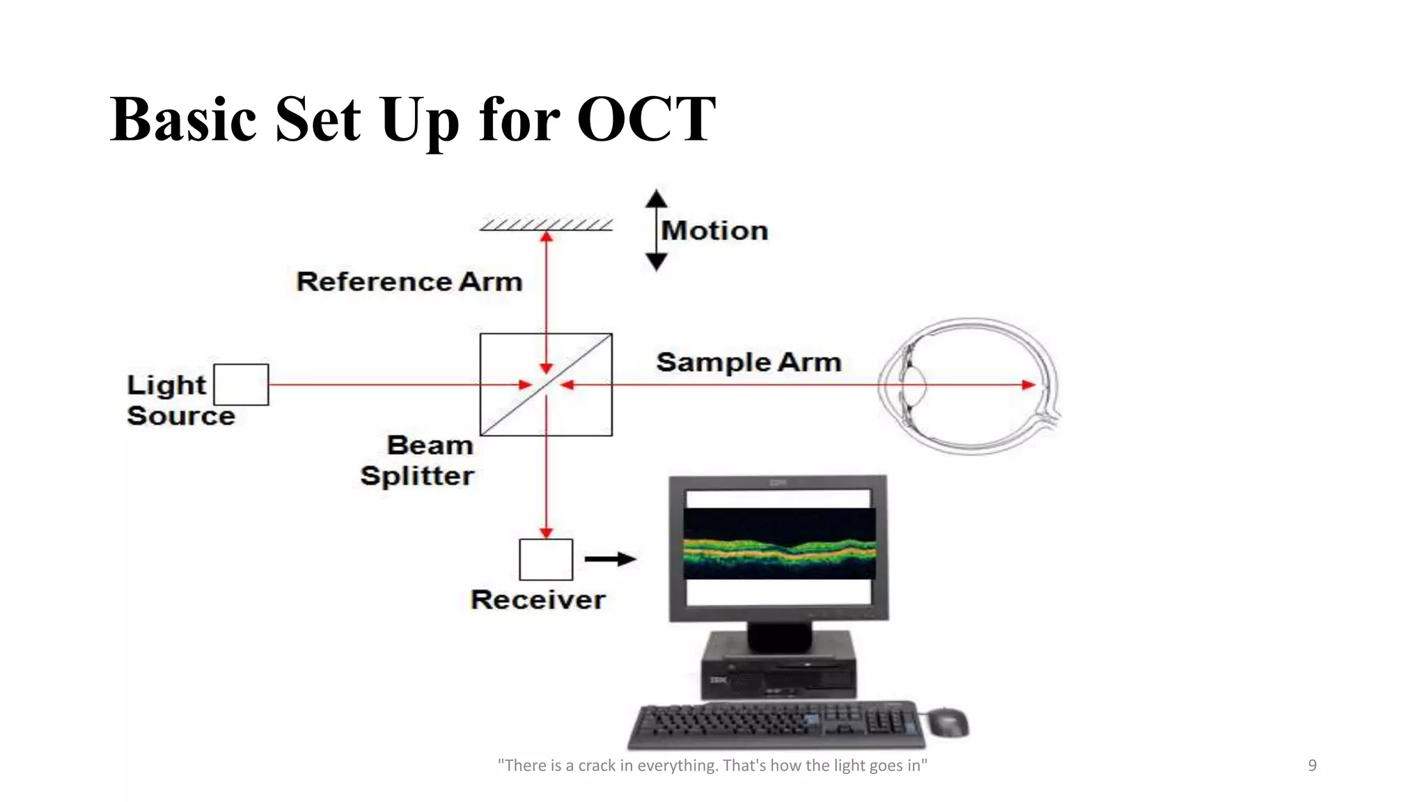
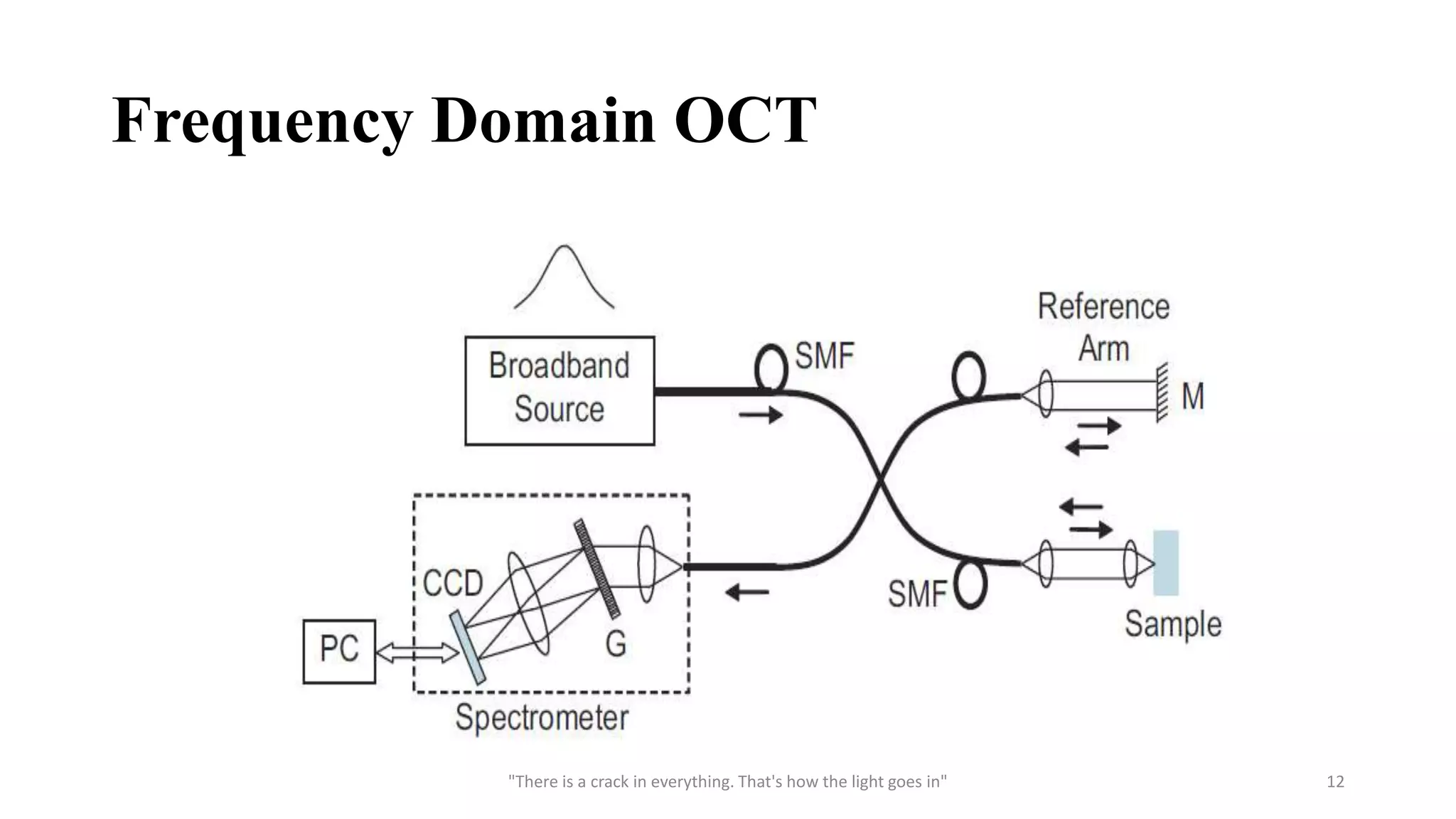
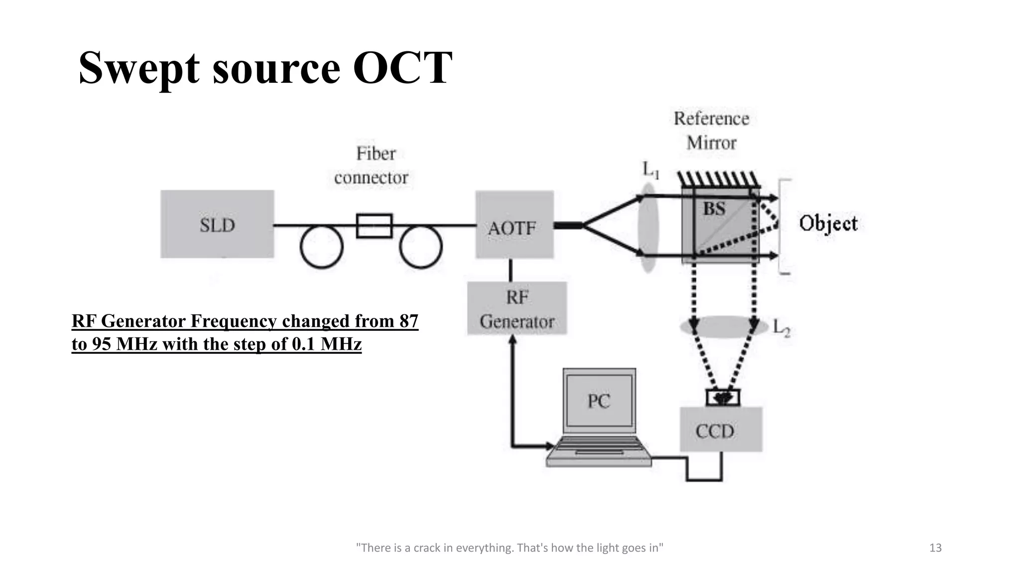
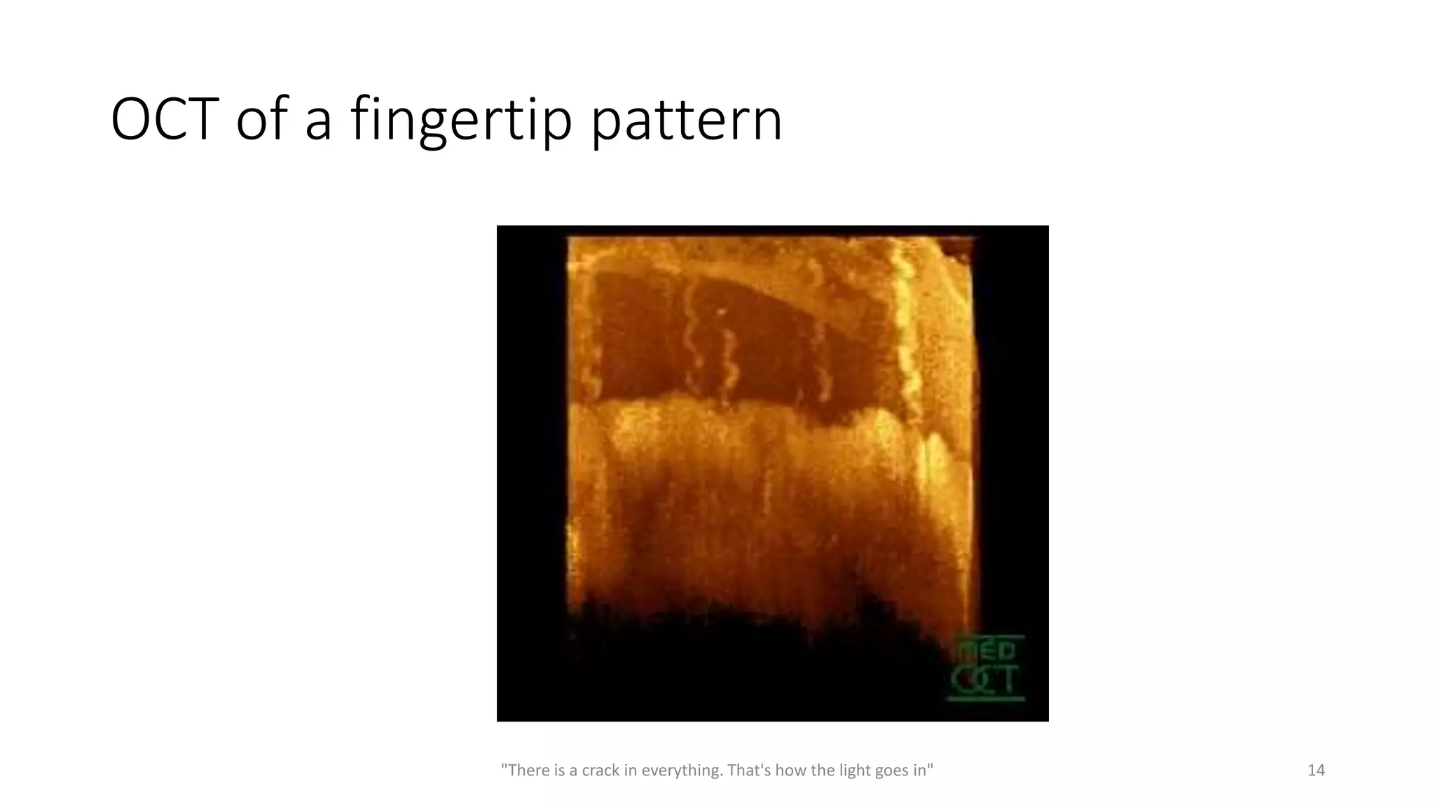
A PRESENTATION ON
OCT
(OPTICAL COHERENCE TOMOGRAPHY)
PREPARED BY
PUNEET SINGH THAKUR
IET DAVV (INDORE)
DIGITAL INSTRUMENTATION
ROLL NO.- DI15719
"There is a crack in everything. That's how the light goes in" 1
What is OCT ?
OCT basically refers to Optical coherence tomography. It is a non
contact, non destructive technique for 3D imaging of engineering and
bio medical samples.
OCT is a new type of optical imaging technology perform high
resolution cross section tomography imaging of the internal
microstructure in a material and biological system by measuring
backscattered or beck reflected light.
"There is a crack in everything. That's how the light goes in" 2
Basic Principle
• For biomedical engineering OCT use low-coherence interferometry to
produce a two or three dimensional image of optical scattering from
internal tissue microstructures.
• OCT is basically based on the interference phenomenon of light.
• For the basic setup of OCT we use “Michaelson Interferometer”.
Sample is to be analyse is placed on one of the arm of the
interferometer.
• But the source we use for the OCT is SLED, that is a Broad band
source preferred for depth scanning.
"There is a crack in everything. That's how the light goes in" 3
Component
Mirror
CCD or CMOS camera
Superluminescent Emitting Diode (SLED)
(800-1300nm, 5mW)
• The unique property of Superluminescent diodes (SLD) is the
combination of laser-diode-like output power and brightness with
broad LED-like optical spectrum. Such combination is allowed by
high optical gain in semiconductor laser materials and its wide optical
spectrum
"There is a crack in everything. That's how the light goes in" 4
Superluminescent Emitting Diode (SLED)
"There is a crack in everything. That's how the light goes in" 5
 Grating
• Any periodic perturbation is generally refers as grating. Here when we
consider Fourier domain OCT we will deal with Diffraction grating.
• Diffraction grating is an optical component with a periodic structure,
which reflect the light into several beams travelling in different
direction.
"There is a crack in everything. That's how the light goes in" 6
Beam Splitter
"There is a crack in everything. That's how the light goes in" 7
Michaelson interferometer
"There is a crack in everything. That's how the light goes in" 8
Condition for Maxima
Path difference ∆x=nλ
Condition for minima
Path difference ∆x=(2n+1)λ/2
Basic Set Up for OCT
"There is a crack in everything. That's how the light goes in" 9
Type Of OCT
1. Time Domain OCT
2. Frequency domain OCT
3. Swept Source OCT
"There is a crack in everything. That's how the light goes in" 10
Time Domain OCT
"There is a crack in everything. That's how the light goes in" 11
Frequency Domain OCT
"There is a crack in everything. That's how the light goes in" 12
Swept source OCT
"There is a crack in everything. That's how the light goes in" 13
RF Generator Frequency changed from 87
to 95 MHz with the step of 0.1 MHz
OCT of a fingertip pattern
"There is a crack in everything. That's how the light goes in" 14
Important Parameter
• Depth resolution-:
The larger the bandwidth of the source in TD-OCT and SB-OCT and the
wider the tuning bandwidth in SS-OCT, the better the depth resolution
is.
• Penetration depth-:
Due to scattering and absorption in the examined object, the number of
photons in the backscattered wave conserving stable phase relations to
the photons in the incident wave reduces with depth.
"There is a crack in everything. That's how the light goes in" 15
• Wavelength range-:
Biomedical optics requires imaging within the therapeutic window,
situated between 600 and 1000 nm, where the main constituents of the
tissue exhibit low absorption.
• Power to the object -:
In scanning regime, safety standards allow: 0.7 mW at the cornea for
imaging the retina of the human eye at 830 nm and up to 2 mW at 1060
nm ;15 mW in imaging the anterior chamber at 1300 nm and larger
power when imaging internal structures and skin.
"There is a crack in everything. That's how the light goes in" 16
Limitations
• Penetration: 2-3mm Ideal: 4mm
• Acquisition rate: <10frames/second
• Lack of large-scale clinical trials
"There is a crack in everything. That's how the light goes in" 17
Advantages
• Non contact
• Non destructive
• Non invasive
• Broad dynamic range
• No preparation of the sample or subject
• No ionizing radiation
• High resolution (1 to 15 μm)
• It can be implemented compact fiber optic components and integrated
with wide range of medical instrument.
"There is a crack in everything. That's how the light goes in" 18
APPLICATION
Meteorological Application
Defect detection of LCD
Inspection of touch screen panel
"There is a crack in everything. That's how the light goes in" 19
Biomedical application
Detection of malignancy in human cells
Eye imaging
Skin imaging
"There is a crack in everything. That's how the light goes in" 20
References
• https://www.researchgate.net/publication/37438932_Optical_Coherence_Tomogra
phy-Principles_and_Applications
• Smart optical coherence tomography for ultra-deep imaging through highly
scattering media Amaury Badon, Dayan Li, Geoffroy Lerosey, Albert C. Boccara,
Mathias Fink, and Alexandre Aubry Institut Langevin, ESPCI ParisTech, PSL
Research University,CNRS UMR 7587, 1 rue Jussieu, 75005 Paris, France (Dated:
October 19, 2015)
• http://www.sciencedirect.com/science/article/pii/S0939388909001524
• J.G. Fujimoto “Optical Coherence Tomography: Technology and Application”
IEEE 2002
• Simultaneous topography and tomography of latent fingerprints using full-field
swept-source optical coherence tomography by Satish Kumar Dubey1, Dalip
Singh Mehta1, Arun Anand2 and Chandra Shakher1, JOURNAL OF OPTICS
A: PURE AND APPLIED OPTICS , 10 (2008) 015307 (8pp)
"There is a crack in everything. That's how the light goes in" 21
THANK YOU
"There is a crack in everything. That's how the light goes in" 22

OCT

  • 1.
    A PRESENTATION ON OCT (OPTICALCOHERENCE TOMOGRAPHY) PREPARED BY PUNEET SINGH THAKUR IET DAVV (INDORE) DIGITAL INSTRUMENTATION ROLL NO.- DI15719 "There is a crack in everything. That's how the light goes in" 1
  • 2.
    What is OCT? OCT basically refers to Optical coherence tomography. It is a non contact, non destructive technique for 3D imaging of engineering and bio medical samples. OCT is a new type of optical imaging technology perform high resolution cross section tomography imaging of the internal microstructure in a material and biological system by measuring backscattered or beck reflected light. "There is a crack in everything. That's how the light goes in" 2
  • 3.
    Basic Principle • Forbiomedical engineering OCT use low-coherence interferometry to produce a two or three dimensional image of optical scattering from internal tissue microstructures. • OCT is basically based on the interference phenomenon of light. • For the basic setup of OCT we use “Michaelson Interferometer”. Sample is to be analyse is placed on one of the arm of the interferometer. • But the source we use for the OCT is SLED, that is a Broad band source preferred for depth scanning. "There is a crack in everything. That's how the light goes in" 3
  • 4.
    Component Mirror CCD or CMOScamera Superluminescent Emitting Diode (SLED) (800-1300nm, 5mW) • The unique property of Superluminescent diodes (SLD) is the combination of laser-diode-like output power and brightness with broad LED-like optical spectrum. Such combination is allowed by high optical gain in semiconductor laser materials and its wide optical spectrum "There is a crack in everything. That's how the light goes in" 4
  • 5.
    Superluminescent Emitting Diode(SLED) "There is a crack in everything. That's how the light goes in" 5
  • 6.
     Grating • Anyperiodic perturbation is generally refers as grating. Here when we consider Fourier domain OCT we will deal with Diffraction grating. • Diffraction grating is an optical component with a periodic structure, which reflect the light into several beams travelling in different direction. "There is a crack in everything. That's how the light goes in" 6
  • 7.
    Beam Splitter "There isa crack in everything. That's how the light goes in" 7
  • 8.
    Michaelson interferometer "There isa crack in everything. That's how the light goes in" 8 Condition for Maxima Path difference ∆x=nλ Condition for minima Path difference ∆x=(2n+1)λ/2
  • 9.
    Basic Set Upfor OCT "There is a crack in everything. That's how the light goes in" 9
  • 10.
    Type Of OCT 1.Time Domain OCT 2. Frequency domain OCT 3. Swept Source OCT "There is a crack in everything. That's how the light goes in" 10
  • 11.
    Time Domain OCT "Thereis a crack in everything. That's how the light goes in" 11
  • 12.
    Frequency Domain OCT "Thereis a crack in everything. That's how the light goes in" 12
  • 13.
    Swept source OCT "Thereis a crack in everything. That's how the light goes in" 13 RF Generator Frequency changed from 87 to 95 MHz with the step of 0.1 MHz
  • 14.
    OCT of afingertip pattern "There is a crack in everything. That's how the light goes in" 14
  • 15.
    Important Parameter • Depthresolution-: The larger the bandwidth of the source in TD-OCT and SB-OCT and the wider the tuning bandwidth in SS-OCT, the better the depth resolution is. • Penetration depth-: Due to scattering and absorption in the examined object, the number of photons in the backscattered wave conserving stable phase relations to the photons in the incident wave reduces with depth. "There is a crack in everything. That's how the light goes in" 15
  • 16.
    • Wavelength range-: Biomedicaloptics requires imaging within the therapeutic window, situated between 600 and 1000 nm, where the main constituents of the tissue exhibit low absorption. • Power to the object -: In scanning regime, safety standards allow: 0.7 mW at the cornea for imaging the retina of the human eye at 830 nm and up to 2 mW at 1060 nm ;15 mW in imaging the anterior chamber at 1300 nm and larger power when imaging internal structures and skin. "There is a crack in everything. That's how the light goes in" 16
  • 17.
    Limitations • Penetration: 2-3mmIdeal: 4mm • Acquisition rate: <10frames/second • Lack of large-scale clinical trials "There is a crack in everything. That's how the light goes in" 17
  • 18.
    Advantages • Non contact •Non destructive • Non invasive • Broad dynamic range • No preparation of the sample or subject • No ionizing radiation • High resolution (1 to 15 μm) • It can be implemented compact fiber optic components and integrated with wide range of medical instrument. "There is a crack in everything. That's how the light goes in" 18
  • 19.
    APPLICATION Meteorological Application Defect detectionof LCD Inspection of touch screen panel "There is a crack in everything. That's how the light goes in" 19
  • 20.
    Biomedical application Detection ofmalignancy in human cells Eye imaging Skin imaging "There is a crack in everything. That's how the light goes in" 20
  • 21.
    References • https://www.researchgate.net/publication/37438932_Optical_Coherence_Tomogra phy-Principles_and_Applications • Smartoptical coherence tomography for ultra-deep imaging through highly scattering media Amaury Badon, Dayan Li, Geoffroy Lerosey, Albert C. Boccara, Mathias Fink, and Alexandre Aubry Institut Langevin, ESPCI ParisTech, PSL Research University,CNRS UMR 7587, 1 rue Jussieu, 75005 Paris, France (Dated: October 19, 2015) • http://www.sciencedirect.com/science/article/pii/S0939388909001524 • J.G. Fujimoto “Optical Coherence Tomography: Technology and Application” IEEE 2002 • Simultaneous topography and tomography of latent fingerprints using full-field swept-source optical coherence tomography by Satish Kumar Dubey1, Dalip Singh Mehta1, Arun Anand2 and Chandra Shakher1, JOURNAL OF OPTICS A: PURE AND APPLIED OPTICS , 10 (2008) 015307 (8pp) "There is a crack in everything. That's how the light goes in" 21
  • 22.
    THANK YOU "There isa crack in everything. That's how the light goes in" 22