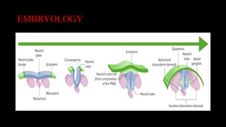
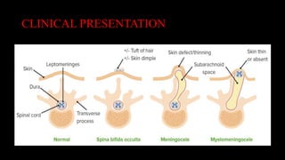
TOPIC: NEURAL TUBE
DEFECTS
PREPARED BY: EMMANUEL R. OLA
MEDICAL STUDENT YEAR – 5
NEUROSURGICAL UNIT – JFK MEMORIAL MEDICAL CENTER
CONTENTS
•Definition
•Epidemiology
•Classification
•Embryology
•Clinical Presentation
•Diagnosis
•Management
DEFINITION
•A neural tube defect, also known as spinal
dysraphism, is a congenital condition where the
covering of the central nervous system does not
fully develop due to a failure of the neural tube
to close during the 3rd or 4th week of embryonic
development.
EPIDEMIOLOGY
• 1 in 1,500 births in the United States
• 2nd-most common major congenital anomalies (most
common are cardiac anomalies)
• Incidence
• Has decreased with prenatal folic acid
supplementation and screening
• Varies by ethnicity and geographic region
• Female neonates are affected more commonly.
EPIDEMIOLOGY
• Maternal risk factors during pregnancy:
• Folate deficiency
• History of NTD in previous children (5%–10%
recurrence with 1–2 affected siblings)
• Folic acid-depleting medications (valproic acid,
triamterene, trimethoprim, sulfasalazine)
• Poorly controlled maternal diabetes
• Maternal obesity
CLASSIFICATION
• Open NTD (80% of cases): a midline defect of the
vertebral bodies with different degrees of protrusion of
the meninges and central nervous system (CNS)
• Defect along the spinal cord:
•Meningocele: Only the meninges protrude.
•Meningomyelocele: Both meninges and spinal
cord protrude (most common NTD).
CLASSIFICATION
Defect of the cranium:
•Cranial meningocele: Meninges protrude.
•Cranial encephalocele: Both the meninges
and brainstem/cerebellum/cerebral cortex
protrude.
•Anencephaly: complete failure of the
cephalic neural tube to close resulting in a fully
exposed fetal brain (not compatible with life)
CLASSIFICATION
• Closed NTD: a midline defect of the vertebral bodies
without protrusion of the meninges or neural tissue
• Without subcutaneous mass: spina bifida occulta
• With subcutaneous mass:
•Lipomyelomeningocele
•Lipomeningocele
EMBRYOLOGY
• Conception to 3rd week of gestation (gastrulation):
• Single cell → single-layered blastula → 3-layer
gastrula: ectoderm, endoderm, and mesoderm
• 3rd–4th week:
• Ectoderm → differentiates into neuroectoderm (creating neural
plate) → cell replication in the neural plate → neural crests +
neural fold
• Mesoderm → differentiates into notochord → signals neural
fold to enlarge/fuse → neural tube (neurulation)
• Neural tube“zips up” from the middle outward → Cranial (head)
and caudal (tail) openings close approximately on days 24 and 28
EMBRYOLOGY
CLINICAL PRESENTATION
• Most NTDs are discovered during prenatal screening
• Open NTDs are evident at birth, but closed NTDs may have more subtle presentations.
Name: Myelomeningocele
Description: Spinal cord and meningeal herniation
Clinical Features: The higher the lesion in the spine, the more severe the symptoms:
Brainstem: type 2 Chiari malformation (cerebellar tonsillar herniation) with hydrocephalus
• Neck: quadriplegia
• Lumbosacral defect:
• Paraplegia/flaccid paralysis
• Loss of touch and pain sensations in lower limbs
• Anesthesia in perianal area
• Bowel + bladder incontinence
CLINICAL PRESENTATION
• Name: Meningocele
• Description: Meningeal herniation without spinal cord
involvement
• Clinical Features: Fluctuant mass, Transilluminates, Covered
by skin
• Name: Encephalocele
• Description: Meninges ± brain tissue protruding from cranial
defect
• Clinical Features: Hydrocephalus, Spasticity, Paralysis,
Seizures, Microcephaly. Developmental delay, Vision
problems, Developmental and growth retardation
CLINICAL PRESENTATION
• Name: Anencephaly
• Description: Major component of the brain and skull is absent.
• Clinical Features: Stillbirth/early death, Spastic with
deafness/blindness
• Name: Spina bifida occulta
• Description: Midline vertebral body fusion defect without protruding
dura or neural tissue
• Clinical Features: Usually asymptomatic, If the spinal cord is involved
(tethered cord syndrome), may develop incontinence, constipation, or
ataxia May be accompanied by abnormal overlying skin with a
hemangioma, discoloration, pit, lump, dermal sinus, or hairy patch
CLINICAL PRESENTATION
DIAGNOSIS
• Prenatal screening
• Serum/amniotic α-fetoprotein (maternal serum α-
fetoprotein; MSAFP): high levels at 15–20 weeks of
gestation suggestive of NTD, but not diagnostic
• Ultrasound: Visualization of defect is 98% specific and
can be used to confirm the diagnosis (closed NTD may
not be detected).
DIAGNOSIS
• Postnatal imaging
• Open NTD: ultrasound is commonly used; magnetic resonance
imaging (MRI) can be used if further detail required
• Closed NTD: ultrasound indicated with following cutaneous signs:
• Palpable subcutaneous mass, Hairy patch, overlying spine, Dermal
sinus, Dimples > 5 mm deep or > 25 mm from the anal verge
• Skin tags or tail-like appendages
• Hyperpigmented patches or deviation of the gluteal cleft
MANAGEMENT
• Prenatal
• Prevention with folic acid (vitamin B9) supplementation:
• Decreases the incidence of NTD by around 70%
• Preconception dose: 0.4 mg/day of folic acid for at least 1 month
prior to conception and throughout pregnancy
• A higher dose (usually 4 mg/day) is indicated for the following: A
previous pregnancy affected by an NTD, A positive family history
of NTD, Use of folic acid-depleting medications
• Pre-gestational diabetes
• Delivery: cesarean section recommended
MANAGEMENT
• Postnatal
• Closed NTD:
• May require no intervention
• Ongoing follow-up for lower neurological symptoms
(incontinence, constipation difficulty ambulating)
• Open NTD:
• Keep warm and in the prone position.
• Cover NTD with a sterile wet dressing.
• Surgical management:
• Neurosurgical intervention for all open NTDs
• Closed NTD typically does not require surgery
REFERENCES
1.Boon, R. L. (2010). Textbooks of Paediatrics.
2.Winn, H. R. (2011). Youmans Neurological Surgery E-Book
3.Goetzl, L.M. Folic acid supplementation in pregnancy.
UpToDate. Retrieved Nov 2, 2020
4.Dukhovny, S., Wilkins-Haug, L. Open neural tube defects:
Risk factors, prenatal screening and diagnosis, and pregnancy
management. UpToDate. Retrieved Nov 2, 2020
THE END
GALATOMA

NEURAL TUBE DEFECTs.pptx

  • 1.
    TOPIC: NEURAL TUBE DEFECTS PREPAREDBY: EMMANUEL R. OLA MEDICAL STUDENT YEAR – 5 NEUROSURGICAL UNIT – JFK MEMORIAL MEDICAL CENTER
  • 2.
  • 3.
    DEFINITION •A neural tubedefect, also known as spinal dysraphism, is a congenital condition where the covering of the central nervous system does not fully develop due to a failure of the neural tube to close during the 3rd or 4th week of embryonic development.
  • 4.
    EPIDEMIOLOGY • 1 in1,500 births in the United States • 2nd-most common major congenital anomalies (most common are cardiac anomalies) • Incidence • Has decreased with prenatal folic acid supplementation and screening • Varies by ethnicity and geographic region • Female neonates are affected more commonly.
  • 5.
    EPIDEMIOLOGY • Maternal riskfactors during pregnancy: • Folate deficiency • History of NTD in previous children (5%–10% recurrence with 1–2 affected siblings) • Folic acid-depleting medications (valproic acid, triamterene, trimethoprim, sulfasalazine) • Poorly controlled maternal diabetes • Maternal obesity
  • 6.
    CLASSIFICATION • Open NTD(80% of cases): a midline defect of the vertebral bodies with different degrees of protrusion of the meninges and central nervous system (CNS) • Defect along the spinal cord: •Meningocele: Only the meninges protrude. •Meningomyelocele: Both meninges and spinal cord protrude (most common NTD).
  • 7.
    CLASSIFICATION Defect of thecranium: •Cranial meningocele: Meninges protrude. •Cranial encephalocele: Both the meninges and brainstem/cerebellum/cerebral cortex protrude. •Anencephaly: complete failure of the cephalic neural tube to close resulting in a fully exposed fetal brain (not compatible with life)
  • 8.
    CLASSIFICATION • Closed NTD:a midline defect of the vertebral bodies without protrusion of the meninges or neural tissue • Without subcutaneous mass: spina bifida occulta • With subcutaneous mass: •Lipomyelomeningocele •Lipomeningocele
  • 9.
    EMBRYOLOGY • Conception to3rd week of gestation (gastrulation): • Single cell → single-layered blastula → 3-layer gastrula: ectoderm, endoderm, and mesoderm • 3rd–4th week: • Ectoderm → differentiates into neuroectoderm (creating neural plate) → cell replication in the neural plate → neural crests + neural fold • Mesoderm → differentiates into notochord → signals neural fold to enlarge/fuse → neural tube (neurulation) • Neural tube“zips up” from the middle outward → Cranial (head) and caudal (tail) openings close approximately on days 24 and 28
  • 10.
  • 11.
    CLINICAL PRESENTATION • MostNTDs are discovered during prenatal screening • Open NTDs are evident at birth, but closed NTDs may have more subtle presentations. Name: Myelomeningocele Description: Spinal cord and meningeal herniation Clinical Features: The higher the lesion in the spine, the more severe the symptoms: Brainstem: type 2 Chiari malformation (cerebellar tonsillar herniation) with hydrocephalus • Neck: quadriplegia • Lumbosacral defect: • Paraplegia/flaccid paralysis • Loss of touch and pain sensations in lower limbs • Anesthesia in perianal area • Bowel + bladder incontinence
  • 12.
    CLINICAL PRESENTATION • Name:Meningocele • Description: Meningeal herniation without spinal cord involvement • Clinical Features: Fluctuant mass, Transilluminates, Covered by skin • Name: Encephalocele • Description: Meninges ± brain tissue protruding from cranial defect • Clinical Features: Hydrocephalus, Spasticity, Paralysis, Seizures, Microcephaly. Developmental delay, Vision problems, Developmental and growth retardation
  • 13.
    CLINICAL PRESENTATION • Name:Anencephaly • Description: Major component of the brain and skull is absent. • Clinical Features: Stillbirth/early death, Spastic with deafness/blindness • Name: Spina bifida occulta • Description: Midline vertebral body fusion defect without protruding dura or neural tissue • Clinical Features: Usually asymptomatic, If the spinal cord is involved (tethered cord syndrome), may develop incontinence, constipation, or ataxia May be accompanied by abnormal overlying skin with a hemangioma, discoloration, pit, lump, dermal sinus, or hairy patch
  • 14.
  • 15.
    DIAGNOSIS • Prenatal screening •Serum/amniotic α-fetoprotein (maternal serum α- fetoprotein; MSAFP): high levels at 15–20 weeks of gestation suggestive of NTD, but not diagnostic • Ultrasound: Visualization of defect is 98% specific and can be used to confirm the diagnosis (closed NTD may not be detected).
  • 16.
    DIAGNOSIS • Postnatal imaging •Open NTD: ultrasound is commonly used; magnetic resonance imaging (MRI) can be used if further detail required • Closed NTD: ultrasound indicated with following cutaneous signs: • Palpable subcutaneous mass, Hairy patch, overlying spine, Dermal sinus, Dimples > 5 mm deep or > 25 mm from the anal verge • Skin tags or tail-like appendages • Hyperpigmented patches or deviation of the gluteal cleft
  • 17.
    MANAGEMENT • Prenatal • Preventionwith folic acid (vitamin B9) supplementation: • Decreases the incidence of NTD by around 70% • Preconception dose: 0.4 mg/day of folic acid for at least 1 month prior to conception and throughout pregnancy • A higher dose (usually 4 mg/day) is indicated for the following: A previous pregnancy affected by an NTD, A positive family history of NTD, Use of folic acid-depleting medications • Pre-gestational diabetes • Delivery: cesarean section recommended
  • 18.
    MANAGEMENT • Postnatal • ClosedNTD: • May require no intervention • Ongoing follow-up for lower neurological symptoms (incontinence, constipation difficulty ambulating) • Open NTD: • Keep warm and in the prone position. • Cover NTD with a sterile wet dressing. • Surgical management: • Neurosurgical intervention for all open NTDs • Closed NTD typically does not require surgery
  • 19.
    REFERENCES 1.Boon, R. L.(2010). Textbooks of Paediatrics. 2.Winn, H. R. (2011). Youmans Neurological Surgery E-Book 3.Goetzl, L.M. Folic acid supplementation in pregnancy. UpToDate. Retrieved Nov 2, 2020 4.Dukhovny, S., Wilkins-Haug, L. Open neural tube defects: Risk factors, prenatal screening and diagnosis, and pregnancy management. UpToDate. Retrieved Nov 2, 2020
  • 20.