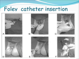
Group A  16th Batch Faculty Of Medicine USJP. Group A,16th Batch,FMS,USJP.
Group A,16th Batch,FMS,USJP.
Labour Process by which regular  contractions bring about effacement and dilatation of the cervix and descent of the presenting part ultimately leading to expulsion of the foetus and the placenta from the mother  Group A,16th Batch,FMS,USJP.
Group A,16th Batch,FMS,USJP.
The  Pelvis The  Pelvic  Brim  or  Inlet Group A,16th Batch,FMS,USJP.
The  Pelvic  Brim Group A,16th Batch,FMS,USJP.
Sagittal  Section  of  Pelvis  Demonstrating  Anterior-Posterior  Diameters  of  Inlet  &  Outlet Group A,16th Batch,FMS,USJP.
The  Pelvic  Outlet Group A,16th Batch,FMS,USJP.
Musculofascial  Gutter  of  Levator  Sling Group A,16th Batch,FMS,USJP.
The  Perineum , Perineal  Body  &  Pelvic  Floor  from  below ; superficial  view  Group A,16th Batch,FMS,USJP.
Deeper  view Group A,16th Batch,FMS,USJP.
Group A,16th Batch,FMS,USJP.
Anatomy of the fetal skull Vault  mainly consists of 2 parietal bones, parts of occipital, frontal and temporal bones. Bones are joined to each other by soft  unossified membranes, known as sutures.  Group A,16th Batch,FMS,USJP. skull vault face base
Superior and lateral view of fetal skull Face and base  of the skull are firmly united. Group A,16th Batch,FMS,USJP. 1. Coronal Suture 2. Anterior Fontanelle 3. Anterolateral Fontanelle 4. Squamosal Suture 5. Posterolateral Fontanelle 6. Lambdoidal Suture 7. External Acoustic Meatus 8. Sagittal Suture
ANTERIOR FONTANELLE Diamond shape Junction of sagittal frontal and coronal sutures POSTERIOR FONTANELLE Triangular shape Junction between sagittal and lambdoid sutures Fontanelles Junctions between  sutures are known as Fontanelles   Group A,16th Batch,FMS,USJP.
Importance of Fontanelles Fontanelles effectively reduce the diameter of the fetal skull during labour without harming the underlying brain. This is known as moulding. Group A,16th Batch,FMS,USJP.
Lateral and posterior view of moulding of the foetal skull Group A,16th Batch,FMS,USJP.
The diameters of the skull The foetal head is ovoid in shape. The attitude of the fetal head refers to the degree of flexion and extension at the upper cervical spine.  Group A,16th Batch,FMS,USJP.
Exact physiological mechanism of initiation of parturition is still unknown. But there are some processes that are of particular importance. Group A,16th Batch,FMS,USJP.
What happens prior to initiation of labour? That means last 4-5 weeks of gestation. A) Cervical ripening. Here the collagen concentrations of the cervix will be reduced. The collagen lysis by collagenases will be increased with the increment of water content. Collagenase activity is enhanced by relaxin hormone. And also the changes in ground substances, like proteoglycans occur. Group A,16th Batch,FMS,USJP.
Cervix becomes soft and decreases the resistance to dilatation.  That helps in cervical effacement and dilatation. B) Myometrial excitement . Here the irregular uterine contractions are initiated and the myometrial excitement will be enhanced. Group A,16th Batch,FMS,USJP.
Onset of labour. What should happen? Uterine contractions. Cervical dilatation. So there are 3 main mechanisms that occur at the onset of labour as;  1)  Increase prostaglandin synthesis in  decidua and foetal membrane.  11) Sensitize myometrium to Oxytocin action.  111) Activation of hypothalamo-pituitary axis  of foetus. Group A,16th Batch,FMS,USJP.
So both mother and foetus make contributions towards this. Also there are certain changes that occur in labour. They are;  1)Hormonal factors . –  Oestrogen.    Oxytocin.    CRH.    Relaxin.    Prostaglandin. Group A,16th Batch,FMS,USJP.
2) Myometrial changes. 3)Changes of cervix (pre labour) How the physiological mechanisms take place in pre and onset of labour ? Reduction in progesterone receptors. Increase in the oestrogen concentration relative to progesterone.  Due to increased oestrogen.  Group A,16th Batch,FMS,USJP.
Decrease membrane potential of myometrium. Increase formation of gap junctions ,creating a functional syncytium. Stimulate prostaglandin production by the chorion & decidua.  Because of that increase Ca 2+  influx into myometrial cells. Increase number of oxytocin receptors & increase release of oxytocin from posterior pituitary. Increase myometrial sensitivity. Maternal CRH increase towards term & potentiates with oxytocin & prostaglandin action on myometrial contractility.  Group A,16th Batch,FMS,USJP.
Progesterone   oestrogen    foetally produced cortisol    Increase CRF in foetus DHEA Increase ACTH Oestradiol increase androgen  secretion from foetal  adrenal cortex Increase oestrogens in placenta Group A,16th Batch,FMS,USJP.
During 1 st  stage of labour. This is the stage of cervical dilatation. Uterine contractions also occur.-retraction is a major feature of uterine contractility. That means the progressive shortening of uterine smooth muscle cells in the upper portion of uterus as labour progresses. As the contractions originate at fundus sweep down Force the foetal head against cervix cervical dilatation & thinning Group A,16th Batch,FMS,USJP.
Stimulation of afferent nerves. Increase oxytocin secretion via reflex arc.( + feed back ) Uterine contraction waves spread in myometrium & upper part contract strongly, due to active interaction of actin & myosin filaments of myometrium. Smooth muscle fibers become shorter & thicker. Stretching and thinning of lower segment & draws the lower  part. Cervical dilatation. Group A,16th Batch,FMS,USJP.
Uterine contractions are involuntary & there’s relatively minimal extra uterine neuronal control. The contraction frequency vary during labour & with parity.  In the 2 nd  stage.   Foetus forced out of uterine cavity & delivered through vagina. Resistance offered by the lower segment & cervix is overcome. Spinal reflexes & voluntary contraction of muscle of abdominal wall & diaphragm. (Bearing down) Delivery. (Sometimes labour can occur without bearing down & a reflex.) Group A,16th Batch,FMS,USJP.
In the 3 rd  stage. Placenta separated from decidual tissue of uterus & evacuated. Retraction of uterus. Myometrial contraction. Constriction of blood vessels. Prevent excessive bleeding. Group A,16th Batch,FMS,USJP.
Assessment is based on History Abdominal examination Vaginal examination Group A,16th Batch,FMS,USJP.
HISTORY 01. LABOUR  PAIN Intermittent in nature Originates in the lower lumbar region & radiates to the lower abdomen & inner aspect of the thighs up to the level of knees Progressive in ; intensity Frequency- (>2 contractions per 10mins) Duration (lasting for >40seconds) Strength of contractions Group A,16th Batch,FMS,USJP.
ABDOMINAL EXAMINATION Lie of the foetus – longitudinal / transverse / oblique  Presentation – Cephalic / breech Head not engaged / head engaged 5/5 , 4/5 , 3/5 , 2/5 , 1/5  , 0/5  Group A,16th Batch,FMS,USJP.
Group A,16th Batch,FMS,USJP.
VAGINAL EXAMINATION Os – closed / dilated ( 1 – 10 cm) Length of cervical canal / Effacement - (cervix being taken up into the lower asegment of the uterus forming a continuum with the lower uterine segment ) >2 / 2 -1 / 1 – 0.5 / <0.5 Group A,16th Batch,FMS,USJP.
Group A,16th Batch,FMS,USJP.
The cervix at 36 weeks is about 3cm long At about 3cm of dilation , the cervix should be fully effaced Position of cervix – Posterior/ Central / anterior Station ( relating to the ischial spines) -3, -2, -1 , 0 , +1 , +2 , +3 , on perineum Group A,16th Batch,FMS,USJP.
Presenting  part – vertex in normal labour  ( determined by locating the occiput by feeling for the triangular posterior fontanelle) Occiput – Transverse ( OT position )    Anterior ( OA position) – Favourable   for NVD Group A,16th Batch,FMS,USJP.
Group A,16th Batch,FMS,USJP.
Cardiotocography is a  method of monitoring   fetal heart rate  (indirect method)  by using ultrasound technique. Group A,16th Batch,FMS,USJP.
Method   Position of mother- Left lateral or semi recumbent position(to avoid compression of IVC)   Two external transducers are used to monitor fetal heart rate record uterine contractions (tocodynometer)   The recording should be done for at least 30 minutes Group A,16th Batch,FMS,USJP.
Fetal cardio physiology   Activity of fetal heart is regulated by, Sympathetic nervous system Parasympathetic nervous system Vasomotor mechanisms Chemoreceptor mechanisms Baroreceptor mechanisms Various pathological effects can modify these signals  e.g.- hypoxia Group A,16th Batch,FMS,USJP.
Analyzing a CTG report Mainly concerned about 4 outcomes   1. Fetal heart rate 2. Baseline variability of Fetal heart rate 3. Accelerations 4. Decelerations Group A,16th Batch,FMS,USJP.
1. Foetal heart rate Foetal heart rate decreases with the advancing of the gestational age. (maturing of parasympathetic nervous system of the foetus)   At term Foetal Heart Rate is within 100 – 150 bpm   Foetal heart rate below 100 bpm ( foetal bradycardia ) *foetal hypoxia   Foetal heart rate above 150 bpm ( foetal tachycardia ) *congenital tachycardia *maternal or fetal infection *acute foetal hypoxia *foetal anaemia *some drugs  e.g. - adrenoceptor agonists Group A,16th Batch,FMS,USJP.
Group A,16th Batch,FMS,USJP.
2.Baseline variability of Foetal heart rate Under normal physiological conditions, interval between successive heart beats varies. “ short term variability” This increases with gestational age  Cannot be seen on standard CTG   In addition Longer term fluctuations in heart rate occur between 2 – 6 times per minute. The term for this variability is  “ Baseline Variability” It  reflects autonomic nervous system function Group A,16th Batch,FMS,USJP.
Baseline variability is considered  abnormal  when it is  below 10 bpm  for a longer duration. Baseline variability varies with gestational age foetal  activity status hypoxia foetal infection drugs suppressing foetal CNS E.g. – Opioids Hypnotics Group A,16th Batch,FMS,USJP.
3. Accelerations Increase in baseline foetal heart rate at least 15 bpm lasting for at least 15 seconds. Reactive CTG  : presence of  two or more accelerations on a 20 – 30 min CTG Importance sign of foetal health foetal hypoxia foetal tachycardia Group A,16th Batch,FMS,USJP.
Group A,16th Batch,FMS,USJP.
4.Decelerations 1)  Early (Type 1) Early decelerations occur at the same time as uterine contractions and are usually due to foetal head compression.   Occur in first and second stage labour with descent of the head. They are normally perfectly benign.  Group A,16th Batch,FMS,USJP.
2)Late (Type 2) Late decelerations persist after the contraction has finished and suggest foetal distress.    May be indicative of hypoxia or cord  compression  Group A,16th Batch,FMS,USJP.
3) Variant (Type3) Group A,16th Batch,FMS,USJP.
In Summary Normal CTG baseline rate 110 – 150 baseline variability 10 – 25 bpm two accelerations in 20 mins no decelerations Group A,16th Batch,FMS,USJP.
Suspicious CTG abnormal heart rate (<110 or >150bpm) reduced  baseline variability absence of accelerations variable decelerations Abnormal CTG No accelerations  and two or more of the following abnormal base line rate abnormal variability repetitive late decelerations Group A,16th Batch,FMS,USJP.
THE PARTOGRAM Group A,16th Batch,FMS,USJP.
THE PARTOGRAM A partogram is the key record of events in labour on a single sheet of paper. Its most important feature is a graphical plot of progress in labour. This allows an instant visual assessment of the rate of cervical dilatation & comparison with an expected normal labour . Group A,16th Batch,FMS,USJP.
THE PARTOGRAM Group A,16th Batch,FMS,USJP. Foetal Conditions Progress of Labour Maternal Conditions
The Components Assessed by the Partogram Foetal Conditions Auscultation of foetal heart Liquor colour Meconium in liquor Group A,16th Batch,FMS,USJP.
contd… Maternal Conditions Pulse rate, Blood Pressure, Respiratory rate, Temperature, Hydration Evaluation of drugs (Oxytocin, Antibiotics, Antihypertensives, Analgesics) Undistended bladder - catheterize if indicated Group A,16th Batch,FMS,USJP.
contd… Progress of Labour Cervical dilatation  Descent of the presenting part  Uterine contractions Group A,16th Batch,FMS,USJP.
contd… There are three lines drawn in the record Alert line Action line Plot of the progress Group A,16th Batch,FMS,USJP. Demonstrating lines
PARTOGRAM Group A,16th Batch,FMS,USJP. ALERT LINE ACTION LINE LSCS
The Importance of the Partogram Slow progress can be recognized early & appropriate actions taken to correct it. Group A,16th Batch,FMS,USJP.
Partogram is a, A graphic representation of the progress of labour –  Cervicograph –  Descent of Head [ moulding] –  Uterine contractions –  Features that assist progress [membranes/augmentation/drugs] –  Maternal condition [heart rate, BP, urinalysis] –  Foetal condition [heart rate, liquor] Group A,16th Batch,FMS,USJP.
Group A,16th Batch,FMS,USJP. Observation of the colour of the liqour Mainly - fresh meconium staining If Meconium is present – watch more closely for signs of fetal distress  Thin meconium -  not much of a risk Thick meconium - more dangerous Fetal heart rate   -checked every 15 minutes If suspicious / eg- fetal bradicardia Apply CTG and monitor Maternal information write here; Date EDD POA BP Blood Gp Blood taken for DT
The lines in the cervical dilated section are the expected patterns of cervical dilation in labour showing a slow latent phase & faster active phase.if dilation crosses the action line then the patient should be reviewed and/or an ARM & Syntocinon infusion started to accelerate labour. Group A,16th Batch,FMS,USJP.
Prolonged latent phase in labour & possible outcomes; 1&2-Vaginal delivery 3- Caesarean section ,ARM or Synto infusion Group A,16th Batch,FMS,USJP.
Secondary arrest of cervical dilation & outcomes 1-Vaginal delivery 2-Caeserean section ,ARM or Synto Group A,16th Batch,FMS,USJP.
Group A,16th Batch,FMS,USJP.
Secondary Arrest of Active Phase •   Definition  – No change in cervical dilatation over a period of 2hrs.   Cervix becomes oedematous. Can occur at 4-7 cm dilatation   or as a protracted Deceleration phase    • Aetiology  – CephaloPelvic Disproportion [often absolute]  – Foetal head malposition or malpresentation    [breech]   – Insufficient uterine action  – Excessive sedation  • Outcome  – Will require LSCS. If protracted deceleration  beware   of  shoulder impaction  Group A,16th Batch,FMS,USJP.
 
Aim The aim of preparation is to facilitate the process of labour and to ensure the safety  and wellbeing of both mother and the baby.
Preparation in the ward  Patient is considered to be favourable for delivery  when  the Os is 3cm  When the delivery is planned next day , on the previous day  Ask the patient  to take bath  Give Clean Enema around  10.00 pm  Keep her fasting from 10.00pm  Shave pubic hair  Take patient’s informed consent for a LSCS in an emergency  In the morning of the delivery  Dress the patient with labour room attire Send the patient to the labour room with baby’s clothes
Preparation in the labour room Admit the patient to  the labour room and do the necessary registration. Reassure the patient . Listen to Foetal Heart Sound. House officer performs Vaginal Examination. If Os (3cm) and cervix (effacement 70%) are  favourable do Artificial Rupture of the membrane. Review FHS in 30 minutes to look for any risk of cord prolapse. 17G cannula is inserted.
Preparation in the labour room…. Start a Normal Saline drip with Syntocinon. DO CTG  Heart rate  contractions Start recording Partogram. Review the patient every four hours. Special  attention is needed in cases of  Gestational Diabetes Mellitus  Hypertension  Heart diseases
Group A,16th Batch,FMS,USJP.
DEFINITION Planned  initiation  of  labour  prior  to  its  spontaneous  onset.
INDICATIONS  FOR  INDUCTION Post  maturity  Prolonged  prelabour  rupture  of  membrane Fetal  growth  restriction Evidence  of  placental  insufficiency / oligohydroamniosis Maternal  complications .pre eclampsia .PIH .GDM Intra  uterine  death  close  to  term  in  history Unexplained  antepartum  haemorrhage
BISHOPS SCORE  IN  INDUCTION Used  to  determine  favorability / ripening  of  cervix  in  vaginal  examination.  If  high  score  Cervix  favorable. Associated with an  easier  shorter  induction. If  low  score  Cervix unfavourable. Induction takes more longer period & more likely  to  fail. May ends  in a  emergency  LSCS .  score 0 1 2 3  Dilation of cervix 0 1 or 2 3 or 4 5 or more Consistency  of  cervix firm medium soft - Length  of  cervical  canal >2 2 _1 1_ 0.5 <0.5 Position  of  cervix posterior central anterior - Station  of  presenting  part -3 -2 -1 or 0  Below  spines
METHODS   Medical  methods   Surgical  methods syntocinon  Amniotomy  (ARM)   prostaglandin  Foley  catheter insertion separation  of  membrane   In every induction  CTG is  performed at start. Normally it  is continued throughout procedure
SYNTOCINON Synthetic hormone which contracts the pregnant uterus Started with minimal dose and never double the dose before 30 mins Always start with 5 units of syntocinon as IV infusion with 500ml of dextrose Rate is increased according to the progression of labour Once 3-5 contractions achieved do not increase rate further Should  stop when contractions >5 per 10 mins  or  prolonged  contraction >2 mins
PROSTAGLANDIN PG tablets are inserted vaginally into posterior  fornix when the cervix is unfavorable for delivery - firm cervix  - os closed  - unsatisfied effacement (Bishop score <4 ) Used in both term and post term women  with adequate pelvis and when membranes are intact.
AMNIOTOMY  (ARM) Surgical method for accelerate or initiate labour. Ideal instrument is sinous  forcep Performed in aseptic condition &  colour, volume of liquor should be noted. Umbilical cord prolaps has to  exclude  before & after  the procedure.
FOLEY  CATHETER Mechanical  dilatation  of  cervix. Inserts  into  cervix through a  speculum &  inflate  bulb  with  20ml  of  distil water. Application  of  traction  to  bulb &  tapping  of  tube  to  leg. Cause  separation  of  amnion  from  uterine  wall  &  release  of  PG. Falling  of  catheter  indicates  dilatation / favorability  of  cervix .
Foley  catheter insertion
COMPLICATIONS IN  INDUCTION Past  LSCS  scar – uterine  rupture Placenta  praevia / abruption Hyperstimulation  Asthma / allergies  - prostaglandin High  doppler/ low AFI Cord  prolapse Obstructed  foetus Infections
Group A,16th Batch,FMS,USJP.
Series of the changes in position and attitude that the foetus undergoes during its passage through birth canal. The relation of the foetal head and body to the maternal pelvis changes as the foetus descends through the pelvis. The optimal diameters of the foetal skull should be present at each stage of descent. Group A,16th Batch,FMS,USJP.
ENGAGEMENT The head normally enters the pelvis in transverse position,  taking advantage of widest diameter.  Engagement occurs when  the widest part of the presenting part passes  through the inlet. The number of fifths of foetal head palpable abdominally is used to describe whether engagement has been taken place.  Group A,16th Batch,FMS,USJP.
Group A,16th Batch,FMS,USJP.
Descent of the foetal head is needed before flexion, internal rotation and extension. During the first stage and first phase of the second stage of labour, descent of the fetus is  secondary to uterine contraction. In the active phase of second stage of labour , descent of the fetus is helped by  voluntary use of abdominal musculature and the Valsalva manoeuvre  (‘pushing’). DESCENT Group A,16th Batch,FMS,USJP.
Group A,16th Batch,FMS,USJP.
FLEXION  The foetal head may not always be completely flexed when it enters the pelvis . As the head descends into the narrower mid-cavity , flexion should occur.  It occurs, in part, due to the surrounding structures and is important in  minimizing the presenting diameter of the foetal head. Group A,16th Batch,FMS,USJP.
Group A,16th Batch,FMS,USJP.
INTERNAL ROTATION On reaching the sloping gutterof the levator ani muscles, it will  rotate  anteriorly.  The sagittal suture  now lies in the AP diameter of the pelvic outlet  ( the widest diameter). Group A,16th Batch,FMS,USJP.
Group A,16th Batch,FMS,USJP.
EXTENSION  The occiput is now underneath the symphysis pubis and the bregma is  near the lower border of the sacrum.  Head extends and the occiput escapes from underneath the symphysis pubis and distends the vulva. This is known as  ‘crowning’  of the head. Group A,16th Batch,FMS,USJP.
Group A,16th Batch,FMS,USJP.
RESTITUTION The slight rotation of the occiput through one- eighth of a circle. EXTERNAL ROTATION Now the shoulders have to rotate into the direct AP plane. When this occurs, the occiput rotates through a further one- eighth of a circle to the transverse position.  Group A,16th Batch,FMS,USJP.
Group A,16th Batch,FMS,USJP.
DELIVERY OF THE SHOULDERS AND FETAL   BODY When restitution and external rotation have occurred, the shoulders will be in the AP position.  The  anterior shoulder is under the symphysis pubis and delivers first, and the posterior shoulder delivers subsequently . Normally the rest of the foetal body is delivered easily, with the posterior shoulder guided over the perineum by traction in the opposite direction, so sweeping the baby onto the maternal abdomen. Group A,16th Batch,FMS,USJP.
THE FIRST STAGE OF LABOUR Group A,16th Batch,FMS,USJP.
Group A,16th Batch,FMS,USJP.
1 st  stage of labour  Latent phase Active phase Group A,16th Batch,FMS,USJP.
The latent phase Time between the onset of labour & 3-4cm dilatation of  the cervix During this time the cervix becomes fully  effaced Usually lasts between 3-8 hours Shorter in multiparous woman  Group A,16th Batch,FMS,USJP.
Effacement…..   Process by which the cervix shortens in length as it becomes included into the lower segment of the uterus. Effacement may begin during the weeks preceding the onset labour, but will be complete by the end of the latent phase. The cervical os cannot usually begin to dilate until effacement is completed Effacement and dilatation should be thought of as consecutive events in nulliparous women, but may occur simultaneously in the multiparous women. Group A,16th Batch,FMS,USJP.
The active phase Time between the end of the latent phase(3-4cm) to full dilatation of the cervix Usually lasts between 2-6 hours Shorter in multiparous women Cervical dilation during the active phase usually occurs at 1cm/hour or more in a normal labour Group A,16th Batch,FMS,USJP.
  Cervix dilatation  &    effacement. Group A,16th Batch,FMS,USJP.
Management of first stage of labour Encouragement & reassurance are extremely important. Intervention during this phase is best avoided unless there are identified risk factors Simple analgesics are preferred over N 2 O & epidurals. No need to restrict eating & drinking, although lighter foods & clear fluids may be better tolerated. Vaginal examination are usually performed every 4 hours to determine the active phase Progress of 1cm dilatation per hour is considered normal. descent of the presenting part through the pelvis should be recorded at each vaginal examination Group A,16th Batch,FMS,USJP.
Full dilatation may be reached but if descent is inadequate vaginal delivery will not occur If the membranes are intact not necessary to rupture them if the progress of labour is satisfactory Mother should undergo intermittent monitoring of Blood pressure Pulse rate Temperature Foetal assessment Heart rate using –Pinard stethoscope Hand held Doppler CTG Group A,16th Batch,FMS,USJP.
Second stage From full dilatation of the cervix to delivery of  the foetus or foetuses.  primiparus <2hrs multiparus <1hr  Group A,16th Batch,FMS,USJP.
Group A,16th Batch,FMS,USJP. 2nd  stage Phase 1( Passive Phase): a) No maternal urge to push b) Fetal head is high c) Sagittal suture is in the  transverse position  Phase 2( Active Phase): a) Maternal urge to push is present b) Fetal head is low c) Sagittal suture is in the anterior-posterior position d) bearing down sensation
Bearing Down Its an additional voluntary effort that appears in the active phase. 2 nd  stage of labour is often diagnosed at this point, when mother is urge to push. This is just prior to full dilation of the cervix. This is initiated by a nerve reflex due to pressure of the pelvic flow by presenting part. In this phase mother takes a deep breath, close her glottis, hold her breath and strain down.  Group A,16th Batch,FMS,USJP.
Maternal signs Respiration starts to slow down with increased  perspiration Face becomes congested with prominent neck  veins Foetal efforts Bradycardia during contraction is very prominent Group A,16th Batch,FMS,USJP.
H x :  - Bearing down        sensation /urge to push    - Urge to defecate  E x :  - full dilatation of the      cervix.    -  Anal dilatation Diagnosis of 2nd stage Group A,16th Batch,FMS,USJP.
  Management General measures  Preparation   Conduction of delivery   for delivery   Head  Shoulders  Rest of the body Group A,16th Batch,FMS,USJP.
General Measures Patient should lie down in bed Constant supervision a. Fetal heart rate – 5 min interval b. Maternal BP/Pulse/temp – 15 min interval c. Give assurance (Advice, instruction) Administration of analgesics Vaginal examination: To confirm the onset of 2 nd  stage Oral fluid should be withheld & sips of water can be given to moisturize the mother. Group A,16th Batch,FMS,USJP.
Management of second stage of labour Until head is visible left lateral position is adopted as it    removes the weight of the uterus from IVC & aorta Placed in - Lateral dorsal position (asked to hold her feet and pull towards her) While pushing she is asked to prop up her head Group A,16th Batch,FMS,USJP.
Descent & delivery of head Bulging of the perineum with contractions Anal dilatation - as the head stretches the perineum Baby’s  head seen at vulva, at height of each strain Between contractions head is pushed back into the pelvic cavity When head has passed through the pelvic floor, under the subpubic arch, head does not recede between contractions Head should be held to prevent it being delivered suddenly Group A,16th Batch,FMS,USJP.
Group A,16th Batch,FMS,USJP.
Once head has crowned, discourage the mother from bearing down & ask her to take rapid, shallow breaths. Place the thumb & a finger on either side of the anus, to apply pressure through perineum on to the forehead. Push the head forward slowly & control the rate of escape with other the hand. An episiotomy is done if the perineum does not stretch adequately and is obstructing delivery.  A local anesthetic is infiltrated. Episiotomy prevents excessive stretching and possible tearing of the perineal tissues, including anterior tears. Group A,16th Batch,FMS,USJP.
Episiotomy Group A,16th Batch,FMS,USJP.
What is an Episiotomy ? Group A,16th Batch,FMS,USJP. Definition An episiotomy is a surgical incision made in the perineum, the area between the vagina and anus. Episiotomies are done during the second stage of labor to expand the opening of the vagina to prevent tearing of the area during the delivery of the baby.
Why ? An episiotomy is usually done during the labour process in order to deliver a baby without tearing the perineum and surrounding tissue. Reasons for an episiotomy include: Evidence of maternal or fetal distress (i.e. no time to allow perineum to stretch).  The baby is premature or in breech position, and his/her head could be damaged by a tight perineum. The baby is too large to be delivered without causing extensive tearing.  The delivery is being assisted by forceps.  The mother is too tired or unable to push.  Existing trauma to the perineum. Group A,16th Batch,FMS,USJP.
Types of episiotomy An episiotomy may be classified into two types: midline or median  -  refers to a vertical incision that is made from the lower opening of the vagina toward the rectum. This type of episiotomy usually heals well but may be more likely to tear and extend into the rectal area, called a third or fourth degree laceration. mediolateral  -  refers to an incision that is made at a 45-degree angle from the lower opening of the vagina to either side. This type of episiotomy does not tend to tear or extend, but is associated with greater blood loss and may not heal as well. Group A,16th Batch,FMS,USJP.
Group A,16th Batch,FMS,USJP.
How to do it ? Anesthesia Prior to an episiotomy being performed adequate anesthesia must be administered  If the women has an epidural it must be popped-up accordingly or the perineum must be infiltrated with local anesthetic  Review general care principles  and apply antiseptic solution to the perineal area Provide emotional support and encouragement. Use local infiltration with lignocaine or a pudendal block Make sure there are no known allergies to  Lignocaine  or related drugs. Infiltrate beneath the vaginal mucosa, beneath the skin of the perineum and deeply into the perineal muscle using about  10 mL 0.5% Lignocaine solution.  Group A,16th Batch,FMS,USJP.
Infiltration of perineal tissue with local anaesthetic  Group A,16th Batch,FMS,USJP.
Performing Large sharp straight scissors are the instrument of choice  If the episiotomy performed too far laterally it will not increase the diameter of the vulval outlet but may cause damage to the right Bartholin’s glands.  This could predispose to a decrease in vaginal lubrications or cyst formation .  If it is too small, it will not increase the diameter of the vulval outlet sufficiently to facilitate delivery and it may form a weak point in the perineal tissues from which a tear could extend  The episiotomy must be made in one single cut if it is enlarged by several  small cut, a zig-zag incision line will be produced which will be difficult to repairer  The episiotomy should begin in the mid line at the  Fourchette Group A,16th Batch,FMS,USJP.
REPAIR OF EPISIOTOMY   Apply antiseptic solution to the area around the episiotomy. If the episiotomy is extended through the anal sphincter or rectal mucosa, manage as third or fourth degree tears, respectively. Close the vaginal mucosa using continuous 2-0 suture Start the repair about 1 cm above the apex (top) of the episiotomy. Continue the suture to the level of the vaginal opening; At the opening of the vagina, bring together the cut edges of the vaginal opening;  Bring the needle under the vaginal opening and out through the incision and tie. Close the perineal muscle using interrupted 2-0 sutures . Close the skin using interrupted (or subcuticular) 2-0 sutures  Group A,16th Batch,FMS,USJP.
Repair of episiotomy    Group A,16th Batch,FMS,USJP.
Complications Bleeding /hemorrhage  Infections  Postpartum pain and dyspareunia Severe posterior perineal trauma Endometrioses and scar  Faecal and urinary incontinence   Group A,16th Batch,FMS,USJP.
Aim The aim as obstetrician  is to safely deliver a healthy baby with minimum complications for both mother & child . Group A,16th Batch,FMS,USJP.
Third Stage of Labour This is the time from delivery of the foetus or foetuses until delivery of the placenta and membranes. This normally takes 5-10 minutes. If longer than 30 minutes, it is prolonged. Separation of the placenta occurs due to uterine contractions and retraction of myometrial muscle fibres. Signs of separation are- - Lengthening of the cord protruding from the vulva. -A small gush of blood from the placental bed which normally stops.  -Rising of the uterine fundus to above the umbilicus. -The fundus becomes hard and globular. Group A,16th Batch,FMS,USJP.
Third stage Active management The vulva of the mother should be inspected for tears and repaired Mother should be closely monitored in the labour room for 2 hours. 3. Controlled cord traction 5. Observation for signs of Haemorrhage Uterine fundal level Evidence of collapse Respiratory difficulty Unusual behaviour Abdominal pain 4. Examine the placenta for missing cotelydons 1. Oxytocics-10IU of syntocinon Given after delivery of anterior shoulder. 2.Cord clamp-close to vulva Group A,16th Batch,FMS,USJP.
Controlled cord traction Clamp the cord close to the perineum using sponge forceps. Hold the clamped cord and the end of forceps with one hand. Place the other hand just above the woman’s pubic bone and stabilize the uterus by applying counter traction during controlled cord traction. This helps prevent inversion of the uterus. Keep slight tension on the cord and await a strong uterine contraction (2–3 minutes).  Group A,16th Batch,FMS,USJP.
When the uterus becomes rounded or the cord lengthens, very gently pull downward on the cord to deliver the placenta. Do not wait for a gush of blood before applying traction on the cord. Continue to apply counter traction to the uterus with the other hand.  If the placenta does not descend during 30–40 seconds of controlled cord traction (i.e. there are no signs of placental separation), do not continue to pull on the cord:  Gently hold the cord and wait until the uterus is well contracted again. If necessary, use a sponge forceps to clamp the cord closer to the perineum as it lengthens; With the next contraction, repeat controlled cord traction with counter traction.  Never apply cord traction (pull) without applying counter traction (push) above the pubic bone with the other hand.  Group A,16th Batch,FMS,USJP.
1 2 3 4 5 6 Group A,16th Batch,FMS,USJP.
7 8 Group A,16th Batch,FMS,USJP.
Group A,16th Batch,FMS,USJP.
DEFINITION Delivery of a baby vaginally using an instrument for assistance. Instruments used are; ventouse forceps Indications for instrumental delivery; Maternal medical complications such as heart diseases, severe pre eclampsia Prolong second stage of labour Fetal distress in second stage of labour Maternal distress in labour (dehydration,ketoacidosis,etc) Group A,16th Batch,FMS,USJP.
Forceps delivery It is used to apply traction to facilitate the process of expulsion of foetal head during labour Basic structure;- Group A,16th Batch,FMS,USJP. handle lock blade fenestra shank Cephalic curve Pelvic curve
Classification of forceps deliveries Classified according to the position of the fetal head in relation to the maternal pelvis at the time of application of forceps.  Group A,16th Batch,FMS,USJP. High cavity forceps Biparietal diameter is still above the inlet .head not engaged
Only outlet & low cavity forceps are safe for routine use. Forceps sometimes uses to deliver of head in caesarian section. Also use in after coming head in breech deliveries. Some cases of preterm deliveries.  Group A,16th Batch,FMS,USJP.
Group A,16th Batch,FMS,USJP. Most widely used type Use for outlet&low cavity deliveries  Used in outlet & low cavity deliveries There is a sliing lock No pelvic curve so it allows fetal head to rotate inside the pelvis Uses for mid cavity deliveries Types of obstetric forceps Wrigley’s forceps Simpson’s forceps kieeland’s forceps
Pre-requesites for forceps delivery Fetal head must be fully engaged. Favourable presentation. Cervix should be fully dilated. Adequete pelvimetry and cephalopelvic propotion. Bladder should be emptied to minimize the risk of damage . Uterus should be contracting. Membranes should already be rupured.  Some form of analgesia Group A,16th Batch,FMS,USJP.
Complications after forceps delivery  Maternal  complications Perineal damage-tears & lacerations Post partum heamorrhage  Pueperal sepsis due to instrumentation Urine retention due to bladder denervation Trauma to bladder and urethra Neonatal complecations Traumatic intracranial heamorrhage Cephalohaematoma Transient facial nerve palsy   Group A,16th Batch,FMS,USJP.
Ventouse delivery   First introduced by Simpson in 1849. Earlier vacuum extractors were metal cap and caused traumatic lesions to fetal head. Currently used extractors are made of silastic cup which has minimize the trauma to fetal head. The currently used extractors consist of a vacuum pump or a suction apparatus. There are different cup sizes available to be apply at different cervical dilatations .  Group A,16th Batch,FMS,USJP.
Indications for vacuum dlivery prolong second stage of the labour. Fetal distress in the second stage of   labour. Maternal conditions requiring a short 2 nd   stage. It does not require fully dilitation of the cervix. Group A,16th Batch,FMS,USJP.
Contraindications for ventouse delivery Face presentation Breech presentation Gestation less than 34 weeks Marked active bleeding from a fetal blood sampling site Group A,16th Batch,FMS,USJP.
Basic rules for delivery with ventous Head should be fully engage. Good uterine contractions sholud be present. Patient should be examined to asses position of the vertex,amount of caput &station of the fetal head. Delivery should be complete within 15 mins. The head not just the scalp should descend with each pull the cup should be re-applied no more than twice If failure with correctly placed ventous occurs, forceps should not be tried as well Group A,16th Batch,FMS,USJP.
Appropriate cup size should be selected Traction should be applied along the pelvic axis Group A,16th Batch,FMS,USJP.
Only analgesia required is local infiltration of 1% Lignocane One hand should be on the bell of the cup while the other hand apply traction Maternal effort is also need Traction should be made at intermittent intervals coincides with uterine contractions Group A,16th Batch,FMS,USJP.
Complicatons of ventouse delivery Maternal-trauma to genital tract (cervix & vaginal wall) Neonatal-cephalo haematoma Oedematous skin bump “chignon’’ at the site of the cup application Intra cranial haemorrhage esp if multiple attempts have made Group A,16th Batch,FMS,USJP.
PAIN RELIEF IN LABOUR
PAIN IN LABOUR IS NOT BENEFICIAL EITHER TO MOTHER OR FETUS PAIN DYSFUNCTIONAL LABOUR UTERINE  VASOCONSTRICTION FETAL ACIDOSIS HYPERVENTILATION Pco 2 MATERNAL  CATECHOLAMINES PLACENTA PERFUSION
WHAT ARE THE METHODS AVAILABLE? PHARMACOLOGICAL METHODS INHALATIONAL  ANALGESIA EPIDURAL ANALGESIA SPINAL ANALGESIA
PHARMACOLOGICAL METHODS We use opiates such as PETHIDINE(75 mg\IM) BUT in conventional  doses, opiates cuase nausea and vomiting. So we use an  anti emetic  together. Eg\  PHENERGAN (25 mg\IM) ADVANTAGES:- These drugs can be administered even by midwives without involvement of medical staff DISADVANTAGES:- NEONATAL RESPIRATORY DEPRESSION  (can be reversed by  injecting  naloxone  20 µg into the umbilical cord vein) MATERNAL RESPIRATORY DEPRESSION DELAY IN MATERNAL GASTRIC EMPTYING  ( ranitidine  can be given)
INHALATIONAL ANALGESIA ENTONOX  is used  (50% nitrus oxide+50%  oxygen) It is self administered It has no effect in  uterine contractility It is used towards the  end of the first  stage and during the second stage  of  labour ADVANTADGES:- It has quick onset It has a short duration of effect It is more effective than drugs DISADVANTAGES:- It is not suitable for prolong  use from early labour
EPIDURAL ANALGESIA This is the  best method  of  relieving labour pain It is done once labour is well established A catheter is inserted and a local anesthesia is administered  (bupivacaine 0.25%)  either  intermittently or in the form of continuous infusion COMPLICATIONS OF EPIDURAL BLOCKADE:- Hypotention  is the most common Accidental dural puncture resulting “  spinal headache” Accidental total spinal anaesthesia Drug toxicity  can occur with  accidental placement of a catheter within a blood vessel CONTRAINDICATIONS FOR EPIDURAL ANALGESIA:- COAGULATON DISODERS LOCAL OR SYSTEMIC SEPSIS HYPOVOLAEMIA LACK OF TRAINED STAFF
Lower segment caesarean section. Definition. Is the operation by which a potentially viable fetus is delivered through an incision in the abdominal wall & in the lower segment of the uterus.   Indications. Broadly divided into 2 categories. ABSOLUTE. RELATIVE. Group A,16th Batch,FMS,USJP.
Absolute indications. Placenta praevia. Severe degree of contracted pelvis. Cervical or broad ligament fibroid. Advanced cancer of cervix. Vaginal atresia. Relative indications. Cephalopelvic disproportion. Previous uterine scar. Foetal distress during 1 st  stage of labour. Abnormal uterine contraction. Ante partum hemorrhage.  Placenta previa.   Placental abruption. Group A,16th Batch,FMS,USJP.
Foetal malpresentation.( Brow presentation, shoulder presentation, face presentation ) Bad obstetric history. Hypertensive disorders. Fail surgical induction. Elderly primi gravidae. Medical problems complicating pregnancy. Diabetes. Heart disease. Time of operation. Elective. (Pre planned) Emergency. Group A,16th Batch,FMS,USJP.
Pre operative preparation. Abdomen is prepared as for laparotomy. Pre medication sedation must not be given. 30 ml of 0.3 molar Sodium citrate given orally before sending to operating theatre. Ranitidine 150 mg  is given orally previous night and it is repeated 50 mg IM or IV 1hour before surgery. Metaclopramide 10mg  IV in the theatre. Stomach should be emptied if necessary by a gastric tube in emergency  LSCS. Bladder should be emptied using soft catheter  and kept till the end of operation. An IV glucose/ saline drip is set up. A litre of cross match blood should be available. Group A,16th Batch,FMS,USJP.
Immediate risk to mother Risk due to anesthesia. Pulmonary embolism (obese and anemic women) Remote risks Rupture of caesarean scar (rare). Burst abdomen (rare). Risk to the fetus  Respiratory depression of newborn. Group A,16th Batch,FMS,USJP.
Group A,16th Batch,FMS,USJP.
Introduction. Postpartum hemorrhage is a life threatening condition with excessive PV bleeding which can occur both after normal vaginal delivery as well as LSCS. It is the most common type of obstetric hemorrhage. 2 types :-  a) Primary PPH  – Occurs within 24 hrs. after delivery. > 500ml blood loss in NVD > 1l blood loss in LSCS b) Secondary PPH  -  Occurs from 24 hrs. – 6 wk.s after delivery.  Group A,16th Batch,FMS,USJP.
Causes of maternal deaths worldwide. Group A,16th Batch,FMS,USJP.
Etiology… Primary PPH uterine atony.  80%  Retained placenta or fragments of placenta Vulval or vaginal lacerations or haematoma  –  20% Cervical lacerations uterine rupture  Rare broad ligament haematoma extra genital bleeding  Group A,16th Batch,FMS,USJP.
Uterine atony & PPH with fundal massage. Group A,16th Batch,FMS,USJP.
Etiology. Secondary PPH. Infection- Endometritis – Occurs in 1-3% after SVD. It is the most common cause of post natal morbidity between  day 2 – day 10. Retained products of conception. Group A,16th Batch,FMS,USJP.
Risk Factors  –  Primary PPH. Factors relating to the pregnancy : Antepartum hemorrhage in this pregnancy Placenta praevia (15x risk) Multiple pregnancy (5x risk) – over distended uterus & large placenta attached to the uterus Pre-eclampsia  or pregnancy induced hypertension (4x risk) Primipara (3x risk) Previous PPH (3x risk) Aspirin usage. Still births Asian ethnic origin (2x risk) Maternal obesity (2x risk) Factors relating to delivery:   Emergency Caesarean section (9x risk) Elective CS (4x risk) - especially if >3 repeat procedures Group A,16th Batch,FMS,USJP.
Risk Factors –  Primary PPH. Retained placenta (5x risk) Forcep or vacuum deliveries. Epidural anesthesia. Mediolateral episiotomy (5x risk) Induced labor. Labor of >12 hours (2x risk) >4kg baby (2x risk) Maternal pyrexia in labor (2x risk) Pre-existing maternal hemorrhagic conditions : Factor 8 deficiency - Haemophilia A carrier Factor 9 deficiency - Haemophilia B carrier Von Willebrands disease Group A,16th Batch,FMS,USJP.
Risk Factors  – Secondary PPH. For Endometritis,  Caesarean section Prolonged rupture of membranes Severe meconium staining in liquor Long labor with multiple examinations Manual removal of placenta Mothers age at extremes of reproductive span Low socio-economic status Maternal anemia Prolonged surgery Internal fetal monitoring  For RPOC, Improper management of 3 rd  stage of labor Group A,16th Batch,FMS,USJP.
Management.  – Primary PPH. General examination. Inform the seniors. Asses blood loss – No. of towels / mackintoshes soaked with blood & the rate of loss. Clinical signs – Pallor , PR- tachycardia & low volume pulse , low BP etc. Palpate the abdomen to feel the uterine tone – Whether it is contracted or not. Thorough examination of the lower genital tract. This may require theatre/anesthesia. Examine the placenta for completeness, if it is expelled out. 2) Resuscitation.  Put the mother in head down position [Trendalenberg position] A , B , C   approach . Group A,16th Batch,FMS,USJP.
Management.  – Primary PPH contd. A [Air way] – head tilt , chin lift & jaw thrust. B [Breathing] – if breathless , give oxygen through the mask. C [ Circulation] - Continuous pulse/BP or CVP monitoring. ECG, pulse oximetry.  Insert 2 wide bore cannulae & take blood for FBC , DT & clotting screen. Set up IV drip of normal saline or Hartmann’s solution. [ Adjust the rate according to the rate of blood loss.  Insert urinary catheter & hourly urine output measurement. If bleeding is heavy, transfuse FFP until blood is available. Transfuse 4 pints of blood Balloon tamponade .- Foley catheter with a condom is used in our set up. [  Bakri balloon catheter  is the ideal.] Temporary method to control bleeding until other effective approaches are taken. Group A,16th Batch,FMS,USJP.
Bakri balloon catheter. Group A,16th Batch,FMS,USJP.
Condom attached to a catheter which can be inflated with saline after introducing in to the uterine cavity. Group A,16th Batch,FMS,USJP.
Management.  – Primary PPH contd.   Placenta  not expelled  Placenta expelled Controlled cord traction  Inspect the placenta Manual removal of the placenta  Incomplete  Complete Palpate the uterus If not hard  If hard Uterine massage  Inspect the perineum under good light for lesions Do PV – Remove clots  Ergometrine 5mg IV  Tears +  No tears Oxytocin 20U drip Suture immediately under  Ix for DIC & other GA or LA  clotting problems Group A,16th Batch,FMS,USJP.
Management.  – Primary PPH contd. If the uterus is still not well contracted 2 nd  dose of Ergometrine Continue Oxytocin drip Continue bimanual compression Carboprost 500µg IM If no response Laparatomy &  Direct injection of Carboprost in to the myometrium. Uterine Brace suturing [B-lynch suture] – to the ant. & post. Uterine  walls. [ Vertical uterine compression sutures or Cho multiple square  compression suture are other options.] B/L ligation of uterine arteries or B/L ligation of internal iliac  arteries Hysterectomy – lastly [consider early if placenta accreta or uterine  rupture is suspected.] Group A,16th Batch,FMS,USJP.
B-Lynch suture. Group A,16th Batch,FMS,USJP.
Management  – Secondary PPH. Assessment History:   As above, also extended labor, difficult 3rd stage, ragged placenta, primary PPH. Examination :  Systemic illness, fever, rigors, tachycardia, tissue visible within loss. Suprapubic area may be tender, with elevated fundus that feels boggy in RPOC. Investigation FBC Blood cultures are positive in 10-30% Check MSU High vaginal swab, also gonorrhoea/chlamydia Ultrasound; may be used if RPOC suspected, although there may be  difficulty distinguishing between clot and products. RPOC are unlikely if a normal endometrial stripe is seen. Group A,16th Batch,FMS,USJP.
Management  –  Secondary PPH contd. Speculum examination will allow visualization of cervix and lower genital tract to exclude lacerations. If clot is visible within the cervical os, it may be removed with tissue forceps (though few GP regularly carry these), allowing the cervix to close. Treatment If infection suspected, combinations of broad spectrum  e.g. amoxicillin, gentamicin and metronidazole, can be given. Patient may need to be referred if too unwell to tolerate oral medication; IV clindamycin and gentamicin tds until afebrile for greater than 24 hours. Oral follow up treatment is not required.  Use doxycyline if chlamydia is suspected. If retained products of conception are suspected elective curettage with antibiotic cover may be required. Patient may require iron supplementation if Hb has fallen. Group A,16th Batch,FMS,USJP.
Prevention of PPH. Asses the risk factors & take appropriate action prior to labor. Active management of 3 rd  stage of labor. Prophylactic Syntocinon Syntometrine –  not for hypertensive mothers. within 1 min. after the birth of the baby. Group A,16th Batch,FMS,USJP.
Controlled cord traction. Group A,16th Batch,FMS,USJP.
Manual removal of the placenta. explain the procedure to the mother & get the consent.  Empty the bladder. Administer Pethidine 50mg & Promethasine 25mg IV slowly. Lubricate the gloved hand well with antiseptic cream or solution. Gently introduce your whole hand in to the vagina by keeping the fingers together like a cone while supporting the uterus with the other hand on the abdomen Follow the umbilical cord & gradually enter the uterine cavity through the partially closed cervix. Keep talking to the mother to get her support & cooperation. Once your hand is inside the uterus gently open the palm & feel around to get to the edge of the placenta. Gently push your fingers between the placenta & the uterine wall so that the placenta is on the palmer side of your hand. Now start moving the hand sideways so that it creeps between the uterine wall & the placenta. Always get the hand on the abdomen to support the hand inside the uterus. Once whole placenta is separated, remove it & enter the uterine cavity again & check whether it is empty. Administer Ergometrine 0.5mg IV & Oxytocin 20U in a drip. Hartmann’s solution or normal saline drip  Group A,16th Batch,FMS,USJP.
PRETERM  LABOUR Group A,16th Batch,FMS,USJP.
PRETERM  LABOUR Labour occuring before the 37 th  week of pregnancy .  In practice this means labour occuring between 24 and 37 week. About 7% of labour are preterm  Group A,16th Batch,FMS,USJP.
RISK FACTORS Group A,16th Batch,FMS,USJP.
Malnutrition  Smoking  Poor – socio economic status Genital infection  Multiple gestation Uterine abnormalities Drug abuse Previous preterm labour Previous prelabour premature rupture Group A,16th Batch,FMS,USJP.
Aetiology Idiopathic -75% Local infection with intact membrane Systemic infection Antepartum haemorrhage Degenerating fibroids Multiple gestation Cervical incompetence Drug abuse Trauma to the abdomen &abdominal operation Group A,16th Batch,FMS,USJP.
Clinical features This also happen as normal labour. Diagnosis of PTL is made with regular uterine contractions accompanied by effacement & dilatation of the cervix occuring between 24 & 37 week gestations. May or may not rupture of membrane. Some time it may present just abdominal pain or backache.  Group A,16th Batch,FMS,USJP.
History Regular contraction or just vague back ache  Exclude gastro enteritis or UTI Nausea, vomiting, diarrhea, urinary frequency, dysuria or haematuria Amniocentesis, bleeding in early pregnancy, uterine anomalies Group A,16th Batch,FMS,USJP.
ALWAYS CHECK ; Menstrual dates agree with ultrasound dates , to realize is this preterm labour or term. Is the baby moving , to concern about IUD. Has there any bleeding. Have the membrane ruptured.  Group A,16th Batch,FMS,USJP.
SIGNS TACHYCARIA MILD PYREXIA PALPABLE CONTRACTION CERVICAL EFFACEMENT & DILATATION MEMBRANE MAY BEINTACT OR RUPTURED Group A,16th Batch,FMS,USJP.
GENARAL EXAMINATION Facial flushing or sweating. Vital signs : temperature, pulse rate , blood pressure  Hydration : skin turgor , dry mouth , tongue , sunken eyes Group A,16th Batch,FMS,USJP.
ABDOMINAL EXAMINATION Rebound, guarding, localized tenderness Exclude pyelonephritis & appendicitis Palpate the uterus for tenderness, irritability & mass Measure symphysis-fundal height Determine lie, presentation, engagement of the presenting parts Listen to fetal heart sound & its base line rate Group A,16th Batch,FMS,USJP.
VAGINAL EXAMINATION If membrane are intact perform digital  VE  Cervical dilatation  Effacement Presence or absence of membrane Bleeding If membrane is ruptured speculum examination under aseptic condition Group A,16th Batch,FMS,USJP.
TREATEMENT Supportive treatment  Give antibiotics  Correct dehydration  Analgesia  Steroids  Tocolytics Group A,16th Batch,FMS,USJP.
MANAGEMENT OF LABOUR Mother must fully aware of what is likely to happen in her labour. Good analgesia & hydration must be maintained. CTG monitoring should be continuous. Membrane should not be ruptured until as late as possible in labour. If the baby is breech or twin or multiple gestation ; caesarean section may be indicated. Caesarean section may be performed classical (longitudinal) rather than lower segment (transverse) uterine incision.  Group A,16th Batch,FMS,USJP.
Group A,16th Batch,FMS,USJP.

Normal Labour

  • 1.
    Group A 16th Batch Faculty Of Medicine USJP. Group A,16th Batch,FMS,USJP.
  • 2.
  • 3.
    Labour Process bywhich regular contractions bring about effacement and dilatation of the cervix and descent of the presenting part ultimately leading to expulsion of the foetus and the placenta from the mother Group A,16th Batch,FMS,USJP.
  • 4.
  • 5.
    The PelvisThe Pelvic Brim or Inlet Group A,16th Batch,FMS,USJP.
  • 6.
    The Pelvic Brim Group A,16th Batch,FMS,USJP.
  • 7.
    Sagittal Section of Pelvis Demonstrating Anterior-Posterior Diameters of Inlet & Outlet Group A,16th Batch,FMS,USJP.
  • 8.
    The Pelvic Outlet Group A,16th Batch,FMS,USJP.
  • 9.
    Musculofascial Gutter of Levator Sling Group A,16th Batch,FMS,USJP.
  • 10.
    The Perineum, Perineal Body & Pelvic Floor from below ; superficial view Group A,16th Batch,FMS,USJP.
  • 11.
    Deeper viewGroup A,16th Batch,FMS,USJP.
  • 12.
  • 13.
  • 14.
    Anatomy of thefetal skull Vault mainly consists of 2 parietal bones, parts of occipital, frontal and temporal bones. Bones are joined to each other by soft unossified membranes, known as sutures. Group A,16th Batch,FMS,USJP. skull vault face base
  • 15.
    Superior and lateralview of fetal skull Face and base of the skull are firmly united. Group A,16th Batch,FMS,USJP. 1. Coronal Suture 2. Anterior Fontanelle 3. Anterolateral Fontanelle 4. Squamosal Suture 5. Posterolateral Fontanelle 6. Lambdoidal Suture 7. External Acoustic Meatus 8. Sagittal Suture
  • 16.
    ANTERIOR FONTANELLE Diamondshape Junction of sagittal frontal and coronal sutures POSTERIOR FONTANELLE Triangular shape Junction between sagittal and lambdoid sutures Fontanelles Junctions between sutures are known as Fontanelles Group A,16th Batch,FMS,USJP.
  • 17.
    Importance of FontanellesFontanelles effectively reduce the diameter of the fetal skull during labour without harming the underlying brain. This is known as moulding. Group A,16th Batch,FMS,USJP.
  • 18.
    Lateral and posteriorview of moulding of the foetal skull Group A,16th Batch,FMS,USJP.
  • 19.
    The diameters ofthe skull The foetal head is ovoid in shape. The attitude of the fetal head refers to the degree of flexion and extension at the upper cervical spine. Group A,16th Batch,FMS,USJP.
  • 20.
    Exact physiological mechanismof initiation of parturition is still unknown. But there are some processes that are of particular importance. Group A,16th Batch,FMS,USJP.
  • 21.
    What happens priorto initiation of labour? That means last 4-5 weeks of gestation. A) Cervical ripening. Here the collagen concentrations of the cervix will be reduced. The collagen lysis by collagenases will be increased with the increment of water content. Collagenase activity is enhanced by relaxin hormone. And also the changes in ground substances, like proteoglycans occur. Group A,16th Batch,FMS,USJP.
  • 22.
    Cervix becomes softand decreases the resistance to dilatation. That helps in cervical effacement and dilatation. B) Myometrial excitement . Here the irregular uterine contractions are initiated and the myometrial excitement will be enhanced. Group A,16th Batch,FMS,USJP.
  • 23.
    Onset of labour.What should happen? Uterine contractions. Cervical dilatation. So there are 3 main mechanisms that occur at the onset of labour as; 1) Increase prostaglandin synthesis in decidua and foetal membrane. 11) Sensitize myometrium to Oxytocin action. 111) Activation of hypothalamo-pituitary axis of foetus. Group A,16th Batch,FMS,USJP.
  • 24.
    So both motherand foetus make contributions towards this. Also there are certain changes that occur in labour. They are; 1)Hormonal factors . – Oestrogen. Oxytocin. CRH. Relaxin. Prostaglandin. Group A,16th Batch,FMS,USJP.
  • 25.
    2) Myometrial changes.3)Changes of cervix (pre labour) How the physiological mechanisms take place in pre and onset of labour ? Reduction in progesterone receptors. Increase in the oestrogen concentration relative to progesterone. Due to increased oestrogen. Group A,16th Batch,FMS,USJP.
  • 26.
    Decrease membrane potentialof myometrium. Increase formation of gap junctions ,creating a functional syncytium. Stimulate prostaglandin production by the chorion & decidua. Because of that increase Ca 2+ influx into myometrial cells. Increase number of oxytocin receptors & increase release of oxytocin from posterior pituitary. Increase myometrial sensitivity. Maternal CRH increase towards term & potentiates with oxytocin & prostaglandin action on myometrial contractility. Group A,16th Batch,FMS,USJP.
  • 27.
    Progesterone oestrogen foetally produced cortisol Increase CRF in foetus DHEA Increase ACTH Oestradiol increase androgen secretion from foetal adrenal cortex Increase oestrogens in placenta Group A,16th Batch,FMS,USJP.
  • 28.
    During 1 st stage of labour. This is the stage of cervical dilatation. Uterine contractions also occur.-retraction is a major feature of uterine contractility. That means the progressive shortening of uterine smooth muscle cells in the upper portion of uterus as labour progresses. As the contractions originate at fundus sweep down Force the foetal head against cervix cervical dilatation & thinning Group A,16th Batch,FMS,USJP.
  • 29.
    Stimulation of afferentnerves. Increase oxytocin secretion via reflex arc.( + feed back ) Uterine contraction waves spread in myometrium & upper part contract strongly, due to active interaction of actin & myosin filaments of myometrium. Smooth muscle fibers become shorter & thicker. Stretching and thinning of lower segment & draws the lower part. Cervical dilatation. Group A,16th Batch,FMS,USJP.
  • 30.
    Uterine contractions areinvoluntary & there’s relatively minimal extra uterine neuronal control. The contraction frequency vary during labour & with parity. In the 2 nd stage. Foetus forced out of uterine cavity & delivered through vagina. Resistance offered by the lower segment & cervix is overcome. Spinal reflexes & voluntary contraction of muscle of abdominal wall & diaphragm. (Bearing down) Delivery. (Sometimes labour can occur without bearing down & a reflex.) Group A,16th Batch,FMS,USJP.
  • 31.
    In the 3rd stage. Placenta separated from decidual tissue of uterus & evacuated. Retraction of uterus. Myometrial contraction. Constriction of blood vessels. Prevent excessive bleeding. Group A,16th Batch,FMS,USJP.
  • 32.
    Assessment is basedon History Abdominal examination Vaginal examination Group A,16th Batch,FMS,USJP.
  • 33.
    HISTORY 01. LABOUR PAIN Intermittent in nature Originates in the lower lumbar region & radiates to the lower abdomen & inner aspect of the thighs up to the level of knees Progressive in ; intensity Frequency- (>2 contractions per 10mins) Duration (lasting for >40seconds) Strength of contractions Group A,16th Batch,FMS,USJP.
  • 34.
    ABDOMINAL EXAMINATION Lieof the foetus – longitudinal / transverse / oblique Presentation – Cephalic / breech Head not engaged / head engaged 5/5 , 4/5 , 3/5 , 2/5 , 1/5 , 0/5 Group A,16th Batch,FMS,USJP.
  • 35.
  • 36.
    VAGINAL EXAMINATION Os– closed / dilated ( 1 – 10 cm) Length of cervical canal / Effacement - (cervix being taken up into the lower asegment of the uterus forming a continuum with the lower uterine segment ) >2 / 2 -1 / 1 – 0.5 / <0.5 Group A,16th Batch,FMS,USJP.
  • 37.
  • 38.
    The cervix at36 weeks is about 3cm long At about 3cm of dilation , the cervix should be fully effaced Position of cervix – Posterior/ Central / anterior Station ( relating to the ischial spines) -3, -2, -1 , 0 , +1 , +2 , +3 , on perineum Group A,16th Batch,FMS,USJP.
  • 39.
    Presenting part– vertex in normal labour ( determined by locating the occiput by feeling for the triangular posterior fontanelle) Occiput – Transverse ( OT position ) Anterior ( OA position) – Favourable for NVD Group A,16th Batch,FMS,USJP.
  • 40.
  • 41.
  • 42.
    Cardiotocography is a method of monitoring fetal heart rate (indirect method) by using ultrasound technique. Group A,16th Batch,FMS,USJP.
  • 43.
    Method   Positionof mother- Left lateral or semi recumbent position(to avoid compression of IVC)   Two external transducers are used to monitor fetal heart rate record uterine contractions (tocodynometer)   The recording should be done for at least 30 minutes Group A,16th Batch,FMS,USJP.
  • 44.
    Fetal cardio physiology  Activity of fetal heart is regulated by, Sympathetic nervous system Parasympathetic nervous system Vasomotor mechanisms Chemoreceptor mechanisms Baroreceptor mechanisms Various pathological effects can modify these signals e.g.- hypoxia Group A,16th Batch,FMS,USJP.
  • 45.
    Analyzing a CTGreport Mainly concerned about 4 outcomes 1. Fetal heart rate 2. Baseline variability of Fetal heart rate 3. Accelerations 4. Decelerations Group A,16th Batch,FMS,USJP.
  • 46.
    1. Foetal heartrate Foetal heart rate decreases with the advancing of the gestational age. (maturing of parasympathetic nervous system of the foetus)   At term Foetal Heart Rate is within 100 – 150 bpm   Foetal heart rate below 100 bpm ( foetal bradycardia ) *foetal hypoxia   Foetal heart rate above 150 bpm ( foetal tachycardia ) *congenital tachycardia *maternal or fetal infection *acute foetal hypoxia *foetal anaemia *some drugs e.g. - adrenoceptor agonists Group A,16th Batch,FMS,USJP.
  • 47.
  • 48.
    2.Baseline variability ofFoetal heart rate Under normal physiological conditions, interval between successive heart beats varies. “ short term variability” This increases with gestational age Cannot be seen on standard CTG   In addition Longer term fluctuations in heart rate occur between 2 – 6 times per minute. The term for this variability is “ Baseline Variability” It reflects autonomic nervous system function Group A,16th Batch,FMS,USJP.
  • 49.
    Baseline variability isconsidered abnormal when it is below 10 bpm for a longer duration. Baseline variability varies with gestational age foetal activity status hypoxia foetal infection drugs suppressing foetal CNS E.g. – Opioids Hypnotics Group A,16th Batch,FMS,USJP.
  • 50.
    3. Accelerations Increasein baseline foetal heart rate at least 15 bpm lasting for at least 15 seconds. Reactive CTG : presence of two or more accelerations on a 20 – 30 min CTG Importance sign of foetal health foetal hypoxia foetal tachycardia Group A,16th Batch,FMS,USJP.
  • 51.
  • 52.
    4.Decelerations 1) Early (Type 1) Early decelerations occur at the same time as uterine contractions and are usually due to foetal head compression. Occur in first and second stage labour with descent of the head. They are normally perfectly benign. Group A,16th Batch,FMS,USJP.
  • 53.
    2)Late (Type 2)Late decelerations persist after the contraction has finished and suggest foetal distress. May be indicative of hypoxia or cord compression Group A,16th Batch,FMS,USJP.
  • 54.
    3) Variant (Type3)Group A,16th Batch,FMS,USJP.
  • 55.
    In Summary NormalCTG baseline rate 110 – 150 baseline variability 10 – 25 bpm two accelerations in 20 mins no decelerations Group A,16th Batch,FMS,USJP.
  • 56.
    Suspicious CTG abnormalheart rate (<110 or >150bpm) reduced baseline variability absence of accelerations variable decelerations Abnormal CTG No accelerations and two or more of the following abnormal base line rate abnormal variability repetitive late decelerations Group A,16th Batch,FMS,USJP.
  • 57.
    THE PARTOGRAM GroupA,16th Batch,FMS,USJP.
  • 58.
    THE PARTOGRAM Apartogram is the key record of events in labour on a single sheet of paper. Its most important feature is a graphical plot of progress in labour. This allows an instant visual assessment of the rate of cervical dilatation & comparison with an expected normal labour . Group A,16th Batch,FMS,USJP.
  • 59.
    THE PARTOGRAM GroupA,16th Batch,FMS,USJP. Foetal Conditions Progress of Labour Maternal Conditions
  • 60.
    The Components Assessedby the Partogram Foetal Conditions Auscultation of foetal heart Liquor colour Meconium in liquor Group A,16th Batch,FMS,USJP.
  • 61.
    contd… Maternal ConditionsPulse rate, Blood Pressure, Respiratory rate, Temperature, Hydration Evaluation of drugs (Oxytocin, Antibiotics, Antihypertensives, Analgesics) Undistended bladder - catheterize if indicated Group A,16th Batch,FMS,USJP.
  • 62.
    contd… Progress ofLabour Cervical dilatation Descent of the presenting part Uterine contractions Group A,16th Batch,FMS,USJP.
  • 63.
    contd… There arethree lines drawn in the record Alert line Action line Plot of the progress Group A,16th Batch,FMS,USJP. Demonstrating lines
  • 64.
    PARTOGRAM Group A,16thBatch,FMS,USJP. ALERT LINE ACTION LINE LSCS
  • 65.
    The Importance ofthe Partogram Slow progress can be recognized early & appropriate actions taken to correct it. Group A,16th Batch,FMS,USJP.
  • 66.
    Partogram is a,A graphic representation of the progress of labour – Cervicograph – Descent of Head [ moulding] – Uterine contractions – Features that assist progress [membranes/augmentation/drugs] – Maternal condition [heart rate, BP, urinalysis] – Foetal condition [heart rate, liquor] Group A,16th Batch,FMS,USJP.
  • 67.
    Group A,16th Batch,FMS,USJP.Observation of the colour of the liqour Mainly - fresh meconium staining If Meconium is present – watch more closely for signs of fetal distress Thin meconium - not much of a risk Thick meconium - more dangerous Fetal heart rate -checked every 15 minutes If suspicious / eg- fetal bradicardia Apply CTG and monitor Maternal information write here; Date EDD POA BP Blood Gp Blood taken for DT
  • 68.
    The lines inthe cervical dilated section are the expected patterns of cervical dilation in labour showing a slow latent phase & faster active phase.if dilation crosses the action line then the patient should be reviewed and/or an ARM & Syntocinon infusion started to accelerate labour. Group A,16th Batch,FMS,USJP.
  • 69.
    Prolonged latent phasein labour & possible outcomes; 1&2-Vaginal delivery 3- Caesarean section ,ARM or Synto infusion Group A,16th Batch,FMS,USJP.
  • 70.
    Secondary arrest ofcervical dilation & outcomes 1-Vaginal delivery 2-Caeserean section ,ARM or Synto Group A,16th Batch,FMS,USJP.
  • 71.
  • 72.
    Secondary Arrest ofActive Phase • Definition – No change in cervical dilatation over a period of 2hrs. Cervix becomes oedematous. Can occur at 4-7 cm dilatation or as a protracted Deceleration phase • Aetiology – CephaloPelvic Disproportion [often absolute] – Foetal head malposition or malpresentation [breech] – Insufficient uterine action – Excessive sedation • Outcome – Will require LSCS. If protracted deceleration beware of shoulder impaction Group A,16th Batch,FMS,USJP.
  • 73.
  • 74.
    Aim The aimof preparation is to facilitate the process of labour and to ensure the safety and wellbeing of both mother and the baby.
  • 75.
    Preparation in theward Patient is considered to be favourable for delivery when the Os is 3cm When the delivery is planned next day , on the previous day Ask the patient to take bath Give Clean Enema around 10.00 pm Keep her fasting from 10.00pm Shave pubic hair Take patient’s informed consent for a LSCS in an emergency In the morning of the delivery Dress the patient with labour room attire Send the patient to the labour room with baby’s clothes
  • 76.
    Preparation in thelabour room Admit the patient to the labour room and do the necessary registration. Reassure the patient . Listen to Foetal Heart Sound. House officer performs Vaginal Examination. If Os (3cm) and cervix (effacement 70%) are favourable do Artificial Rupture of the membrane. Review FHS in 30 minutes to look for any risk of cord prolapse. 17G cannula is inserted.
  • 77.
    Preparation in thelabour room…. Start a Normal Saline drip with Syntocinon. DO CTG Heart rate contractions Start recording Partogram. Review the patient every four hours. Special attention is needed in cases of Gestational Diabetes Mellitus Hypertension Heart diseases
  • 78.
  • 79.
    DEFINITION Planned initiation of labour prior to its spontaneous onset.
  • 80.
    INDICATIONS FOR INDUCTION Post maturity Prolonged prelabour rupture of membrane Fetal growth restriction Evidence of placental insufficiency / oligohydroamniosis Maternal complications .pre eclampsia .PIH .GDM Intra uterine death close to term in history Unexplained antepartum haemorrhage
  • 81.
    BISHOPS SCORE IN INDUCTION Used to determine favorability / ripening of cervix in vaginal examination. If high score Cervix favorable. Associated with an easier shorter induction. If low score Cervix unfavourable. Induction takes more longer period & more likely to fail. May ends in a emergency LSCS . score 0 1 2 3 Dilation of cervix 0 1 or 2 3 or 4 5 or more Consistency of cervix firm medium soft - Length of cervical canal >2 2 _1 1_ 0.5 <0.5 Position of cervix posterior central anterior - Station of presenting part -3 -2 -1 or 0 Below spines
  • 82.
    METHODS Medical methods Surgical methods syntocinon Amniotomy (ARM) prostaglandin Foley catheter insertion separation of membrane In every induction CTG is performed at start. Normally it is continued throughout procedure
  • 83.
    SYNTOCINON Synthetic hormonewhich contracts the pregnant uterus Started with minimal dose and never double the dose before 30 mins Always start with 5 units of syntocinon as IV infusion with 500ml of dextrose Rate is increased according to the progression of labour Once 3-5 contractions achieved do not increase rate further Should stop when contractions >5 per 10 mins or prolonged contraction >2 mins
  • 84.
    PROSTAGLANDIN PG tabletsare inserted vaginally into posterior fornix when the cervix is unfavorable for delivery - firm cervix - os closed - unsatisfied effacement (Bishop score <4 ) Used in both term and post term women with adequate pelvis and when membranes are intact.
  • 85.
    AMNIOTOMY (ARM)Surgical method for accelerate or initiate labour. Ideal instrument is sinous forcep Performed in aseptic condition & colour, volume of liquor should be noted. Umbilical cord prolaps has to exclude before & after the procedure.
  • 86.
    FOLEY CATHETERMechanical dilatation of cervix. Inserts into cervix through a speculum & inflate bulb with 20ml of distil water. Application of traction to bulb & tapping of tube to leg. Cause separation of amnion from uterine wall & release of PG. Falling of catheter indicates dilatation / favorability of cervix .
  • 87.
    Foley catheterinsertion
  • 88.
    COMPLICATIONS IN INDUCTION Past LSCS scar – uterine rupture Placenta praevia / abruption Hyperstimulation Asthma / allergies - prostaglandin High doppler/ low AFI Cord prolapse Obstructed foetus Infections
  • 89.
  • 90.
    Series of thechanges in position and attitude that the foetus undergoes during its passage through birth canal. The relation of the foetal head and body to the maternal pelvis changes as the foetus descends through the pelvis. The optimal diameters of the foetal skull should be present at each stage of descent. Group A,16th Batch,FMS,USJP.
  • 91.
    ENGAGEMENT The headnormally enters the pelvis in transverse position, taking advantage of widest diameter. Engagement occurs when the widest part of the presenting part passes through the inlet. The number of fifths of foetal head palpable abdominally is used to describe whether engagement has been taken place. Group A,16th Batch,FMS,USJP.
  • 92.
  • 93.
    Descent of thefoetal head is needed before flexion, internal rotation and extension. During the first stage and first phase of the second stage of labour, descent of the fetus is secondary to uterine contraction. In the active phase of second stage of labour , descent of the fetus is helped by voluntary use of abdominal musculature and the Valsalva manoeuvre (‘pushing’). DESCENT Group A,16th Batch,FMS,USJP.
  • 94.
  • 95.
    FLEXION Thefoetal head may not always be completely flexed when it enters the pelvis . As the head descends into the narrower mid-cavity , flexion should occur. It occurs, in part, due to the surrounding structures and is important in minimizing the presenting diameter of the foetal head. Group A,16th Batch,FMS,USJP.
  • 96.
  • 97.
    INTERNAL ROTATION Onreaching the sloping gutterof the levator ani muscles, it will rotate anteriorly. The sagittal suture now lies in the AP diameter of the pelvic outlet ( the widest diameter). Group A,16th Batch,FMS,USJP.
  • 98.
  • 99.
    EXTENSION Theocciput is now underneath the symphysis pubis and the bregma is near the lower border of the sacrum. Head extends and the occiput escapes from underneath the symphysis pubis and distends the vulva. This is known as ‘crowning’ of the head. Group A,16th Batch,FMS,USJP.
  • 100.
  • 101.
    RESTITUTION The slightrotation of the occiput through one- eighth of a circle. EXTERNAL ROTATION Now the shoulders have to rotate into the direct AP plane. When this occurs, the occiput rotates through a further one- eighth of a circle to the transverse position. Group A,16th Batch,FMS,USJP.
  • 102.
  • 103.
    DELIVERY OF THESHOULDERS AND FETAL BODY When restitution and external rotation have occurred, the shoulders will be in the AP position. The anterior shoulder is under the symphysis pubis and delivers first, and the posterior shoulder delivers subsequently . Normally the rest of the foetal body is delivered easily, with the posterior shoulder guided over the perineum by traction in the opposite direction, so sweeping the baby onto the maternal abdomen. Group A,16th Batch,FMS,USJP.
  • 104.
    THE FIRST STAGEOF LABOUR Group A,16th Batch,FMS,USJP.
  • 105.
  • 106.
    1 st stage of labour Latent phase Active phase Group A,16th Batch,FMS,USJP.
  • 107.
    The latent phaseTime between the onset of labour & 3-4cm dilatation of the cervix During this time the cervix becomes fully effaced Usually lasts between 3-8 hours Shorter in multiparous woman Group A,16th Batch,FMS,USJP.
  • 108.
    Effacement….. Process by which the cervix shortens in length as it becomes included into the lower segment of the uterus. Effacement may begin during the weeks preceding the onset labour, but will be complete by the end of the latent phase. The cervical os cannot usually begin to dilate until effacement is completed Effacement and dilatation should be thought of as consecutive events in nulliparous women, but may occur simultaneously in the multiparous women. Group A,16th Batch,FMS,USJP.
  • 109.
    The active phaseTime between the end of the latent phase(3-4cm) to full dilatation of the cervix Usually lasts between 2-6 hours Shorter in multiparous women Cervical dilation during the active phase usually occurs at 1cm/hour or more in a normal labour Group A,16th Batch,FMS,USJP.
  • 110.
    Cervixdilatation & effacement. Group A,16th Batch,FMS,USJP.
  • 111.
    Management of firststage of labour Encouragement & reassurance are extremely important. Intervention during this phase is best avoided unless there are identified risk factors Simple analgesics are preferred over N 2 O & epidurals. No need to restrict eating & drinking, although lighter foods & clear fluids may be better tolerated. Vaginal examination are usually performed every 4 hours to determine the active phase Progress of 1cm dilatation per hour is considered normal. descent of the presenting part through the pelvis should be recorded at each vaginal examination Group A,16th Batch,FMS,USJP.
  • 112.
    Full dilatation maybe reached but if descent is inadequate vaginal delivery will not occur If the membranes are intact not necessary to rupture them if the progress of labour is satisfactory Mother should undergo intermittent monitoring of Blood pressure Pulse rate Temperature Foetal assessment Heart rate using –Pinard stethoscope Hand held Doppler CTG Group A,16th Batch,FMS,USJP.
  • 113.
    Second stage Fromfull dilatation of the cervix to delivery of the foetus or foetuses. primiparus <2hrs multiparus <1hr Group A,16th Batch,FMS,USJP.
  • 114.
    Group A,16th Batch,FMS,USJP.2nd stage Phase 1( Passive Phase): a) No maternal urge to push b) Fetal head is high c) Sagittal suture is in the transverse position Phase 2( Active Phase): a) Maternal urge to push is present b) Fetal head is low c) Sagittal suture is in the anterior-posterior position d) bearing down sensation
  • 115.
    Bearing Down Itsan additional voluntary effort that appears in the active phase. 2 nd stage of labour is often diagnosed at this point, when mother is urge to push. This is just prior to full dilation of the cervix. This is initiated by a nerve reflex due to pressure of the pelvic flow by presenting part. In this phase mother takes a deep breath, close her glottis, hold her breath and strain down. Group A,16th Batch,FMS,USJP.
  • 116.
    Maternal signs Respirationstarts to slow down with increased perspiration Face becomes congested with prominent neck veins Foetal efforts Bradycardia during contraction is very prominent Group A,16th Batch,FMS,USJP.
  • 117.
    H x : - Bearing down sensation /urge to push - Urge to defecate E x : - full dilatation of the cervix. - Anal dilatation Diagnosis of 2nd stage Group A,16th Batch,FMS,USJP.
  • 118.
    ManagementGeneral measures Preparation Conduction of delivery for delivery Head Shoulders Rest of the body Group A,16th Batch,FMS,USJP.
  • 119.
    General Measures Patientshould lie down in bed Constant supervision a. Fetal heart rate – 5 min interval b. Maternal BP/Pulse/temp – 15 min interval c. Give assurance (Advice, instruction) Administration of analgesics Vaginal examination: To confirm the onset of 2 nd stage Oral fluid should be withheld & sips of water can be given to moisturize the mother. Group A,16th Batch,FMS,USJP.
  • 120.
    Management of secondstage of labour Until head is visible left lateral position is adopted as it removes the weight of the uterus from IVC & aorta Placed in - Lateral dorsal position (asked to hold her feet and pull towards her) While pushing she is asked to prop up her head Group A,16th Batch,FMS,USJP.
  • 121.
    Descent & deliveryof head Bulging of the perineum with contractions Anal dilatation - as the head stretches the perineum Baby’s head seen at vulva, at height of each strain Between contractions head is pushed back into the pelvic cavity When head has passed through the pelvic floor, under the subpubic arch, head does not recede between contractions Head should be held to prevent it being delivered suddenly Group A,16th Batch,FMS,USJP.
  • 122.
  • 123.
    Once head hascrowned, discourage the mother from bearing down & ask her to take rapid, shallow breaths. Place the thumb & a finger on either side of the anus, to apply pressure through perineum on to the forehead. Push the head forward slowly & control the rate of escape with other the hand. An episiotomy is done if the perineum does not stretch adequately and is obstructing delivery. A local anesthetic is infiltrated. Episiotomy prevents excessive stretching and possible tearing of the perineal tissues, including anterior tears. Group A,16th Batch,FMS,USJP.
  • 124.
    Episiotomy Group A,16thBatch,FMS,USJP.
  • 125.
    What is anEpisiotomy ? Group A,16th Batch,FMS,USJP. Definition An episiotomy is a surgical incision made in the perineum, the area between the vagina and anus. Episiotomies are done during the second stage of labor to expand the opening of the vagina to prevent tearing of the area during the delivery of the baby.
  • 126.
    Why ? Anepisiotomy is usually done during the labour process in order to deliver a baby without tearing the perineum and surrounding tissue. Reasons for an episiotomy include: Evidence of maternal or fetal distress (i.e. no time to allow perineum to stretch). The baby is premature or in breech position, and his/her head could be damaged by a tight perineum. The baby is too large to be delivered without causing extensive tearing. The delivery is being assisted by forceps. The mother is too tired or unable to push. Existing trauma to the perineum. Group A,16th Batch,FMS,USJP.
  • 127.
    Types of episiotomyAn episiotomy may be classified into two types: midline or median - refers to a vertical incision that is made from the lower opening of the vagina toward the rectum. This type of episiotomy usually heals well but may be more likely to tear and extend into the rectal area, called a third or fourth degree laceration. mediolateral - refers to an incision that is made at a 45-degree angle from the lower opening of the vagina to either side. This type of episiotomy does not tend to tear or extend, but is associated with greater blood loss and may not heal as well. Group A,16th Batch,FMS,USJP.
  • 128.
  • 129.
    How to doit ? Anesthesia Prior to an episiotomy being performed adequate anesthesia must be administered If the women has an epidural it must be popped-up accordingly or the perineum must be infiltrated with local anesthetic Review general care principles  and apply antiseptic solution to the perineal area Provide emotional support and encouragement. Use local infiltration with lignocaine or a pudendal block Make sure there are no known allergies to Lignocaine or related drugs. Infiltrate beneath the vaginal mucosa, beneath the skin of the perineum and deeply into the perineal muscle using about 10 mL 0.5% Lignocaine solution. Group A,16th Batch,FMS,USJP.
  • 130.
    Infiltration of perinealtissue with local anaesthetic Group A,16th Batch,FMS,USJP.
  • 131.
    Performing Large sharpstraight scissors are the instrument of choice If the episiotomy performed too far laterally it will not increase the diameter of the vulval outlet but may cause damage to the right Bartholin’s glands. This could predispose to a decrease in vaginal lubrications or cyst formation . If it is too small, it will not increase the diameter of the vulval outlet sufficiently to facilitate delivery and it may form a weak point in the perineal tissues from which a tear could extend The episiotomy must be made in one single cut if it is enlarged by several small cut, a zig-zag incision line will be produced which will be difficult to repairer The episiotomy should begin in the mid line at the Fourchette Group A,16th Batch,FMS,USJP.
  • 132.
    REPAIR OF EPISIOTOMY Apply antiseptic solution to the area around the episiotomy. If the episiotomy is extended through the anal sphincter or rectal mucosa, manage as third or fourth degree tears, respectively. Close the vaginal mucosa using continuous 2-0 suture Start the repair about 1 cm above the apex (top) of the episiotomy. Continue the suture to the level of the vaginal opening; At the opening of the vagina, bring together the cut edges of the vaginal opening; Bring the needle under the vaginal opening and out through the incision and tie. Close the perineal muscle using interrupted 2-0 sutures . Close the skin using interrupted (or subcuticular) 2-0 sutures Group A,16th Batch,FMS,USJP.
  • 133.
    Repair of episiotomy  Group A,16th Batch,FMS,USJP.
  • 134.
    Complications Bleeding /hemorrhage Infections Postpartum pain and dyspareunia Severe posterior perineal trauma Endometrioses and scar Faecal and urinary incontinence Group A,16th Batch,FMS,USJP.
  • 135.
    Aim The aimas obstetrician is to safely deliver a healthy baby with minimum complications for both mother & child . Group A,16th Batch,FMS,USJP.
  • 136.
    Third Stage ofLabour This is the time from delivery of the foetus or foetuses until delivery of the placenta and membranes. This normally takes 5-10 minutes. If longer than 30 minutes, it is prolonged. Separation of the placenta occurs due to uterine contractions and retraction of myometrial muscle fibres. Signs of separation are- - Lengthening of the cord protruding from the vulva. -A small gush of blood from the placental bed which normally stops. -Rising of the uterine fundus to above the umbilicus. -The fundus becomes hard and globular. Group A,16th Batch,FMS,USJP.
  • 137.
    Third stage Activemanagement The vulva of the mother should be inspected for tears and repaired Mother should be closely monitored in the labour room for 2 hours. 3. Controlled cord traction 5. Observation for signs of Haemorrhage Uterine fundal level Evidence of collapse Respiratory difficulty Unusual behaviour Abdominal pain 4. Examine the placenta for missing cotelydons 1. Oxytocics-10IU of syntocinon Given after delivery of anterior shoulder. 2.Cord clamp-close to vulva Group A,16th Batch,FMS,USJP.
  • 138.
    Controlled cord tractionClamp the cord close to the perineum using sponge forceps. Hold the clamped cord and the end of forceps with one hand. Place the other hand just above the woman’s pubic bone and stabilize the uterus by applying counter traction during controlled cord traction. This helps prevent inversion of the uterus. Keep slight tension on the cord and await a strong uterine contraction (2–3 minutes). Group A,16th Batch,FMS,USJP.
  • 139.
    When the uterusbecomes rounded or the cord lengthens, very gently pull downward on the cord to deliver the placenta. Do not wait for a gush of blood before applying traction on the cord. Continue to apply counter traction to the uterus with the other hand. If the placenta does not descend during 30–40 seconds of controlled cord traction (i.e. there are no signs of placental separation), do not continue to pull on the cord: Gently hold the cord and wait until the uterus is well contracted again. If necessary, use a sponge forceps to clamp the cord closer to the perineum as it lengthens; With the next contraction, repeat controlled cord traction with counter traction. Never apply cord traction (pull) without applying counter traction (push) above the pubic bone with the other hand. Group A,16th Batch,FMS,USJP.
  • 140.
    1 2 34 5 6 Group A,16th Batch,FMS,USJP.
  • 141.
    7 8 GroupA,16th Batch,FMS,USJP.
  • 142.
  • 143.
    DEFINITION Delivery ofa baby vaginally using an instrument for assistance. Instruments used are; ventouse forceps Indications for instrumental delivery; Maternal medical complications such as heart diseases, severe pre eclampsia Prolong second stage of labour Fetal distress in second stage of labour Maternal distress in labour (dehydration,ketoacidosis,etc) Group A,16th Batch,FMS,USJP.
  • 144.
    Forceps delivery Itis used to apply traction to facilitate the process of expulsion of foetal head during labour Basic structure;- Group A,16th Batch,FMS,USJP. handle lock blade fenestra shank Cephalic curve Pelvic curve
  • 145.
    Classification of forcepsdeliveries Classified according to the position of the fetal head in relation to the maternal pelvis at the time of application of forceps. Group A,16th Batch,FMS,USJP. High cavity forceps Biparietal diameter is still above the inlet .head not engaged
  • 146.
    Only outlet &low cavity forceps are safe for routine use. Forceps sometimes uses to deliver of head in caesarian section. Also use in after coming head in breech deliveries. Some cases of preterm deliveries. Group A,16th Batch,FMS,USJP.
  • 147.
    Group A,16th Batch,FMS,USJP.Most widely used type Use for outlet&low cavity deliveries Used in outlet & low cavity deliveries There is a sliing lock No pelvic curve so it allows fetal head to rotate inside the pelvis Uses for mid cavity deliveries Types of obstetric forceps Wrigley’s forceps Simpson’s forceps kieeland’s forceps
  • 148.
    Pre-requesites for forcepsdelivery Fetal head must be fully engaged. Favourable presentation. Cervix should be fully dilated. Adequete pelvimetry and cephalopelvic propotion. Bladder should be emptied to minimize the risk of damage . Uterus should be contracting. Membranes should already be rupured. Some form of analgesia Group A,16th Batch,FMS,USJP.
  • 149.
    Complications after forcepsdelivery Maternal complications Perineal damage-tears & lacerations Post partum heamorrhage Pueperal sepsis due to instrumentation Urine retention due to bladder denervation Trauma to bladder and urethra Neonatal complecations Traumatic intracranial heamorrhage Cephalohaematoma Transient facial nerve palsy Group A,16th Batch,FMS,USJP.
  • 150.
    Ventouse delivery First introduced by Simpson in 1849. Earlier vacuum extractors were metal cap and caused traumatic lesions to fetal head. Currently used extractors are made of silastic cup which has minimize the trauma to fetal head. The currently used extractors consist of a vacuum pump or a suction apparatus. There are different cup sizes available to be apply at different cervical dilatations . Group A,16th Batch,FMS,USJP.
  • 151.
    Indications for vacuumdlivery prolong second stage of the labour. Fetal distress in the second stage of labour. Maternal conditions requiring a short 2 nd stage. It does not require fully dilitation of the cervix. Group A,16th Batch,FMS,USJP.
  • 152.
    Contraindications for ventousedelivery Face presentation Breech presentation Gestation less than 34 weeks Marked active bleeding from a fetal blood sampling site Group A,16th Batch,FMS,USJP.
  • 153.
    Basic rules fordelivery with ventous Head should be fully engage. Good uterine contractions sholud be present. Patient should be examined to asses position of the vertex,amount of caput &station of the fetal head. Delivery should be complete within 15 mins. The head not just the scalp should descend with each pull the cup should be re-applied no more than twice If failure with correctly placed ventous occurs, forceps should not be tried as well Group A,16th Batch,FMS,USJP.
  • 154.
    Appropriate cup sizeshould be selected Traction should be applied along the pelvic axis Group A,16th Batch,FMS,USJP.
  • 155.
    Only analgesia requiredis local infiltration of 1% Lignocane One hand should be on the bell of the cup while the other hand apply traction Maternal effort is also need Traction should be made at intermittent intervals coincides with uterine contractions Group A,16th Batch,FMS,USJP.
  • 156.
    Complicatons of ventousedelivery Maternal-trauma to genital tract (cervix & vaginal wall) Neonatal-cephalo haematoma Oedematous skin bump “chignon’’ at the site of the cup application Intra cranial haemorrhage esp if multiple attempts have made Group A,16th Batch,FMS,USJP.
  • 157.
  • 158.
    PAIN IN LABOURIS NOT BENEFICIAL EITHER TO MOTHER OR FETUS PAIN DYSFUNCTIONAL LABOUR UTERINE VASOCONSTRICTION FETAL ACIDOSIS HYPERVENTILATION Pco 2 MATERNAL CATECHOLAMINES PLACENTA PERFUSION
  • 159.
    WHAT ARE THEMETHODS AVAILABLE? PHARMACOLOGICAL METHODS INHALATIONAL ANALGESIA EPIDURAL ANALGESIA SPINAL ANALGESIA
  • 160.
    PHARMACOLOGICAL METHODS Weuse opiates such as PETHIDINE(75 mg\IM) BUT in conventional doses, opiates cuase nausea and vomiting. So we use an anti emetic together. Eg\ PHENERGAN (25 mg\IM) ADVANTAGES:- These drugs can be administered even by midwives without involvement of medical staff DISADVANTAGES:- NEONATAL RESPIRATORY DEPRESSION (can be reversed by injecting naloxone 20 µg into the umbilical cord vein) MATERNAL RESPIRATORY DEPRESSION DELAY IN MATERNAL GASTRIC EMPTYING ( ranitidine can be given)
  • 161.
    INHALATIONAL ANALGESIA ENTONOX is used (50% nitrus oxide+50% oxygen) It is self administered It has no effect in uterine contractility It is used towards the end of the first stage and during the second stage of labour ADVANTADGES:- It has quick onset It has a short duration of effect It is more effective than drugs DISADVANTAGES:- It is not suitable for prolong use from early labour
  • 162.
    EPIDURAL ANALGESIA Thisis the best method of relieving labour pain It is done once labour is well established A catheter is inserted and a local anesthesia is administered (bupivacaine 0.25%) either intermittently or in the form of continuous infusion COMPLICATIONS OF EPIDURAL BLOCKADE:- Hypotention is the most common Accidental dural puncture resulting “ spinal headache” Accidental total spinal anaesthesia Drug toxicity can occur with accidental placement of a catheter within a blood vessel CONTRAINDICATIONS FOR EPIDURAL ANALGESIA:- COAGULATON DISODERS LOCAL OR SYSTEMIC SEPSIS HYPOVOLAEMIA LACK OF TRAINED STAFF
  • 163.
    Lower segment caesareansection. Definition. Is the operation by which a potentially viable fetus is delivered through an incision in the abdominal wall & in the lower segment of the uterus. Indications. Broadly divided into 2 categories. ABSOLUTE. RELATIVE. Group A,16th Batch,FMS,USJP.
  • 164.
    Absolute indications. Placentapraevia. Severe degree of contracted pelvis. Cervical or broad ligament fibroid. Advanced cancer of cervix. Vaginal atresia. Relative indications. Cephalopelvic disproportion. Previous uterine scar. Foetal distress during 1 st stage of labour. Abnormal uterine contraction. Ante partum hemorrhage. Placenta previa. Placental abruption. Group A,16th Batch,FMS,USJP.
  • 165.
    Foetal malpresentation.( Browpresentation, shoulder presentation, face presentation ) Bad obstetric history. Hypertensive disorders. Fail surgical induction. Elderly primi gravidae. Medical problems complicating pregnancy. Diabetes. Heart disease. Time of operation. Elective. (Pre planned) Emergency. Group A,16th Batch,FMS,USJP.
  • 166.
    Pre operative preparation.Abdomen is prepared as for laparotomy. Pre medication sedation must not be given. 30 ml of 0.3 molar Sodium citrate given orally before sending to operating theatre. Ranitidine 150 mg is given orally previous night and it is repeated 50 mg IM or IV 1hour before surgery. Metaclopramide 10mg IV in the theatre. Stomach should be emptied if necessary by a gastric tube in emergency LSCS. Bladder should be emptied using soft catheter and kept till the end of operation. An IV glucose/ saline drip is set up. A litre of cross match blood should be available. Group A,16th Batch,FMS,USJP.
  • 167.
    Immediate risk tomother Risk due to anesthesia. Pulmonary embolism (obese and anemic women) Remote risks Rupture of caesarean scar (rare). Burst abdomen (rare). Risk to the fetus Respiratory depression of newborn. Group A,16th Batch,FMS,USJP.
  • 168.
  • 169.
    Introduction. Postpartum hemorrhageis a life threatening condition with excessive PV bleeding which can occur both after normal vaginal delivery as well as LSCS. It is the most common type of obstetric hemorrhage. 2 types :- a) Primary PPH – Occurs within 24 hrs. after delivery. > 500ml blood loss in NVD > 1l blood loss in LSCS b) Secondary PPH - Occurs from 24 hrs. – 6 wk.s after delivery. Group A,16th Batch,FMS,USJP.
  • 170.
    Causes of maternaldeaths worldwide. Group A,16th Batch,FMS,USJP.
  • 171.
    Etiology… Primary PPHuterine atony. 80% Retained placenta or fragments of placenta Vulval or vaginal lacerations or haematoma – 20% Cervical lacerations uterine rupture Rare broad ligament haematoma extra genital bleeding Group A,16th Batch,FMS,USJP.
  • 172.
    Uterine atony &PPH with fundal massage. Group A,16th Batch,FMS,USJP.
  • 173.
    Etiology. Secondary PPH.Infection- Endometritis – Occurs in 1-3% after SVD. It is the most common cause of post natal morbidity between day 2 – day 10. Retained products of conception. Group A,16th Batch,FMS,USJP.
  • 174.
    Risk Factors – Primary PPH. Factors relating to the pregnancy : Antepartum hemorrhage in this pregnancy Placenta praevia (15x risk) Multiple pregnancy (5x risk) – over distended uterus & large placenta attached to the uterus Pre-eclampsia or pregnancy induced hypertension (4x risk) Primipara (3x risk) Previous PPH (3x risk) Aspirin usage. Still births Asian ethnic origin (2x risk) Maternal obesity (2x risk) Factors relating to delivery: Emergency Caesarean section (9x risk) Elective CS (4x risk) - especially if >3 repeat procedures Group A,16th Batch,FMS,USJP.
  • 175.
    Risk Factors – Primary PPH. Retained placenta (5x risk) Forcep or vacuum deliveries. Epidural anesthesia. Mediolateral episiotomy (5x risk) Induced labor. Labor of >12 hours (2x risk) >4kg baby (2x risk) Maternal pyrexia in labor (2x risk) Pre-existing maternal hemorrhagic conditions : Factor 8 deficiency - Haemophilia A carrier Factor 9 deficiency - Haemophilia B carrier Von Willebrands disease Group A,16th Batch,FMS,USJP.
  • 176.
    Risk Factors – Secondary PPH. For Endometritis, Caesarean section Prolonged rupture of membranes Severe meconium staining in liquor Long labor with multiple examinations Manual removal of placenta Mothers age at extremes of reproductive span Low socio-economic status Maternal anemia Prolonged surgery Internal fetal monitoring For RPOC, Improper management of 3 rd stage of labor Group A,16th Batch,FMS,USJP.
  • 177.
    Management. –Primary PPH. General examination. Inform the seniors. Asses blood loss – No. of towels / mackintoshes soaked with blood & the rate of loss. Clinical signs – Pallor , PR- tachycardia & low volume pulse , low BP etc. Palpate the abdomen to feel the uterine tone – Whether it is contracted or not. Thorough examination of the lower genital tract. This may require theatre/anesthesia. Examine the placenta for completeness, if it is expelled out. 2) Resuscitation. Put the mother in head down position [Trendalenberg position] A , B , C approach . Group A,16th Batch,FMS,USJP.
  • 178.
    Management. –Primary PPH contd. A [Air way] – head tilt , chin lift & jaw thrust. B [Breathing] – if breathless , give oxygen through the mask. C [ Circulation] - Continuous pulse/BP or CVP monitoring. ECG, pulse oximetry. Insert 2 wide bore cannulae & take blood for FBC , DT & clotting screen. Set up IV drip of normal saline or Hartmann’s solution. [ Adjust the rate according to the rate of blood loss. Insert urinary catheter & hourly urine output measurement. If bleeding is heavy, transfuse FFP until blood is available. Transfuse 4 pints of blood Balloon tamponade .- Foley catheter with a condom is used in our set up. [ Bakri balloon catheter is the ideal.] Temporary method to control bleeding until other effective approaches are taken. Group A,16th Batch,FMS,USJP.
  • 179.
    Bakri balloon catheter.Group A,16th Batch,FMS,USJP.
  • 180.
    Condom attached toa catheter which can be inflated with saline after introducing in to the uterine cavity. Group A,16th Batch,FMS,USJP.
  • 181.
    Management. –Primary PPH contd. Placenta not expelled Placenta expelled Controlled cord traction Inspect the placenta Manual removal of the placenta Incomplete Complete Palpate the uterus If not hard If hard Uterine massage Inspect the perineum under good light for lesions Do PV – Remove clots Ergometrine 5mg IV Tears + No tears Oxytocin 20U drip Suture immediately under Ix for DIC & other GA or LA clotting problems Group A,16th Batch,FMS,USJP.
  • 182.
    Management. –Primary PPH contd. If the uterus is still not well contracted 2 nd dose of Ergometrine Continue Oxytocin drip Continue bimanual compression Carboprost 500µg IM If no response Laparatomy & Direct injection of Carboprost in to the myometrium. Uterine Brace suturing [B-lynch suture] – to the ant. & post. Uterine walls. [ Vertical uterine compression sutures or Cho multiple square compression suture are other options.] B/L ligation of uterine arteries or B/L ligation of internal iliac arteries Hysterectomy – lastly [consider early if placenta accreta or uterine rupture is suspected.] Group A,16th Batch,FMS,USJP.
  • 183.
    B-Lynch suture. GroupA,16th Batch,FMS,USJP.
  • 184.
    Management –Secondary PPH. Assessment History: As above, also extended labor, difficult 3rd stage, ragged placenta, primary PPH. Examination : Systemic illness, fever, rigors, tachycardia, tissue visible within loss. Suprapubic area may be tender, with elevated fundus that feels boggy in RPOC. Investigation FBC Blood cultures are positive in 10-30% Check MSU High vaginal swab, also gonorrhoea/chlamydia Ultrasound; may be used if RPOC suspected, although there may be difficulty distinguishing between clot and products. RPOC are unlikely if a normal endometrial stripe is seen. Group A,16th Batch,FMS,USJP.
  • 185.
    Management – Secondary PPH contd. Speculum examination will allow visualization of cervix and lower genital tract to exclude lacerations. If clot is visible within the cervical os, it may be removed with tissue forceps (though few GP regularly carry these), allowing the cervix to close. Treatment If infection suspected, combinations of broad spectrum e.g. amoxicillin, gentamicin and metronidazole, can be given. Patient may need to be referred if too unwell to tolerate oral medication; IV clindamycin and gentamicin tds until afebrile for greater than 24 hours. Oral follow up treatment is not required. Use doxycyline if chlamydia is suspected. If retained products of conception are suspected elective curettage with antibiotic cover may be required. Patient may require iron supplementation if Hb has fallen. Group A,16th Batch,FMS,USJP.
  • 186.
    Prevention of PPH.Asses the risk factors & take appropriate action prior to labor. Active management of 3 rd stage of labor. Prophylactic Syntocinon Syntometrine – not for hypertensive mothers. within 1 min. after the birth of the baby. Group A,16th Batch,FMS,USJP.
  • 187.
    Controlled cord traction.Group A,16th Batch,FMS,USJP.
  • 188.
    Manual removal ofthe placenta. explain the procedure to the mother & get the consent. Empty the bladder. Administer Pethidine 50mg & Promethasine 25mg IV slowly. Lubricate the gloved hand well with antiseptic cream or solution. Gently introduce your whole hand in to the vagina by keeping the fingers together like a cone while supporting the uterus with the other hand on the abdomen Follow the umbilical cord & gradually enter the uterine cavity through the partially closed cervix. Keep talking to the mother to get her support & cooperation. Once your hand is inside the uterus gently open the palm & feel around to get to the edge of the placenta. Gently push your fingers between the placenta & the uterine wall so that the placenta is on the palmer side of your hand. Now start moving the hand sideways so that it creeps between the uterine wall & the placenta. Always get the hand on the abdomen to support the hand inside the uterus. Once whole placenta is separated, remove it & enter the uterine cavity again & check whether it is empty. Administer Ergometrine 0.5mg IV & Oxytocin 20U in a drip. Hartmann’s solution or normal saline drip Group A,16th Batch,FMS,USJP.
  • 189.
    PRETERM LABOURGroup A,16th Batch,FMS,USJP.
  • 190.
    PRETERM LABOURLabour occuring before the 37 th week of pregnancy . In practice this means labour occuring between 24 and 37 week. About 7% of labour are preterm Group A,16th Batch,FMS,USJP.
  • 191.
    RISK FACTORS GroupA,16th Batch,FMS,USJP.
  • 192.
    Malnutrition Smoking Poor – socio economic status Genital infection Multiple gestation Uterine abnormalities Drug abuse Previous preterm labour Previous prelabour premature rupture Group A,16th Batch,FMS,USJP.
  • 193.
    Aetiology Idiopathic -75%Local infection with intact membrane Systemic infection Antepartum haemorrhage Degenerating fibroids Multiple gestation Cervical incompetence Drug abuse Trauma to the abdomen &abdominal operation Group A,16th Batch,FMS,USJP.
  • 194.
    Clinical features Thisalso happen as normal labour. Diagnosis of PTL is made with regular uterine contractions accompanied by effacement & dilatation of the cervix occuring between 24 & 37 week gestations. May or may not rupture of membrane. Some time it may present just abdominal pain or backache. Group A,16th Batch,FMS,USJP.
  • 195.
    History Regular contractionor just vague back ache Exclude gastro enteritis or UTI Nausea, vomiting, diarrhea, urinary frequency, dysuria or haematuria Amniocentesis, bleeding in early pregnancy, uterine anomalies Group A,16th Batch,FMS,USJP.
  • 196.
    ALWAYS CHECK ;Menstrual dates agree with ultrasound dates , to realize is this preterm labour or term. Is the baby moving , to concern about IUD. Has there any bleeding. Have the membrane ruptured. Group A,16th Batch,FMS,USJP.
  • 197.
    SIGNS TACHYCARIA MILDPYREXIA PALPABLE CONTRACTION CERVICAL EFFACEMENT & DILATATION MEMBRANE MAY BEINTACT OR RUPTURED Group A,16th Batch,FMS,USJP.
  • 198.
    GENARAL EXAMINATION Facialflushing or sweating. Vital signs : temperature, pulse rate , blood pressure Hydration : skin turgor , dry mouth , tongue , sunken eyes Group A,16th Batch,FMS,USJP.
  • 199.
    ABDOMINAL EXAMINATION Rebound,guarding, localized tenderness Exclude pyelonephritis & appendicitis Palpate the uterus for tenderness, irritability & mass Measure symphysis-fundal height Determine lie, presentation, engagement of the presenting parts Listen to fetal heart sound & its base line rate Group A,16th Batch,FMS,USJP.
  • 200.
    VAGINAL EXAMINATION Ifmembrane are intact perform digital VE Cervical dilatation Effacement Presence or absence of membrane Bleeding If membrane is ruptured speculum examination under aseptic condition Group A,16th Batch,FMS,USJP.
  • 201.
    TREATEMENT Supportive treatment Give antibiotics Correct dehydration Analgesia Steroids Tocolytics Group A,16th Batch,FMS,USJP.
  • 202.
    MANAGEMENT OF LABOURMother must fully aware of what is likely to happen in her labour. Good analgesia & hydration must be maintained. CTG monitoring should be continuous. Membrane should not be ruptured until as late as possible in labour. If the baby is breech or twin or multiple gestation ; caesarean section may be indicated. Caesarean section may be performed classical (longitudinal) rather than lower segment (transverse) uterine incision. Group A,16th Batch,FMS,USJP.
  • 203.

Editor's Notes