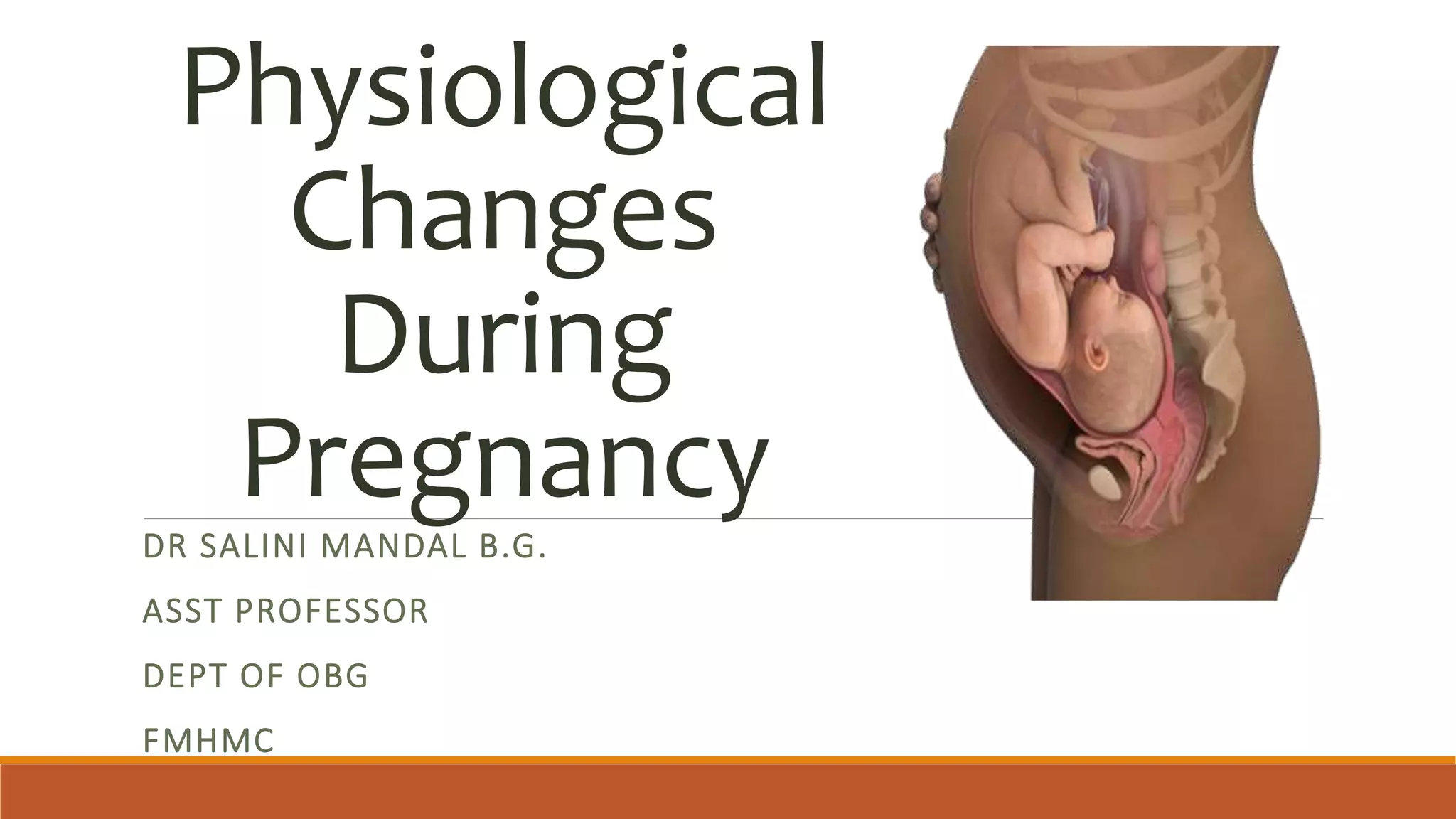
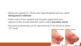
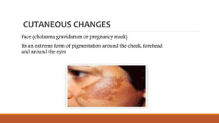
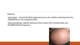
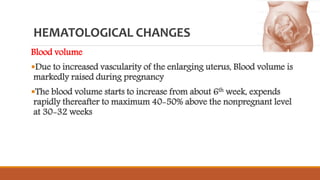
Physiological
Changes
During
Pregnancy
DR SALINI MANDAL B.G.
ASST PROFESSOR
DEPT OF OBG
FMHMC
VULVA
Vulva becomes edematous and more vascular
Superficial varicosities may appear especially in multiparae.
Labia minora are pigmented and hypertrophied.
VAGINA
Vaginal walls become hypertrophied, edematous and more vascular.
Increased blood supply of the venous plexus surrounding the walls gives the
bluish coloration of the mucosa (Jacquemier’s sign)
The length of the anterior vaginal wall is increased.
Secretion:
The secretion becomes copious, thin and curdy white due to marked
exfoliated cells and bacteria.
The pH becomes acidic (3.5–6) due to more conversion of glycogen
into lactic acid by the Lactobacillus acidophilus consequent on high
estrogen level.
The acidic pH prevents multiplication of pathogenic organisms.
UTERUS
BODY OF THE UTERUS:
Enlargement:
Changes in the muscles
Hypertrophy and hyperplasia: These occur under the infl uence of the
hormones—estrogen and progesterone
Stretching: The muscle fibers further elongate beyond 20 weeks due to
distension by the growing fetus. Th e wall becomes thinner and, at term,
measures about 1.5 cm or less. The uterus feels soft and elastic in contrast to
firm feel of the nongravid uterus.
Vascular system—
There is marked spiraling of the arteries, reaching the maximum at
20 weeks; thereafter, they straighten out.
Blood flow increases by eightfold at 20 weeks of pregnancy.
This vasodilatation is mainly due to estradiol and progesterone.
Veins become dilated and are valveless. Numerous lymphatic
channels open up.
Weight:
The increase in weight is due to the increased growth of the uterine
muscles, connective tissues and vascular channels.
Shape
Nonpregnant pyriform shape is maintained in early months. It
becomes globular at 12 weeks. As the uterus enlarges, the shape once
more becomes pyriform or ovoid by 28 weeks and changes to
spherical beyond 36th week.
Isthmus
During the first trimester isthmus hypertrophies and elongates to
about 3 times its original length
Becomes softer
Cervix
Hypertrophy and hyperplasia of the elastic and connective tissues
Vascularity is increased
Softening of the cervix (Goodell’s sign)
Squamous cells also become hyperactive
Mucosal changes simulate basal cell hyperplasia or cervical intraepithelial
neoplasia (CIN)
Secretion is copious and tenacious –physiological leucorrhoea of pregnancy
Becomes bulky
Fallopian Tube
Total length is increased
Tube becomes congested
Muscles undergo hypertrophy
Ovary
Growth and function of the corpus luteum reaches its maximum at 8th week
Hormones-oestrogen and progesterone secreted by the corpus luteum maintain
the environment for the growing ovum
Control the formation and maintenance of decidua of pregnancy
Inhibit ripening of the follicles
BREAST CHANGES
Increased size of the breasts
Marked hypertrophy and proliferation of the ducts (oestrogen
and progesterone)
Vascularity is increased
The nipples become larger, erectile and deeply pigmented
Sebaceous glands (5-15) become hypertrophied and are called
Montgomery’s tubercles
Outer zone of less marked and irregular pigmented area
appears in the second trimester and is called secondary areola
Secretion (colostrum) can be squeezed out of the breast at about
12th week
CUTANEOUS CHANGES
Face (cholasma gravidarum or pregnancy mask)
Its an extreme form of pigmentation around the cheek, forehead
and around the eyes
Abdomen
Linea nigra : a brownish black pigmented area in the midline stretching from the
xiphisternum to the symphysis pubis
Straie graviderum :slightly depressed linear marks with varying length and
breadth found in pregnancy
HEMATOLOGICAL CHANGES
Blood volume
Due to increased vascularity of the enlarging uterus, Blood volume is
markedly raised during pregnancy
The blood volume starts to increase from about 6th week, expends
rapidly thereafter to maximum 40-50% above the nonpregnant level
at 30-32 weeks
Plasma Volume
Starts to increase by 6 weeks
Rate of increase almost parallels to that of blood volume
Reached to the extent of 50%
Total plasma volume increases to the extent of 1.25 litters
RBC And Haemoglobin
The RBC mass is increased to the extent of 20-30%
Increase demand of oxygen transport during pregnancy
Disproportionate increase in plasma and RBC volume produces state
of haemodilution (fall in haemocrit)
Hb fall is about 2 gm.% from the non-pregnant value.
Total plasma protein increases from the normal 180 gm. (non-
pregnant) to 230 gm
Due to haemodilution(increase plasma volume), the plasma protein
concentration falls from 7 gm.% to 6 gm.%
Blood Coagulation Factor Pregnancy is a hypercoagulable state.
Plasma fibrinogen (factor 1) increases from the third month of
pregnancy
METABOLIC CHANGES
General Metabolic Changes
Total metabolism is increased due to the needs of the growing fetus
and the uterus
Basal metabolic rate is increased to the extent of 30%
Protein Metabolism
Positive nitrogenous balance throughout pregnancy
At term, the fetus and the placenta contain about 500 gm. of protein
and the maternal gain is also about 500 gm.
Carbohydrate Metabolism
Hyperplasia and hypertrophy of beta cells of pancreas.
Increased insulin level favours lipogenesis (fat storage).This
mechanism ensures continuously supply of glucose to the fetus
Fat Metabolism
An average of 3-4 kg of fat is stored during pregnancy mostly in the
abdominal wall, breasts, hips and thighs
Iron Metabolism
Total iron requirement during pregnancy is estimated approximately
1000mg
In the absence of iron supplementation, there is drop in
haemoglobin, serum iron and serum ferritin concentration at term
pregnancy
Calcium metabolism and locomotor system
Relaxation of pelvic ligaments and muscles occurs because of the
influence of estrogen and relaxtin reaches maximum during last
weeks of the pregnancy
Increased lumber lordosis during later months of the pregnancy due
to enlarged uterus backache and wadding gait
SYSTEMIC CHANGES
Respiratory System
Progressive increase in oxygen consumption, which is caused by the
increased metabolic needs of the mother and fetus
The mucosa of the nasopharynx becomes hyperaemic and
oedematous
A state of hyperventilation occurs during pregnancy leading to
increase tidal volume
The woman feels shortness of breath
CARDIOVASCULAR CHANGES
The Heart :
The growing uterus pushes the heart upward and to the left
During pregnancy the heart rate and stroke volume (the amount of
the blood pumped by heart with each beat) increases due to the
increase blood volume and oxygen requirement of the maternal
tissues and growing fetus
Cardiac Output :
Increases markedly by the end of the first trimester.
In the third trimester, a rise, fall or no change at all has been showed
to occur, depending on individual variables.
The capacity of veins and venules increases.
Arterial walls relax and dilate due to the effect of progesterone. The
increase production of vasodilator prostaglandin also contributes to
this.
Blood Pressure
During the mid-trimester, changes in blood pressure may occur
causing fainting
In later pregnancy, hypotension may occur in 10% of women in
unsupported supine position. This termed as “supine hypotensive
syndrome”
The pressure of gravid uterus compresses the vena cava, reducing the
venous return
Cardiac output is reduced by 25-30 percent and the blood pressure
may fall by 10-15 percent
Urinary System
Kidney
Dilatation of the ureter, renal pelvis and calyces. The kidneys enlarge
in length by 1 cm.
Glomerular filtration rate (GFR) is increased by 50% all throughout
the pregnancy
Ureter
ureters become atonic due to high progesterone level.
Dilatation of the ureter above the pelvic brim with stasis is marked
on the right side specially in primigravidae.
•Bladder
Increased frequency of micturition is noticed at 6-8 weeks of
pregnancy which subside after 12 weeks and In late pregnancy,
frequency of micturition once more reappears due to pressure on the
bladder as the presenting part descends down the pelvis.
 Stress incontinence may observe in late pregnancy due to urethral
sphincter weakness
Alimentary System
Gums become congested and spongy and may bleed to touch
Risk of peptic ulcer disease is reduced.
Atonicity of the gut leads to constipation
Liver and gall bladder
Liver functions are depressed
High blood cholesterol level during pregnancy, favour stone
formation
NERVOUS SYSTEM
Temperamental changes are found during pregnancy and in the
puerperium
Nausea, vomiting, mental irritability and sleeplessness are probably
due to some psychological background
Weight Gain
In early weeks, the patient may lose weight because of nausea and
vomiting
During subsequent months, the weight gain is progressive until the
last one or two weeks, when the weight remains static
The total weight gain during the course of a singleton pregnancy for
a healthy woman averages 11 kg
Distributed to 1 kg in first trimester and 5 kg each in second and
third trimester
The total weight gain at term is distributed approximately as :
Reproductive weight gain : 6 kg Net maternal weight gain : 6 kg
Fetus – 3.3 kg,
placenta – 0.6 kg and liquor – 0.8 kg
uterus – 0.9 kg and breast -0.4 kg,
accumulation of the fat and protein –
3.5 kg
Increases in blood volume – 1.3 kg
Increases in extracellular fluid – 1.2 kg

Physiological changes during pregnancy

  • 1.
    Physiological Changes During Pregnancy DR SALINI MANDALB.G. ASST PROFESSOR DEPT OF OBG FMHMC
  • 2.
    VULVA Vulva becomes edematousand more vascular Superficial varicosities may appear especially in multiparae. Labia minora are pigmented and hypertrophied.
  • 3.
    VAGINA Vaginal walls becomehypertrophied, edematous and more vascular. Increased blood supply of the venous plexus surrounding the walls gives the bluish coloration of the mucosa (Jacquemier’s sign) The length of the anterior vaginal wall is increased.
  • 4.
    Secretion: The secretion becomescopious, thin and curdy white due to marked exfoliated cells and bacteria. The pH becomes acidic (3.5–6) due to more conversion of glycogen into lactic acid by the Lactobacillus acidophilus consequent on high estrogen level. The acidic pH prevents multiplication of pathogenic organisms.
  • 5.
    UTERUS BODY OF THEUTERUS: Enlargement: Changes in the muscles Hypertrophy and hyperplasia: These occur under the infl uence of the hormones—estrogen and progesterone Stretching: The muscle fibers further elongate beyond 20 weeks due to distension by the growing fetus. Th e wall becomes thinner and, at term, measures about 1.5 cm or less. The uterus feels soft and elastic in contrast to firm feel of the nongravid uterus.
  • 6.
    Vascular system— There ismarked spiraling of the arteries, reaching the maximum at 20 weeks; thereafter, they straighten out. Blood flow increases by eightfold at 20 weeks of pregnancy. This vasodilatation is mainly due to estradiol and progesterone. Veins become dilated and are valveless. Numerous lymphatic channels open up.
  • 7.
    Weight: The increase inweight is due to the increased growth of the uterine muscles, connective tissues and vascular channels. Shape Nonpregnant pyriform shape is maintained in early months. It becomes globular at 12 weeks. As the uterus enlarges, the shape once more becomes pyriform or ovoid by 28 weeks and changes to spherical beyond 36th week.
  • 8.
    Isthmus During the firsttrimester isthmus hypertrophies and elongates to about 3 times its original length Becomes softer
  • 9.
    Cervix Hypertrophy and hyperplasiaof the elastic and connective tissues Vascularity is increased Softening of the cervix (Goodell’s sign) Squamous cells also become hyperactive Mucosal changes simulate basal cell hyperplasia or cervical intraepithelial neoplasia (CIN) Secretion is copious and tenacious –physiological leucorrhoea of pregnancy Becomes bulky
  • 10.
    Fallopian Tube Total lengthis increased Tube becomes congested Muscles undergo hypertrophy
  • 11.
    Ovary Growth and functionof the corpus luteum reaches its maximum at 8th week Hormones-oestrogen and progesterone secreted by the corpus luteum maintain the environment for the growing ovum Control the formation and maintenance of decidua of pregnancy Inhibit ripening of the follicles
  • 12.
    BREAST CHANGES Increased sizeof the breasts Marked hypertrophy and proliferation of the ducts (oestrogen and progesterone) Vascularity is increased The nipples become larger, erectile and deeply pigmented
  • 13.
    Sebaceous glands (5-15)become hypertrophied and are called Montgomery’s tubercles Outer zone of less marked and irregular pigmented area appears in the second trimester and is called secondary areola Secretion (colostrum) can be squeezed out of the breast at about 12th week
  • 14.
    CUTANEOUS CHANGES Face (cholasmagravidarum or pregnancy mask) Its an extreme form of pigmentation around the cheek, forehead and around the eyes
  • 15.
    Abdomen Linea nigra :a brownish black pigmented area in the midline stretching from the xiphisternum to the symphysis pubis Straie graviderum :slightly depressed linear marks with varying length and breadth found in pregnancy
  • 16.
    HEMATOLOGICAL CHANGES Blood volume Dueto increased vascularity of the enlarging uterus, Blood volume is markedly raised during pregnancy The blood volume starts to increase from about 6th week, expends rapidly thereafter to maximum 40-50% above the nonpregnant level at 30-32 weeks
  • 17.
    Plasma Volume Starts toincrease by 6 weeks Rate of increase almost parallels to that of blood volume Reached to the extent of 50% Total plasma volume increases to the extent of 1.25 litters
  • 18.
    RBC And Haemoglobin TheRBC mass is increased to the extent of 20-30% Increase demand of oxygen transport during pregnancy Disproportionate increase in plasma and RBC volume produces state of haemodilution (fall in haemocrit) Hb fall is about 2 gm.% from the non-pregnant value.
  • 19.
    Total plasma proteinincreases from the normal 180 gm. (non- pregnant) to 230 gm Due to haemodilution(increase plasma volume), the plasma protein concentration falls from 7 gm.% to 6 gm.% Blood Coagulation Factor Pregnancy is a hypercoagulable state. Plasma fibrinogen (factor 1) increases from the third month of pregnancy
  • 20.
    METABOLIC CHANGES General MetabolicChanges Total metabolism is increased due to the needs of the growing fetus and the uterus Basal metabolic rate is increased to the extent of 30% Protein Metabolism Positive nitrogenous balance throughout pregnancy At term, the fetus and the placenta contain about 500 gm. of protein and the maternal gain is also about 500 gm.
  • 21.
    Carbohydrate Metabolism Hyperplasia andhypertrophy of beta cells of pancreas. Increased insulin level favours lipogenesis (fat storage).This mechanism ensures continuously supply of glucose to the fetus Fat Metabolism An average of 3-4 kg of fat is stored during pregnancy mostly in the abdominal wall, breasts, hips and thighs
  • 22.
    Iron Metabolism Total ironrequirement during pregnancy is estimated approximately 1000mg In the absence of iron supplementation, there is drop in haemoglobin, serum iron and serum ferritin concentration at term pregnancy
  • 23.
    Calcium metabolism andlocomotor system Relaxation of pelvic ligaments and muscles occurs because of the influence of estrogen and relaxtin reaches maximum during last weeks of the pregnancy Increased lumber lordosis during later months of the pregnancy due to enlarged uterus backache and wadding gait
  • 24.
    SYSTEMIC CHANGES Respiratory System Progressiveincrease in oxygen consumption, which is caused by the increased metabolic needs of the mother and fetus The mucosa of the nasopharynx becomes hyperaemic and oedematous A state of hyperventilation occurs during pregnancy leading to increase tidal volume The woman feels shortness of breath
  • 25.
    CARDIOVASCULAR CHANGES The Heart: The growing uterus pushes the heart upward and to the left During pregnancy the heart rate and stroke volume (the amount of the blood pumped by heart with each beat) increases due to the increase blood volume and oxygen requirement of the maternal tissues and growing fetus
  • 26.
    Cardiac Output : Increasesmarkedly by the end of the first trimester. In the third trimester, a rise, fall or no change at all has been showed to occur, depending on individual variables. The capacity of veins and venules increases. Arterial walls relax and dilate due to the effect of progesterone. The increase production of vasodilator prostaglandin also contributes to this.
  • 27.
    Blood Pressure During themid-trimester, changes in blood pressure may occur causing fainting In later pregnancy, hypotension may occur in 10% of women in unsupported supine position. This termed as “supine hypotensive syndrome” The pressure of gravid uterus compresses the vena cava, reducing the venous return Cardiac output is reduced by 25-30 percent and the blood pressure may fall by 10-15 percent
  • 28.
    Urinary System Kidney Dilatation ofthe ureter, renal pelvis and calyces. The kidneys enlarge in length by 1 cm. Glomerular filtration rate (GFR) is increased by 50% all throughout the pregnancy Ureter ureters become atonic due to high progesterone level. Dilatation of the ureter above the pelvic brim with stasis is marked on the right side specially in primigravidae.
  • 29.
    •Bladder Increased frequency ofmicturition is noticed at 6-8 weeks of pregnancy which subside after 12 weeks and In late pregnancy, frequency of micturition once more reappears due to pressure on the bladder as the presenting part descends down the pelvis.  Stress incontinence may observe in late pregnancy due to urethral sphincter weakness
  • 30.
    Alimentary System Gums becomecongested and spongy and may bleed to touch Risk of peptic ulcer disease is reduced. Atonicity of the gut leads to constipation Liver and gall bladder Liver functions are depressed High blood cholesterol level during pregnancy, favour stone formation
  • 31.
    NERVOUS SYSTEM Temperamental changesare found during pregnancy and in the puerperium Nausea, vomiting, mental irritability and sleeplessness are probably due to some psychological background
  • 32.
    Weight Gain In earlyweeks, the patient may lose weight because of nausea and vomiting During subsequent months, the weight gain is progressive until the last one or two weeks, when the weight remains static The total weight gain during the course of a singleton pregnancy for a healthy woman averages 11 kg Distributed to 1 kg in first trimester and 5 kg each in second and third trimester
  • 33.
    The total weightgain at term is distributed approximately as : Reproductive weight gain : 6 kg Net maternal weight gain : 6 kg Fetus – 3.3 kg, placenta – 0.6 kg and liquor – 0.8 kg uterus – 0.9 kg and breast -0.4 kg, accumulation of the fat and protein – 3.5 kg Increases in blood volume – 1.3 kg Increases in extracellular fluid – 1.2 kg