Download to read offline

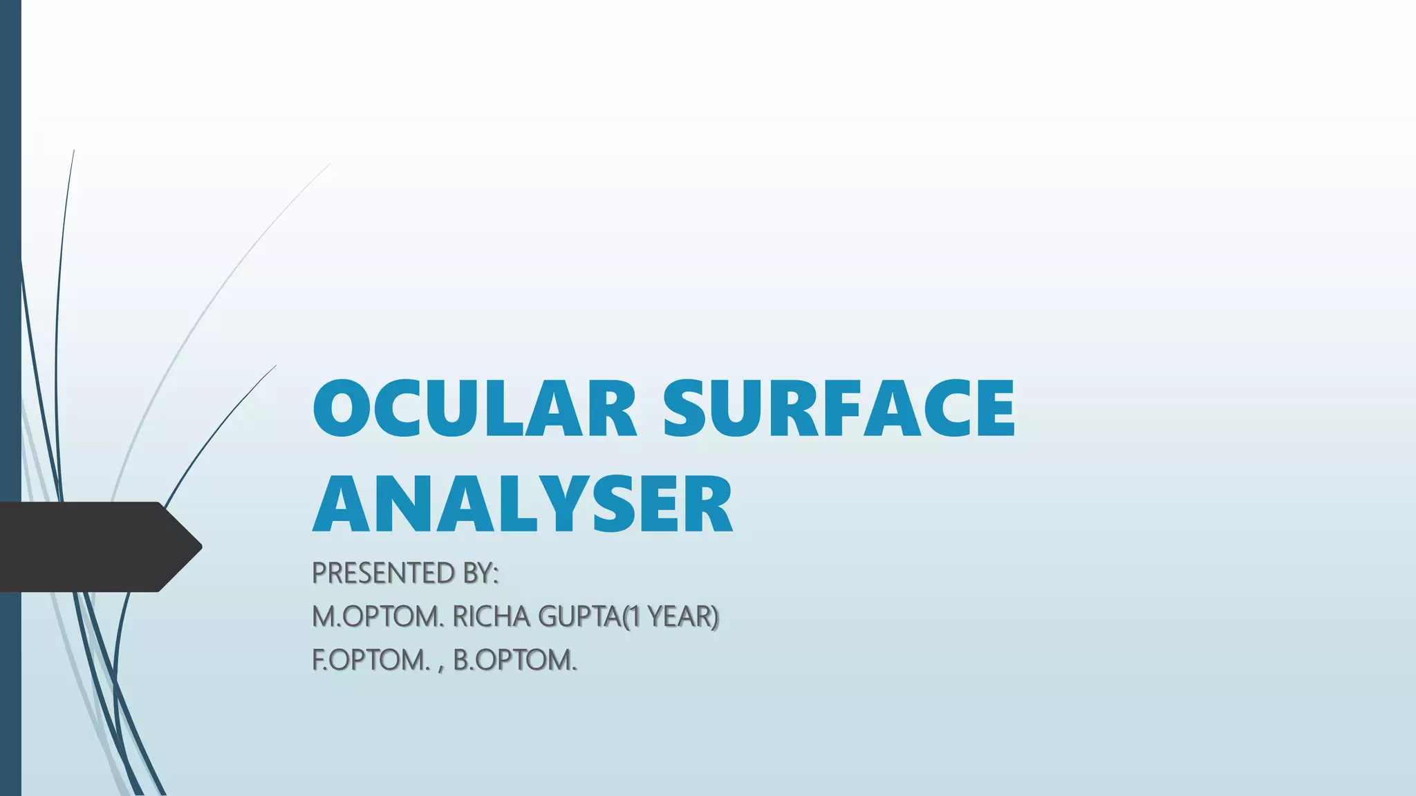
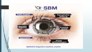
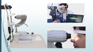
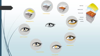
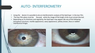
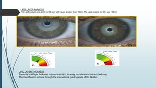
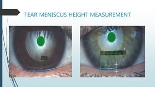
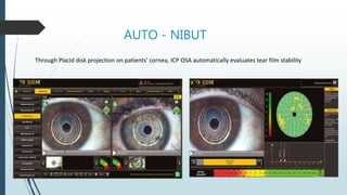
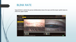
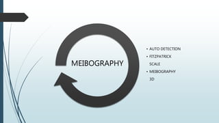
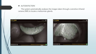
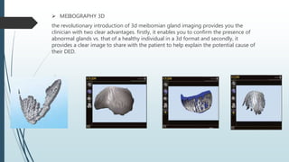
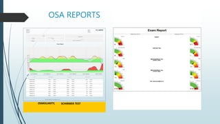
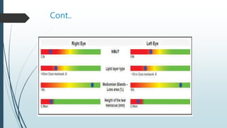
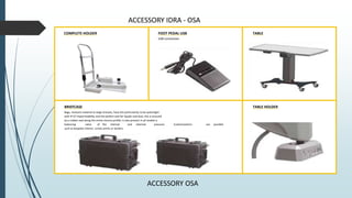
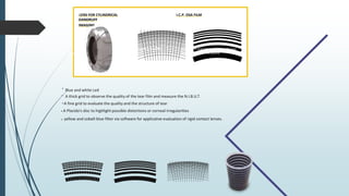


The document summarizes an ocular surface analyzer (OSA) device. It can perform various tear film tests and analyze the meibomian glands using international grading scales. The OSA allows for interferometric analysis of the lipid layer and measurement of the tear meniscus height. It also features automated NIBUT measurement, meibography imaging, and analysis of the lipid layer thickness, redness, and other ocular measurements. The OSA provides reports on examination findings and can be used for screening, diagnosis, and monitoring treatment by optometrists and ophthalmologists.


