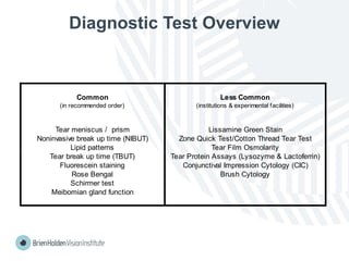
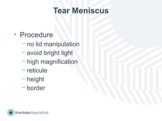
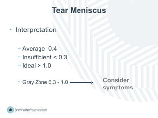
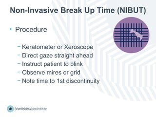
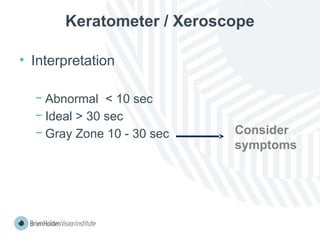
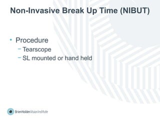
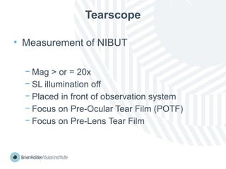
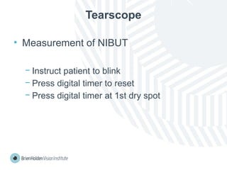
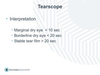
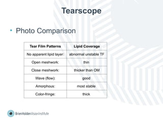
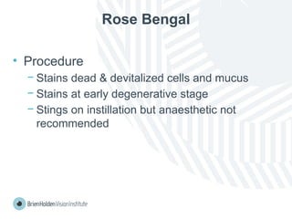
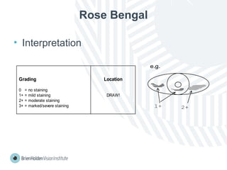
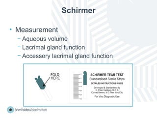
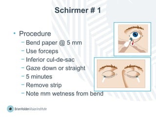
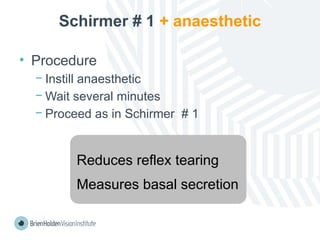
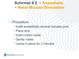
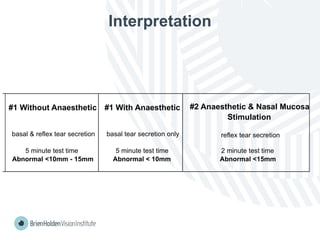
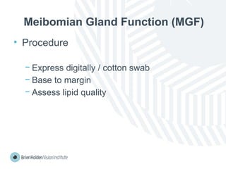
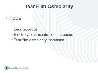
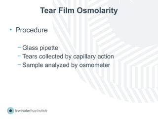
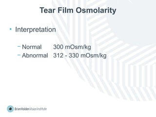
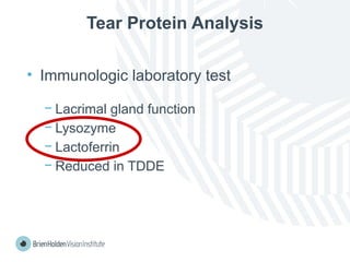
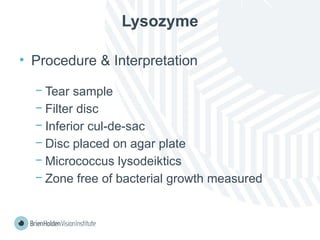
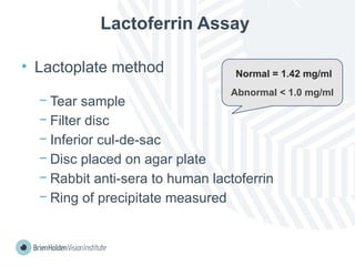
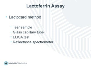
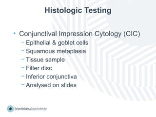
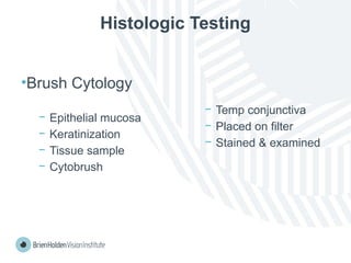
CLINICAL OPTOMETRIC
PROCEDURES 2
Eyelid procedures
Authors
Luigi Bilotto
Brien Holden Vision Institute
Peer Reviewer
Meng Meng Xu
The New England College of Optometry
Editors
Brien Holden Vision Institute, Public Health Division
Brien Holden Vision Institute Foundation (formerly ICEE) is a Public Health division of Brien Holden Vision Institute
COPYRIGHT © 2013 Brien Holden Vision Institute. All rights reserved.
This publication is protected by laws relating to copyright. Except as permitted under applicable legislation, no part of this publication may be adapted, modified, stored in a retrieval system, transmitted or reproduced in any form or by any process, electronic or otherwise, without the Brien Holden Vision
Institute’s (The institute) prior written permission. You may, especially if you are from a not-for-profit organisation, be eligible for a free license to use and make limited copies of parts of this manual in certain limited circumstances. To see if you are eligible for such a license, please visit
http://education.brienholdenvision.org/ .
DISCLAIMER
The material and tools provided in this publication are provided for purposes of general information only. The Institute is not providing specific advice concerning the clinical management of any case or condition that may be mentioned in this publication, and the information must not be used as a
substitute for specific advice from a qualified professional.
The mention of specific companies or certain manufacturers’ products does not imply that those companies or products are endorsed or recommended by the Institute in preference to others of a similar nature that are not mentioned. The Institute makes no representation or warranty that the
information contained in this publication is complete or free of omissions or errors. To the extent permitted by law, the Institute excludes all implied warranties, including regarding merchantability and fitness for purpose, and disclaims any and all liability for any loss or damage incurred as a result of the
use of the material and tools provided.
Diagnostic Test Overview
Common Less Common
(in recommended order) (institutions & experimental facilities)
Tear meniscus / prism Lissamine Green Stain
Noninvasive break up time (NIBUT) Zone Quick Test/Cotton Thread Tear Test
Lipid patterns Tear Film Osmolarity
Tear break up time (TBUT) Tear Protein Assays (Lysozyme & Lactoferrin)
Fluorescein staining Conjunctival Impression Cytology (CIC)
Rose Bengal Brush Cytology
Schirmer test
Meibomian gland function
Tear Meniscus
• Procedure
− no lid manipulation
− avoid bright light
− high magnification
− reticule
− height
− border
Tear Meniscus
• Interpretation
− Average 0.4
− Insufficient < 0.3
− Ideal > 1.0
− Gray Zone 0.3 - 1.0 Consider
symptoms
Non-Invasive Break Up Time (NIBUT)
• Procedure
− Keratometer or Xeroscope
− Direct gaze straight ahead
− Instruct patient to blink
− Observe mires or grid
− Note time to 1st discontinuity
Keratometer / Xeroscope
• Interpretation
− Abnormal < 10 sec
− Ideal > 30 sec
− Gray Zone 10 - 30 sec Consider
symptoms
Non-Invasive Break Up Time (NIBUT)
• Procedure
− Tearscope
− SL mounted or hand held
Tearscope
• Measurement of NIBUT
− Mag > or = 20x
− SL illumination off
− Placed in front of observation system
− Focus on Pre-Ocular Tear Film (POTF)
− Focus on Pre-Lens Tear Film
Tearscope
• Measurement of NIBUT
− Instruct patient to blink
− Press digital timer to reset
− Press digital timer at 1st dry spot
Tearscope
• Interpretation
− Marginal dry eye < 10 sec
− Borderline dry eye < 20 sec
− Stable tear film > 20 sec
Tearscope
• Photo Comparison
Tear Film Patterns Lipid Coverage
No apparent lipid layer: abnormal unstable TF
Open meshwork: thin
Close meshwork: thicker than OM
Wave (flow): good
Amorphous: most stable
Color-fringe: thick
Tear Break Up Time (TBUT)
• Procedure
− Evaluates TF stability
− Place NaFl in eye
− Moderate mag
− Patient blink
− Note time to 1st dry spot
TBUT
• Interpretation
− Abnormal < 5 sec
− Ideal > 0 sec
− Gray Zone 5 - 10 sec Consider
symptoms
Fluorescein Staining
• Procedure
− Erosions
− Abrasions
− Mucus
Fluorescein Staining
• Interpretation
Grading Location
0 = no staining
1+ = mild staining DRAW!
2+ = moderate staining
3+ = marked/severe staining
1+ 2+
e.g.
Rose Bengal
• Procedure
− Stains dead & devitalized cells and mucus
− Stains at early degenerative stage
− Stings on instillation but anaesthetic not
recommended
Rose Bengal
• Interpretation
Grading Location
0 = no staining
1+ = mild staining DRAW!
2+ = moderate staining
3+ = marked/severe staining 1+ 2+
e.g.
Schirmer
• Measurement
− Aqueous volume
− Lacrimal gland function
− Accessory lacrimal gland function
Schirmer # 1
• Procedure
− Bend paper @ 5 mm
− Use forceps
− Inferior cul-de-sac
− Gaze down or straight
− 5 minutes
− Remove strip
− Note mm wetness from bend
Schirmer # 1 + anaesthetic
• Procedure
− Instill anaesthetic
− Wait several minutes
− Proceed as in Schirmer # 1
Reduces reflex tearing
Measures basal secretion
Schirmer # 2 + Anaesthetic
+ Nasal Mucosa Stimulation
• Procedure
− Instill anaesthetic several minutes prior
− Place strip
− Insert cotton swab
− Gently rotate
− Leave in place for 2 minutes
Interpretation
#1 Without Anaesthetic #1 With Anaesthetic #2 Anaesthetic & Nasal Mucosa
Stimulation
basal & reflex tear secretion basal tear secretion only reflex tear secretion
5 minute test time 5 minute test time 2 minute test time
Abnormal <10mm - 15mm Abnormal < 10mm Abnormal <15mm
Meibomian Gland Function (MGF)
• Procedure
− Express digitally / cotton swab
− Base to margin
− Assess lipid quality
MGF
• Interpretation
− Normal = clear oily fluid
− Abnormal = clogging of orifice
milky fluid
cheesy
toothpaste
Lissamine Green Stain
• Procedure & Interpretation
− Similar to RB
• devitalized cells, mucus
− Green Staining
− Does not sting
− Now available commercially
Cotton Thread Test
− Measures aqueous volume
− 70mm sterile yellow cotton thread
− Phenol red impregnated
− Turns red with tears
− Less invasive & irritating
− Reduces reflex tearing
CTT
• Procedure
− Cotton thread bent @ 3mm
− Placed at inferior cul-de-sac
− 15 sec
− Remove thread
− Measure mm from bend
CTT
• Interpretation
− Normal 9 - 20mm
− Abnormal < 9mm
Tear Film Osmolarity
• TDDE
− Less aqueous
− Electrolyte concentration increased
− Tear film osmolarity increased
Tear Film Osmolarity
• Procedure
− Glass pipette
− Tears collected by capillary action
− Sample analyzed by osmometer
Tear Film Osmolarity
• Interpretation
− Normal 300 mOsm/kg
− Abnormal 312 - 330 mOsm/kg
Tear Protein Analysis
• Immunologic laboratory test
− Lacrimal gland function
− Lysozyme
− Lactoferrin
− Reduced in TDDE
Lysozyme
• Procedure & Interpretation
− Tear sample
− Filter disc
− Inferior cul-de-sac
− Disc placed on agar plate
− Micrococcus lysodeiktics
− Zone free of bacterial growth measured
Lactoferrin Assay
• Lactoplate method
− Tear sample
− Filter disc
− Inferior cul-de-sac
− Disc placed on agar plate
− Rabbit anti-sera to human lactoferrin
− Ring of precipitate measured
Normal = 1.42 mg/ml
Abnormal < 1.0 mg/ml
Lactoferrin Assay
• Lactocard method
− Tear sample
− Glass capillary tube
− ELISA test
− Reflectance spectrometer
Histologic Testing
• Conjunctival Impression Cytology (CIC)
− Epithelial & goblet cells
− Squamous metaplasia
− Tissue sample
− Filter disc
− Inferior conjunctiva
− Analysed on slides
Histologic Testing
•Brush Cytology
− Epithelial mucosa
− Keratinization
− Tissue sample
− Cytobrush
− Temp conjunctiva
− Placed on filter
− Stained & examined
11 Eyelid procedures. Kmu peshawar .  .  .  .  .

11 Eyelid procedures. Kmu peshawar . . . . .

  • 1.
  • 2.
    Authors Luigi Bilotto Brien HoldenVision Institute Peer Reviewer Meng Meng Xu The New England College of Optometry Editors Brien Holden Vision Institute, Public Health Division Brien Holden Vision Institute Foundation (formerly ICEE) is a Public Health division of Brien Holden Vision Institute COPYRIGHT © 2013 Brien Holden Vision Institute. All rights reserved. This publication is protected by laws relating to copyright. Except as permitted under applicable legislation, no part of this publication may be adapted, modified, stored in a retrieval system, transmitted or reproduced in any form or by any process, electronic or otherwise, without the Brien Holden Vision Institute’s (The institute) prior written permission. You may, especially if you are from a not-for-profit organisation, be eligible for a free license to use and make limited copies of parts of this manual in certain limited circumstances. To see if you are eligible for such a license, please visit http://education.brienholdenvision.org/ . DISCLAIMER The material and tools provided in this publication are provided for purposes of general information only. The Institute is not providing specific advice concerning the clinical management of any case or condition that may be mentioned in this publication, and the information must not be used as a substitute for specific advice from a qualified professional. The mention of specific companies or certain manufacturers’ products does not imply that those companies or products are endorsed or recommended by the Institute in preference to others of a similar nature that are not mentioned. The Institute makes no representation or warranty that the information contained in this publication is complete or free of omissions or errors. To the extent permitted by law, the Institute excludes all implied warranties, including regarding merchantability and fitness for purpose, and disclaims any and all liability for any loss or damage incurred as a result of the use of the material and tools provided.
  • 3.
    Diagnostic Test Overview CommonLess Common (in recommended order) (institutions & experimental facilities) Tear meniscus / prism Lissamine Green Stain Noninvasive break up time (NIBUT) Zone Quick Test/Cotton Thread Tear Test Lipid patterns Tear Film Osmolarity Tear break up time (TBUT) Tear Protein Assays (Lysozyme & Lactoferrin) Fluorescein staining Conjunctival Impression Cytology (CIC) Rose Bengal Brush Cytology Schirmer test Meibomian gland function
  • 4.
    Tear Meniscus • Procedure −no lid manipulation − avoid bright light − high magnification − reticule − height − border
  • 5.
    Tear Meniscus • Interpretation −Average 0.4 − Insufficient < 0.3 − Ideal > 1.0 − Gray Zone 0.3 - 1.0 Consider symptoms
  • 6.
    Non-Invasive Break UpTime (NIBUT) • Procedure − Keratometer or Xeroscope − Direct gaze straight ahead − Instruct patient to blink − Observe mires or grid − Note time to 1st discontinuity
  • 7.
    Keratometer / Xeroscope •Interpretation − Abnormal < 10 sec − Ideal > 30 sec − Gray Zone 10 - 30 sec Consider symptoms
  • 8.
    Non-Invasive Break UpTime (NIBUT) • Procedure − Tearscope − SL mounted or hand held
  • 9.
    Tearscope • Measurement ofNIBUT − Mag > or = 20x − SL illumination off − Placed in front of observation system − Focus on Pre-Ocular Tear Film (POTF) − Focus on Pre-Lens Tear Film
  • 10.
    Tearscope • Measurement ofNIBUT − Instruct patient to blink − Press digital timer to reset − Press digital timer at 1st dry spot
  • 11.
    Tearscope • Interpretation − Marginaldry eye < 10 sec − Borderline dry eye < 20 sec − Stable tear film > 20 sec
  • 12.
    Tearscope • Photo Comparison TearFilm Patterns Lipid Coverage No apparent lipid layer: abnormal unstable TF Open meshwork: thin Close meshwork: thicker than OM Wave (flow): good Amorphous: most stable Color-fringe: thick
  • 13.
    Tear Break UpTime (TBUT) • Procedure − Evaluates TF stability − Place NaFl in eye − Moderate mag − Patient blink − Note time to 1st dry spot
  • 14.
    TBUT • Interpretation − Abnormal< 5 sec − Ideal > 0 sec − Gray Zone 5 - 10 sec Consider symptoms
  • 15.
    Fluorescein Staining • Procedure −Erosions − Abrasions − Mucus
  • 16.
    Fluorescein Staining • Interpretation GradingLocation 0 = no staining 1+ = mild staining DRAW! 2+ = moderate staining 3+ = marked/severe staining 1+ 2+ e.g.
  • 17.
    Rose Bengal • Procedure −Stains dead & devitalized cells and mucus − Stains at early degenerative stage − Stings on instillation but anaesthetic not recommended
  • 18.
    Rose Bengal • Interpretation GradingLocation 0 = no staining 1+ = mild staining DRAW! 2+ = moderate staining 3+ = marked/severe staining 1+ 2+ e.g.
  • 19.
    Schirmer • Measurement − Aqueousvolume − Lacrimal gland function − Accessory lacrimal gland function
  • 20.
    Schirmer # 1 •Procedure − Bend paper @ 5 mm − Use forceps − Inferior cul-de-sac − Gaze down or straight − 5 minutes − Remove strip − Note mm wetness from bend
  • 21.
    Schirmer # 1+ anaesthetic • Procedure − Instill anaesthetic − Wait several minutes − Proceed as in Schirmer # 1 Reduces reflex tearing Measures basal secretion
  • 22.
    Schirmer # 2+ Anaesthetic + Nasal Mucosa Stimulation • Procedure − Instill anaesthetic several minutes prior − Place strip − Insert cotton swab − Gently rotate − Leave in place for 2 minutes
  • 23.
    Interpretation #1 Without Anaesthetic#1 With Anaesthetic #2 Anaesthetic & Nasal Mucosa Stimulation basal & reflex tear secretion basal tear secretion only reflex tear secretion 5 minute test time 5 minute test time 2 minute test time Abnormal <10mm - 15mm Abnormal < 10mm Abnormal <15mm
  • 24.
    Meibomian Gland Function(MGF) • Procedure − Express digitally / cotton swab − Base to margin − Assess lipid quality
  • 25.
    MGF • Interpretation − Normal= clear oily fluid − Abnormal = clogging of orifice milky fluid cheesy toothpaste
  • 26.
    Lissamine Green Stain •Procedure & Interpretation − Similar to RB • devitalized cells, mucus − Green Staining − Does not sting − Now available commercially
  • 27.
    Cotton Thread Test −Measures aqueous volume − 70mm sterile yellow cotton thread − Phenol red impregnated − Turns red with tears − Less invasive & irritating − Reduces reflex tearing
  • 28.
    CTT • Procedure − Cottonthread bent @ 3mm − Placed at inferior cul-de-sac − 15 sec − Remove thread − Measure mm from bend
  • 29.
    CTT • Interpretation − Normal9 - 20mm − Abnormal < 9mm
  • 30.
    Tear Film Osmolarity •TDDE − Less aqueous − Electrolyte concentration increased − Tear film osmolarity increased
  • 31.
    Tear Film Osmolarity •Procedure − Glass pipette − Tears collected by capillary action − Sample analyzed by osmometer
  • 32.
    Tear Film Osmolarity •Interpretation − Normal 300 mOsm/kg − Abnormal 312 - 330 mOsm/kg
  • 33.
    Tear Protein Analysis •Immunologic laboratory test − Lacrimal gland function − Lysozyme − Lactoferrin − Reduced in TDDE
  • 34.
    Lysozyme • Procedure &Interpretation − Tear sample − Filter disc − Inferior cul-de-sac − Disc placed on agar plate − Micrococcus lysodeiktics − Zone free of bacterial growth measured
  • 35.
    Lactoferrin Assay • Lactoplatemethod − Tear sample − Filter disc − Inferior cul-de-sac − Disc placed on agar plate − Rabbit anti-sera to human lactoferrin − Ring of precipitate measured Normal = 1.42 mg/ml Abnormal < 1.0 mg/ml
  • 36.
    Lactoferrin Assay • Lactocardmethod − Tear sample − Glass capillary tube − ELISA test − Reflectance spectrometer
  • 37.
    Histologic Testing • ConjunctivalImpression Cytology (CIC) − Epithelial & goblet cells − Squamous metaplasia − Tissue sample − Filter disc − Inferior conjunctiva − Analysed on slides
  • 38.
    Histologic Testing •Brush Cytology −Epithelial mucosa − Keratinization − Tissue sample − Cytobrush − Temp conjunctiva − Placed on filter − Stained & examined