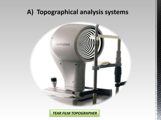
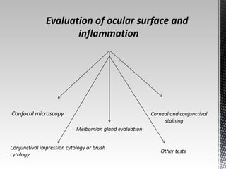
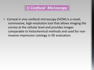
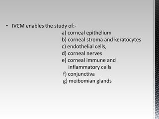
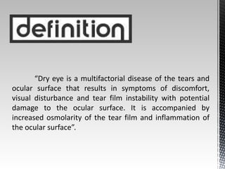
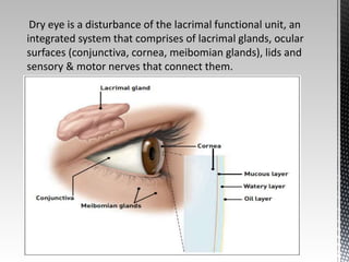
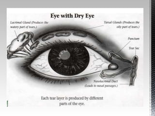
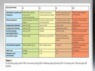
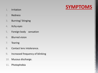
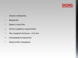
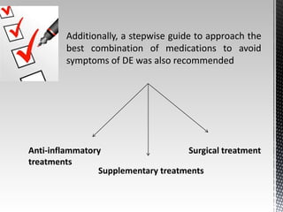
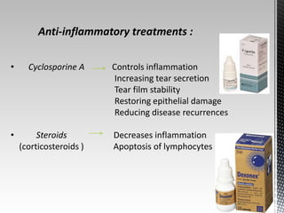
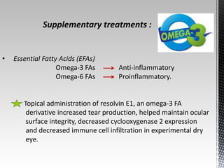
Dry eye disease : A 
review of diagnostic 
approaches and 
Treatments 
Saudi journal of ophthalmology (2014)28,173- 
181 
Hui Lin,MD,PhD;Samuel C.Yiu,MD,PhD 
Presented By – Dr. Dildar Singh 
Moderator - Dr. K.Kanthamani
“Dry eye is a multifactorial disease of the tears and 
ocular surface that results in symptoms of discomfort, 
visual disturbance and tear film instability with potential 
damage to the ocular surface. It is accompanied by 
increased osmolarity of the tear film and inflammation of 
the ocular surface”.
Dry eye is a disturbance of the lacrimal functional unit, an 
integrated system that comprises of lacrimal glands, ocular 
surfaces (conjunctiva, cornea, meibomian glands), lids and 
sensory & motor nerves that connect them.
1. Irritation 
2. Redness 
3. Burning/ Stinging 
4. Itchy eyes 
5. Foreign body sensation 
6. Blurred vision 
7. Tearing 
8. Contact lens intolerance. 
9. Increased frequency of blinking 
10. Mucous discharge. 
11. Photophobia 
SYMPTOMS
1. Chronic meibomitis 
2. Blepharitis 
3. Debris in tear film 
4. Chronic papillary conjunctivitis 
5. Tear marginal meniscus < 0.3 mm 
6. Interpalpebral hyperemia 
7. Gland orifice metaplasia 
SIGNS
- Although both ADDE and EDE present with similar signs of 
reduced stability and increased tear film osmolarity. 
- ADDE chiefly refers to a failure of lacrimal secretion and 
EDE is due to excessive water loss from the exposed ocular 
surface in the presence of normal lacrimal secretory 
function.
Diagnostic assessment 
An ideal diagnostic method should be preferably non-invasive, 
objective, specific, reproducible and sustainable in terms of 
cost and time. 
a) Subjective evaluation 
b) Objective evaluation
Subjective evaluation : 
• The symptoms and history of DE patients vary widely; 
therefore, validated questionnaires have been developed to 
ensure consistency in recording symptomatic information. 
• Previously it was believed that DE can be diagnosed largely on 
the basis of symptoms; however, recent studies have 
questioned this opinion as there is often a lack of correlation 
between the severity of the symptoms and signs of DE.
Objective evaluation : 
a) tear film assessment 
c) the Schirmer test as the most commonly used diagnostic 
tests for initial assessment of dry eye. 
d) Lacrimal river width 
e) Tear ferning test 
f) Lactoferrin contents 
g) Tear penetration pressure test
SCHIRMER TEST 
SCHIRMER'S TEST I 
If wetting >10mm = normal eye 
If wetting <3mm = very severe dry eye 
If wetting 3-5 mm = severe dry eye 
If wetting 5-10mm = moderate dry eye 
If wetting is 10mm = mild dry eye 
SCHIRMER'S TEST II 
If Wetting<10mm -- >irratate the nasal 
mucosa with cotton bud & note the wetting 
after 2 min. 
If no Wetting or <1mm Sjogren's 
syndrome 
If Wetting increases by 1mm Non-Sjogren's 
syndrome 
SCHIRMER'S TEST III – no diagnostic
LACRIMAL RIVER WIDTH TEAR FERN TEST 
A drop of tear is collected from the 
lower meniscus and dropped onto a 
microscope slide and allowed to dry by 
evaporation.
Apart from these traditional clinical tests, we will discuss 
more about the less invasive evaluations based on the 
recently developed technologies related to : 
a) tear hyperosmolarity 
b) tear film instability 
c) inflammation.
Tear osmolarity 
• Increase in tear osmolarity is common to all types of DE 
• Normal Osmolarity value 305 mOsms/l 
> 308 mOsms/l (mild DE) 
>312 mOsms/l (moderate to severe DE)
Tear film osmolarity can be measured in three ways: 
c) electrical conductivity or impedance
Assessment of tear stability 
• Tear break-up time (TBUT), introduced by Norn , remains 
the most frequently used diagnostic test to determine tear 
film instability. 
• Non-invasive tear break-up time (NIBUT) involves the 
observation of an illuminated grid pattern reflected from 
the anterior tear surface.
NIBUT can be measured by: 
a) corneal topography 
b) interferometry 
c) aberrometry 
d) functional visual acuity assessment 
e) confocal microscopy
A) Topographical analysis systems 
TEAR FILM TOPOGRAPHER
Uses : 
• evaluate corneal surface regularity 
• tear film stability 
• evaluation of post-LASIK dry eye
B) Interferometry 
LIPIVIEW INTERFEROMETER
• Coloured fringes that arise from interference between 
light reflected from the surface of the lipid layer and 
from the interface between the lipid layer and the 
aqueous layer of the tear film. 
• used to observe the nature, thickness and rupture of 
the lipid layer.
C) Aberrometry 
WAVEFRONT ABERROMETER
• non-invasive assessment of the visual disturbances 
caused by higher order aberrations arising from tear 
film instability and break-up. 
• Aberrometry could be utilized for not only detecting 
DE but also for monitoring the efficacy of 
treatments.
D) Functional visual acuity 
• measure of visual acuity during sustained eye 
opening without blinking 
• defined as the monocular recognition acuity at a 
specific time point 
• decreases significantly in both non-Sjögren’s 
syndrome (NSS) and Sjögren’s syndrome (SS) dry eye 
patients
Optical coherence tomography 
• Anterior segment OCT can measure the tear film 
thickness and tear meniscus parameters which indicate 
total tear volume. 
• lower tear meniscus height and radius were the best 
indicators of DE with a cutoff meniscus height of 1.64 mm 
and radius of 1.82 mm.
E) Non Contact Confocal Microscopy 
• non-contact, tandem-scanning confocal microscope 
demonstrating real-time images to observe the tear 
film.
Evaluation of ocular surface and 
inflammation 
Confocal microscopy 
Meibomian gland evaluation 
Corneal and conjunctival 
staining 
Conjunctival impression cytology or brush 
cytology 
Other tests
1) Confocal Microscopy 
• Corneal in vivo confocal microscopy (IVCM) is a novel, 
noninvasive, high-resolution tool that allows imaging the 
cornea at the cellular level and provides images 
comparable to histochemical methods and used for non-invasive 
impression cytology in DE evaluation.
• IVCM enables the study of:- 
a) corneal epithelium 
b) corneal stroma and keratocytes 
c) endothelial cells, 
d) corneal nerves 
e) corneal immune and 
inflammatory cells 
f) conjunctiva 
g) meibomian glands
2) Meibomian gland evaluation 
• source of lipids in the lipid layer of the tear film 
• MGD is the most common cause of evaporative dry eye 
• Meibomian glands can be assessed by :- 
a) slit lamp 
b) Meiboscopy 
c) Meibography 
d) Meibometry
3) Corneal and conjunctival staining 
• Invasive procedure which enables the assessment of 
ocular surface damage by instilling a dye: 
a) sodium fluorescein 
b) rose bengal 
c) lissamine green
Sodium fluorescein 
• A vital dye which has no intrinsic toxicity. 
• Detects epithelial defects & irregularities. 
• Filter paper strips or 0.25% - 2% solution. 
• Sodium fluorescein emits green light(520nm) when 
excited with with blue light (490nm).
Rose bengal 
• Rose bengal is a vital stain taken up by dead and devitalised 
epithelial cells. 
• Also stains mucous threads, filaments & strands . 
• Does not diffuse into the epithelial defects or penetrate 
corneal stroma like fluorescein. 
• It is toxic resulting in decreased cell vitality, motility & cell 
death. 
• Causes ocular discomfort –prior anaesthesia is needed. 
Score 0 - absent 
Score 1-just present 
Score 2-moderate staining 
Score 3-gross staining
• Dark green, water soluble 
• Degenerated cells, mucus, dead cells 
• Less irritating 
Lissamine green
4) Conjunctival impression cytology or brush cytology 
• harvesting conjunctival epithelial,Goblet, and inflammatory 
cells from the bulbar mucosa 
• cytokines IL-1a, mature IL-1b, and IL-1Ra are found in a 
significantly greater percentage of conjunctival cytology 
specimens from eyes with SS 
• dry eye group was found to have a significant difference in the 
CD4/CD8 ratio 
• Elevated matrix metalloproteinases levels in DE
Treatment
• Artificial tears provide palliative relief to eye irritation in 
patients with aqueous tear deficiency, but do not 
prevent the underlying inflammation 
• Mild-to-moderate DE disease- Combinations of 
artificial tears, oral omega-3 essential fatty acid 
supplements, mucin secretagogues,short-term steroids, 
and daily cyclosporine A
Severe- autologous serum, oral tetracyclines, prosthetic 
lens, and systemic immunesuppressants 
Severe forms associated with systemic diseases- surgical 
intervention, including tarsorrhaphy and amniotic 
membrane transplant.
Additionally, a stepwise guide to approach the 
best combination of medications to avoid 
symptoms of DE was also recommended 
Anti-inflammatory 
treatments 
Surgical treatment 
Supplementary treatments
Anti-inflammatory treatments : 
• Cyclosporine A Controls inflammation 
Increasing tear secretion 
Tear film stability 
Restoring epithelial damage 
Reducing disease recurrences 
• Steroids Decreases inflammation 
(corticosteroids ) Apoptosis of lymphocytes
• Hormonal therapy 
(androgen and estrogen) 
Increase tear production TBUT, 
lipid layer thickness. 
• Antibiotics 
(azithromycin and tetracycline, 
doxycycline, and minocycline) 
Decrease ocular surface 
inflammation, 
Normalize lipid production by 
the meibomian glands.
Supplementary treatments : 
• Essential Fatty Acids (EFAs) 
Omega-3 FAs Anti-inflammatory 
Omega-6 FAs Proinflammatory. 
Topical administration of resolvin E1, an omega-3 FA 
derivative increased tear production, helped maintain ocular 
surface integrity, decreased cyclooxygenase 2 expression 
and decreased immune cell infiltration in experimental dry 
eye.
• Nerve growth factor (NGF) Increase ocular surface 
sensitivity, 
Inhibit inflammatory reactions, 
Regulate tear film production, 
Reducing the apoptosis 
of corneal epithelial cells 
triggered by hyperosmolarity . 
• Autologous serum Autologous and umbilical cord 
serum contains substances 
that support the proliferation, 
differentiation, and maturation 
of the normal ocular surface 
epithelium and therefore, finds 
application in the treatment of 
severe DE. 
• Acupuncture
Surgical treatment 
• Punctal occlusion Reduces drainage, preserves natural 
tears and prolongs the effect of 
lubricants. 
• Salivary gland procedures 
• Subcutaneous abdominal artificial tear pump-reservoir 
- treatment of severe dry eye.
• Understanding of the pathogenesis and specific cellular 
responses involved in different forms of DE could result in the 
development of other treatment strategies for a better 
management and long lasting results. 
• Development of additional treatment options in the form of 
compounds targeting specific components would provide 
hope for the millions of individuals who daily experience this 
deleterious condition.
Dry eye ppt by dr dildar

Dry eye ppt by dr dildar

  • 1.
    Dry eye disease: A review of diagnostic approaches and Treatments Saudi journal of ophthalmology (2014)28,173- 181 Hui Lin,MD,PhD;Samuel C.Yiu,MD,PhD Presented By – Dr. Dildar Singh Moderator - Dr. K.Kanthamani
  • 2.
    “Dry eye isa multifactorial disease of the tears and ocular surface that results in symptoms of discomfort, visual disturbance and tear film instability with potential damage to the ocular surface. It is accompanied by increased osmolarity of the tear film and inflammation of the ocular surface”.
  • 3.
    Dry eye isa disturbance of the lacrimal functional unit, an integrated system that comprises of lacrimal glands, ocular surfaces (conjunctiva, cornea, meibomian glands), lids and sensory & motor nerves that connect them.
  • 7.
    1. Irritation 2.Redness 3. Burning/ Stinging 4. Itchy eyes 5. Foreign body sensation 6. Blurred vision 7. Tearing 8. Contact lens intolerance. 9. Increased frequency of blinking 10. Mucous discharge. 11. Photophobia SYMPTOMS
  • 8.
    1. Chronic meibomitis 2. Blepharitis 3. Debris in tear film 4. Chronic papillary conjunctivitis 5. Tear marginal meniscus < 0.3 mm 6. Interpalpebral hyperemia 7. Gland orifice metaplasia SIGNS
  • 9.
    - Although bothADDE and EDE present with similar signs of reduced stability and increased tear film osmolarity. - ADDE chiefly refers to a failure of lacrimal secretion and EDE is due to excessive water loss from the exposed ocular surface in the presence of normal lacrimal secretory function.
  • 11.
    Diagnostic assessment Anideal diagnostic method should be preferably non-invasive, objective, specific, reproducible and sustainable in terms of cost and time. a) Subjective evaluation b) Objective evaluation
  • 12.
    Subjective evaluation : • The symptoms and history of DE patients vary widely; therefore, validated questionnaires have been developed to ensure consistency in recording symptomatic information. • Previously it was believed that DE can be diagnosed largely on the basis of symptoms; however, recent studies have questioned this opinion as there is often a lack of correlation between the severity of the symptoms and signs of DE.
  • 13.
    Objective evaluation : a) tear film assessment c) the Schirmer test as the most commonly used diagnostic tests for initial assessment of dry eye. d) Lacrimal river width e) Tear ferning test f) Lactoferrin contents g) Tear penetration pressure test
  • 14.
    SCHIRMER TEST SCHIRMER'STEST I If wetting >10mm = normal eye If wetting <3mm = very severe dry eye If wetting 3-5 mm = severe dry eye If wetting 5-10mm = moderate dry eye If wetting is 10mm = mild dry eye SCHIRMER'S TEST II If Wetting<10mm -- >irratate the nasal mucosa with cotton bud & note the wetting after 2 min. If no Wetting or <1mm Sjogren's syndrome If Wetting increases by 1mm Non-Sjogren's syndrome SCHIRMER'S TEST III – no diagnostic
  • 15.
    LACRIMAL RIVER WIDTHTEAR FERN TEST A drop of tear is collected from the lower meniscus and dropped onto a microscope slide and allowed to dry by evaporation.
  • 16.
    Apart from thesetraditional clinical tests, we will discuss more about the less invasive evaluations based on the recently developed technologies related to : a) tear hyperosmolarity b) tear film instability c) inflammation.
  • 17.
    Tear osmolarity •Increase in tear osmolarity is common to all types of DE • Normal Osmolarity value 305 mOsms/l > 308 mOsms/l (mild DE) >312 mOsms/l (moderate to severe DE)
  • 18.
    Tear film osmolaritycan be measured in three ways: c) electrical conductivity or impedance
  • 19.
    Assessment of tearstability • Tear break-up time (TBUT), introduced by Norn , remains the most frequently used diagnostic test to determine tear film instability. • Non-invasive tear break-up time (NIBUT) involves the observation of an illuminated grid pattern reflected from the anterior tear surface.
  • 20.
    NIBUT can bemeasured by: a) corneal topography b) interferometry c) aberrometry d) functional visual acuity assessment e) confocal microscopy
  • 21.
    A) Topographical analysissystems TEAR FILM TOPOGRAPHER
  • 22.
    Uses : •evaluate corneal surface regularity • tear film stability • evaluation of post-LASIK dry eye
  • 23.
  • 24.
    • Coloured fringesthat arise from interference between light reflected from the surface of the lipid layer and from the interface between the lipid layer and the aqueous layer of the tear film. • used to observe the nature, thickness and rupture of the lipid layer.
  • 25.
  • 26.
    • non-invasive assessmentof the visual disturbances caused by higher order aberrations arising from tear film instability and break-up. • Aberrometry could be utilized for not only detecting DE but also for monitoring the efficacy of treatments.
  • 27.
    D) Functional visualacuity • measure of visual acuity during sustained eye opening without blinking • defined as the monocular recognition acuity at a specific time point • decreases significantly in both non-Sjögren’s syndrome (NSS) and Sjögren’s syndrome (SS) dry eye patients
  • 28.
    Optical coherence tomography • Anterior segment OCT can measure the tear film thickness and tear meniscus parameters which indicate total tear volume. • lower tear meniscus height and radius were the best indicators of DE with a cutoff meniscus height of 1.64 mm and radius of 1.82 mm.
  • 29.
    E) Non ContactConfocal Microscopy • non-contact, tandem-scanning confocal microscope demonstrating real-time images to observe the tear film.
  • 30.
    Evaluation of ocularsurface and inflammation Confocal microscopy Meibomian gland evaluation Corneal and conjunctival staining Conjunctival impression cytology or brush cytology Other tests
  • 31.
    1) Confocal Microscopy • Corneal in vivo confocal microscopy (IVCM) is a novel, noninvasive, high-resolution tool that allows imaging the cornea at the cellular level and provides images comparable to histochemical methods and used for non-invasive impression cytology in DE evaluation.
  • 32.
    • IVCM enablesthe study of:- a) corneal epithelium b) corneal stroma and keratocytes c) endothelial cells, d) corneal nerves e) corneal immune and inflammatory cells f) conjunctiva g) meibomian glands
  • 33.
    2) Meibomian glandevaluation • source of lipids in the lipid layer of the tear film • MGD is the most common cause of evaporative dry eye • Meibomian glands can be assessed by :- a) slit lamp b) Meiboscopy c) Meibography d) Meibometry
  • 34.
    3) Corneal andconjunctival staining • Invasive procedure which enables the assessment of ocular surface damage by instilling a dye: a) sodium fluorescein b) rose bengal c) lissamine green
  • 35.
    Sodium fluorescein •A vital dye which has no intrinsic toxicity. • Detects epithelial defects & irregularities. • Filter paper strips or 0.25% - 2% solution. • Sodium fluorescein emits green light(520nm) when excited with with blue light (490nm).
  • 36.
    Rose bengal •Rose bengal is a vital stain taken up by dead and devitalised epithelial cells. • Also stains mucous threads, filaments & strands . • Does not diffuse into the epithelial defects or penetrate corneal stroma like fluorescein. • It is toxic resulting in decreased cell vitality, motility & cell death. • Causes ocular discomfort –prior anaesthesia is needed. Score 0 - absent Score 1-just present Score 2-moderate staining Score 3-gross staining
  • 37.
    • Dark green,water soluble • Degenerated cells, mucus, dead cells • Less irritating Lissamine green
  • 38.
    4) Conjunctival impressioncytology or brush cytology • harvesting conjunctival epithelial,Goblet, and inflammatory cells from the bulbar mucosa • cytokines IL-1a, mature IL-1b, and IL-1Ra are found in a significantly greater percentage of conjunctival cytology specimens from eyes with SS • dry eye group was found to have a significant difference in the CD4/CD8 ratio • Elevated matrix metalloproteinases levels in DE
  • 39.
  • 40.
    • Artificial tearsprovide palliative relief to eye irritation in patients with aqueous tear deficiency, but do not prevent the underlying inflammation • Mild-to-moderate DE disease- Combinations of artificial tears, oral omega-3 essential fatty acid supplements, mucin secretagogues,short-term steroids, and daily cyclosporine A
  • 41.
    Severe- autologous serum,oral tetracyclines, prosthetic lens, and systemic immunesuppressants Severe forms associated with systemic diseases- surgical intervention, including tarsorrhaphy and amniotic membrane transplant.
  • 42.
    Additionally, a stepwiseguide to approach the best combination of medications to avoid symptoms of DE was also recommended Anti-inflammatory treatments Surgical treatment Supplementary treatments
  • 43.
    Anti-inflammatory treatments : • Cyclosporine A Controls inflammation Increasing tear secretion Tear film stability Restoring epithelial damage Reducing disease recurrences • Steroids Decreases inflammation (corticosteroids ) Apoptosis of lymphocytes
  • 44.
    • Hormonal therapy (androgen and estrogen) Increase tear production TBUT, lipid layer thickness. • Antibiotics (azithromycin and tetracycline, doxycycline, and minocycline) Decrease ocular surface inflammation, Normalize lipid production by the meibomian glands.
  • 45.
    Supplementary treatments : • Essential Fatty Acids (EFAs) Omega-3 FAs Anti-inflammatory Omega-6 FAs Proinflammatory. Topical administration of resolvin E1, an omega-3 FA derivative increased tear production, helped maintain ocular surface integrity, decreased cyclooxygenase 2 expression and decreased immune cell infiltration in experimental dry eye.
  • 46.
    • Nerve growthfactor (NGF) Increase ocular surface sensitivity, Inhibit inflammatory reactions, Regulate tear film production, Reducing the apoptosis of corneal epithelial cells triggered by hyperosmolarity . • Autologous serum Autologous and umbilical cord serum contains substances that support the proliferation, differentiation, and maturation of the normal ocular surface epithelium and therefore, finds application in the treatment of severe DE. • Acupuncture
  • 47.
    Surgical treatment •Punctal occlusion Reduces drainage, preserves natural tears and prolongs the effect of lubricants. • Salivary gland procedures • Subcutaneous abdominal artificial tear pump-reservoir - treatment of severe dry eye.
  • 48.
    • Understanding ofthe pathogenesis and specific cellular responses involved in different forms of DE could result in the development of other treatment strategies for a better management and long lasting results. • Development of additional treatment options in the form of compounds targeting specific components would provide hope for the millions of individuals who daily experience this deleterious condition.