Download as PDF, PPTX


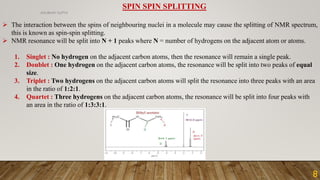


NMR spectroscopy is a technique that uses radio waves and strong magnetic fields to analyze atomic nuclei and their magnetic properties. It provides information about the molecular structure of compounds. The document discusses the basic principles of NMR spectroscopy including nuclear spin, chemical shifts, spin-spin coupling, and instrumentation. It also provides an example 1H NMR spectrum of ethanol to demonstrate how peaks are split based on neighboring hydrogen atoms.


