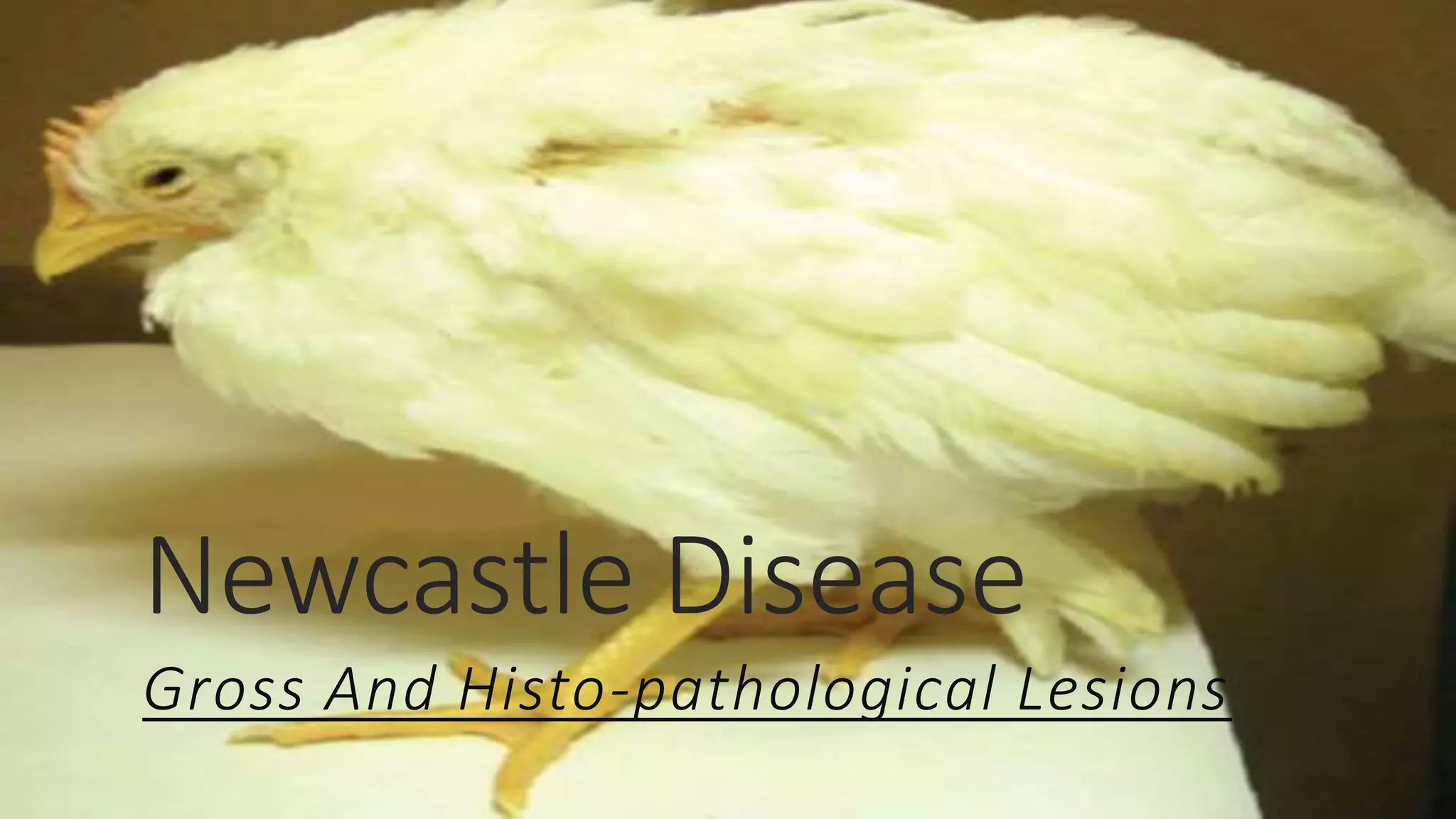
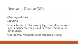
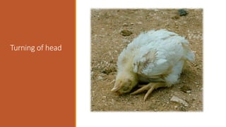
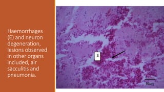
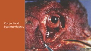
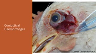
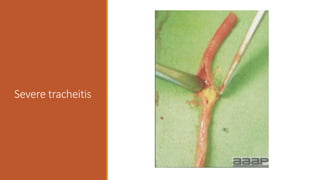
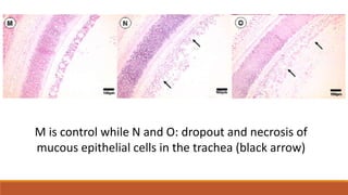
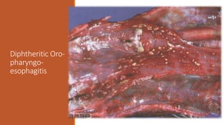
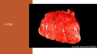
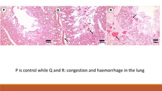
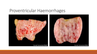
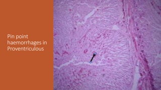
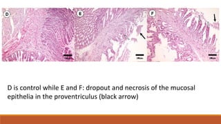
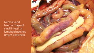
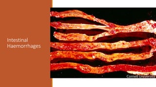
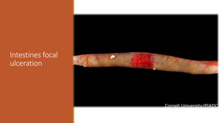
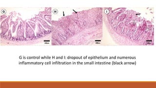
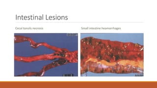
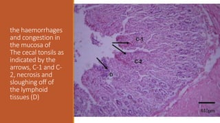
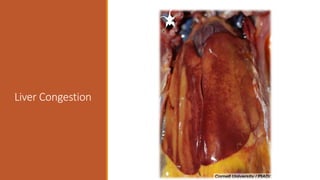
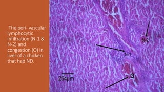
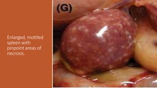
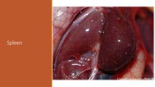
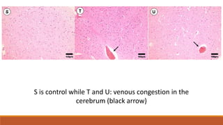
Newcastle Disease
Gross And Histo-pathological Lesions
Newcastle Disease (ND)
Paramyxoviridea
APMV-1
characterized in chickens by high mortality, nervous
signs and haemorrhagic and necrotic lesions in the
gut mucosa.
Lentogenic, Mesogenic and Valogenic strains.
ND Virus
Transmission
Reports of NDV
OIE 1997-2000
Pathogenesis
The virus replicates in the mucosa of the upper
respiratory and intestinal tracts.
Virus spreads via blood to spleen and bone marrow
(viremia) causing infection of other organs: lung
,intestines & C.N.S.
Clinical Signs and Symptoms
Respiratory symptoms.
Nervous signs.
Digestive symptoms.
Drop in egg production with thin, rough-shelled eggs.
Swelling of tissues around eyes and in the neck.
Sudden death.
In human;(Mild conjunctivitis, influenza-like symptoms and
laryngitis).
Turning of head
Turning of head
Haemorrhages
(E) and neuron
degeneration,
lesions observed
in other organs
included, air
sacculitis and
pneumonia.
Conjuctival
Haemorrhages
Conjuctival
Haemorrhages
Facial edema
Severe tracheitis
M is control while N and O: dropout and necrosis of
mucous epithelial cells in the trachea (black arrow)
Diphtheritic Oro-
pharyngo-
esophagitis
Lungs
P is control while Q and R: congestion and haemorrhage in the lung
Proventricular Haemorrhages
Pin point
haemorrhages in
Proventriculous
D is control while E and F: dropout and necrosis of the mucosal
epithelia in the proventriculus (black arrow)
Necrosis and
haemorrhage of
small intestinal
lymphoid patches
(Peyer’s patches).
Intestinal
Haemorrhages
Intestines focal
ulceration
G is control while H and I: dropout of epithelium and numerous
inflammatory cell infiltration in the small intestine (black arrow)
Intestinal Lesions
Cecal tonsils necrosis Small intestine heamorrhages
the haemorrhages
and congestion in
the mucosa of
The cecal tonsils as
indicated by the
arrows, C-1 and C-
2, necrosis and
sloughing off of
the lymphoid
tissues (D)
Liver Congestion
The peri- vascular
lymphocytic
infiltration (N-1 &
N-2) and
congestion (O) in
liver of a chicken
that had ND.
Enlarged, mottled
spleen with
pinpoint areas of
necrosis.
Spleen
Spleen
The necrosis and
lymphocytes
degeneration in
the spleen,
nuclear pyknosis
(K-I & K-2) and
macrophages
infiltration in the
spleen tissue (J)
A is control while B and C: amalgamation of collapsed cell and inflammatory
exudates created the homogeneous and pink-staining appearance in the white
pulps of spleens (black arrow)
Bursa
Embryo- Body as
a whole: Severe
acute
hemorrhage and
congestion
S is control while T and U: venous congestion in the
cerebrum (black arrow)
References
Anandan Paldurai et al. Journal of Virology. 2014;88:8579-8596
A thesis on LESIONS AND PREVALENCE OF NEWCASTLE DISEASE IN CHICKEN
PRESENTED FOR NECROPSY AT FACULTY OF VETERINARY MEDICINE,
MAKERERE UNIVERSITY by Afayoa Mathias.
Avian Disease Manual by AAAP 7th Edition
Diseases of Poultry by David E. Swayne 13th Edition
A presentation by Dennis A. Senne from U.S. Department of Agriculture, Animal and Plant
Health Inspection Service, Veterinary Services, National Veterinary Services Laboratories, Ames,
IA 50010
http://partnersah.vet.cornell.edu/avian-atlas/#/disease/Newcastle_Disease
Cont.….
https://openi.nlm.nih.gov/detailedresult.php?img=PMC3176290_pone.0025000.g003&req=4#
http://www.thepoultrysite.com/publications/6/diseases-of-poultry/199/newcastle-disease/
https://www.ncbi.nlm.nih.gov/pmc/articles/PMC3176290/
 Zhang S, Wang X, Zhao C, Liu D, Hu Y, et al. (2011) Phylogenetic and Pathotypical Analysis of Two
Virulent Newcastle Disease Viruses Isolated from Domestic Ducks in China. PLoS ONE 6(9):
e25000. doi:10.1371/journal.pone.0025000
Author’s contact
Dr. Aamir Farooq
M. Phil. Pathology
dr.aamrifarooq@hotmail.com
Contact cell number:
+92-334-6801514

Newcastle disease (Ranikhet)