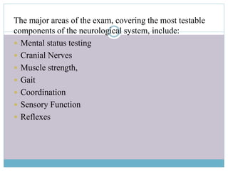
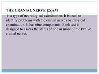
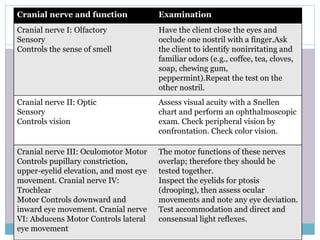
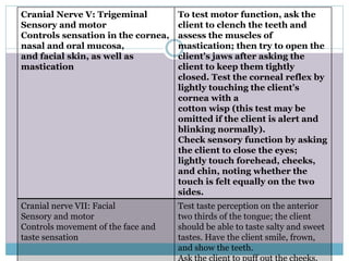
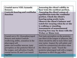
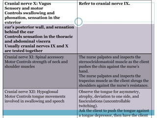
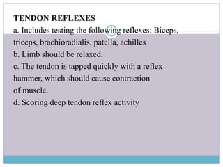
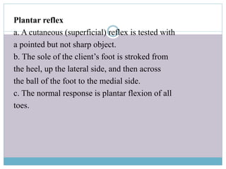
Neurological examination
A neurological examination is the assessment
of sensory neuron and motor responses,
especially reflexes, to determine whether the nervous
system is impaired.
The major areas of the exam, covering the most testable
components of the neurological system, include:
 Mental status testing
 Cranial Nerves
 Muscle strength,
 Gait
 Coordination
 Sensory Function
 Reflexes
MENTAL STATUS EXAMINATION
 mental and emotional status, behaviour and appearance,
language ability, and intellectual functioning, including
memory, knowledge, abstract thinking, association, and
judgment.
 The assessment of consciousness, often using
the Glasgow Coma Scale (GCS),Mental status
examination, often including the abbreviated mental test
score (AMTS) or mini mental state
examination (MMSE)
Glasgow coma scale
1
2 3 4 5 6
Eye
Does not
open eyes
Opens
eyes in
response
to pain
Opens
eyes in
response
to voice
Opens
eyes
spontaneo
usly
N/A N/A
Verbal
Makes no
sounds
Makes
sounds
Words
Confused,
disoriente
d
Oriented,
converses
normally
N/A
Motor
Makes no
movemen
ts
Extension
to painful
stimuli
Abnormal
flexion to
painful
stimuli
Flexion /
Withdraw
al to
painful
stimuli
Localizes
to painful
stimuli
Obeys
command
s
Level of consciousness
a. Assess the client’s behavior to determine
level of consciousness (e.g., alertness, confusion,
delirium, unconsciousness, stupor, coma); assessment
becomes increasingly invasive as the client is less
responsive.
b. Speak to client.
c. Assess appropriateness of behavior and
conversation.
d. Lightly touch the client (as culturally
appropriate).
THE CRANIAL NERVE EXAM
is a type of neurological examination. It is used to
identify problems with the cranial nerves by physical
examination. It has nine components. Each test is
designed to assess the status of one or more of the twelve
cranial nerves
Cranial nerve and function Examination
Cranial nerve I: Olfactory
Sensory
Controls the sense of smell
Have the client close the eyes and
occlude one nostril with a finger.Ask
the client to identify nonirritating and
familiar odors (e.g., coffee, tea, cloves,
soap, chewing gum,
peppermint).Repeat the test on the
other nostril.
Cranial nerve II: Optic
Sensory
Controls vision
Assess visual acuity with a Snellen
chart and perform an ophthalmoscopic
exam. Check peripheral vision by
confrontation. Check color vision.
Cranial nerve III: Oculomotor Motor
Controls pupillary constriction,
upper-eyelid elevation, and most eye
movement. Cranial nerve IV:
Trochlear
Motor Controls downward and
inward eye movement. Cranial nerve
VI: Abducens Motor Controls lateral
eye movement
The motor functions of these nerves
overlap; therefore they should be
tested together.
Inspect the eyelids for ptosis
(drooping), then assess ocular
movements and note any eye deviation.
Test accommodation and direct and
consensual light reflexes.
Cranial Nerve V: Trigeminal
Sensory and motor
Controls sensation in the cornea,
nasal and oral mucosa,
and facial skin, as well as
mastication
To test motor function, ask the
client to clench the teeth and
assess the muscles of
mastication; then try to open the
client’s jaws after asking the
client to keep them tightly
closed. Test the corneal reflex by
lightly touching the client’s
cornea with a
cotton wisp (this test may be
omitted if the client is alert and
blinking normally).
Check sensory function by asking
the client to close the eyes;
lightly touch forehead, cheeks,
and chin, noting whether the
touch is felt equally on the two
sides.
Cranial nerve VII: Facial
Sensory and motor
Controls movement of the face and
taste sensation
Test taste perception on the anterior
two thirds of the tongue; the client
should be able to taste salty and sweet
tastes. Have the client smile, frown,
and show the teeth.
Ask the client to puff out the cheeks.
Cranial nerve VIII: Acoustic
Sensory
Controls hearing and vestibular
function
Assessing the client’s ability to
hear tests the cochlear portion.
Assessing the client’s sense of
equilibrium tests the vestibular
portion. Check the client’s
hearing using acuity tests.
Observe the client’s balance and
watch for swaying when he or she
is walking or standing.
Assessment of sensorineural
hearing loss may be done with the
Weber or Rinne test.
Cranial nerve IX: Glossopharyngeal
Sensory and motor Controls
swallowing ability, sensation in the
pharyngeal soft
palate and tonsillar mucosa, taste
perception on the posterior third of
the tongue, and salivation Usually
cranial nerves IX and X are tested
together
.
Test taste perception on the posterior
one third of the tongue or pharynx; the
client should be able to taste bitter and
sour tastes. Inspect the soft palate and
watch for symmetrical elevation when
the client says “aaah.” Touch the
posterior pharyngeal wall with a tongue
depressor to elicit the gag reflex
Cranial nerve X: Vagus
Sensory and motor
Controls swallowing and
phonation, sensation in the
exterior
ear’s posterior wall, and sensation
behind the ear
Controls sensation in the thoracic
and abdominal viscera
Usually cranial nerves IX and X
are tested together
Refer to cranial nerve IX.
Cranial nerve XI: Spinal accessory
Motor Controls strength of neck and
shoulder muscles
The nurse palpates and inspects the
sternocleidomastoid muscle as the client
pushes the chin against the nurse’s
hand.
The nurse palpates and inspects the
trapezius muscle as the client shrugs the
shoulders against the nurse’s resistance.
Cranial nerve XII: Hypoglossal
Motor Controls tongue movements
involved in swallowing and speech
Observe the tongue for asymmetry,
atrophy, deviation to one side, and
fasciculations (uncontrollable
twitching).
Ask the client to push the tongue against
a tongue depressor, then have the client
MOTOR FUNCTION(MUSCLE STRENGTH)
a. Assess muscle tone, including strength and
equality.
b. Assess for voluntary and involuntary movements and
purposeful and non-purposeful movements.
c. This component of the neurological examination may
be performed during assessment of the musculoskeletal
system.
Grading and Recording Muscle Strength
Functional Level
Lovett
Scale
Grade
Percent
of
Normal
No evidence of contractility Zero (0) 0 0
Evidence of slight contractility Trace (T) 1 10
Complete range of motion with gravity
eliminated
Poor (P) 2 25
Complete range of motion with gravity Fair (F) 3 50
Complete range of motion against gravity with
some resistance
Good (G) 4 75
Complete range of motion against gravity with
full resistance
Normal
(N)
5 100
CEREBELLAR FUNCTION(GAIT)
a. Monitor gait as the client walks in a straight
line, heel to toe (tandem walking).
b. Romberg test: Client is asked to stand with the feet
together and the arms at the sides and to close the eyes
and hold the position; normally the client can maintain
posture and balance.
c. If appropriate, ask the client to perform a
shallow knee bend or to hop in place on one leg and then
the other.
COORDINATION
a. Assess by asking the client to perform rapid
alternating movements of the hands (e.g.,
turning the hands over and patting the
knees continuously).
b. The nurse asks the client to touch the nurse’s
finger, then his or her own nose; the client keeps the eyes
open and the nurse moves the finger to different spots to
ensure that the client’s
movements are smooth and accurate.
c. Heel-to-shin test: Assist the client into a supine
position, then ask the client to place the heel on the
opposite knee and run it down the shin; normally the
client moves the heel down the shin in a straight line.
SENSORY FUNCTION
a. Pain: Assess by applying an object with a
sharp point and one with a dull point to the client’s body
in random order; ask the client to identify the sharp and
dull feelings.
b. Light touch: Brush a piece of cotton over the
client’s skin at various locations in a random
order and ask the client to say when the touch is felt.
c. Vibration: Use a tuning fork to test the client’s
ability to feel vibrations over bony prominences;
ask the client to announce when the vibration starts and
stops.
d. Position sense (kinesthesia): Move the client’s
finger or toe up or down and ask the client which way it
has been moved; this tests the client’s ability to perceive
passive movement.
e. Stereognosis: Tests the client’s ability to recognize
objects placed in his or her hand
f. Graphesthesia: Tests the client’s ability to
identify a number traced on the client’s hand
g. Two-point discrimination: Tests the client’s
ability to discriminate two simultaneous
pinpricks on the skin
TENDON REFLEXES
a. Includes testing the following reflexes: Biceps,
triceps, brachioradialis, patella, achilles
b. Limb should be relaxed.
c. The tendon is tapped quickly with a reflex
hammer, which should cause contraction
of muscle.
d. Scoring deep tendon reflex activity
Plantar reflex
a. A cutaneous (superficial) reflex is tested with
a pointed but not sharp object.
b. The sole of the client’s foot is stroked from
the heel, up the lateral side, and then across
the ball of the foot to the medial side.
c. The normal response is plantar flexion of all
toes.
Scoring Deep Tendon Reflex
Activity
 0 = No response
 1+ = Sluggish or diminished
 2+ = Active or expected response
 3+ = Slightly hyperactive, more brisk than normal; not
necessarily pathologic
 4+ = Brisk, hyperactive with intermittent clonus
associated with disease
Neurological assessment include the following
Also :
Subjective data: Headaches, dizziness or vertigo,
tremors, weakness, incoordination, numbness or tingling
in any area of the body, difficulty speaking or swallowing,
medications being taken, history of seizures, history of
head injury or surgery, exposure to environmental or
occupational hazards (e.g., chemicals, alcohol, drugs)
Objective data: Assessment of cranial nerves,
level of consciousness, pupils, motor function,
cerebellar function, coordination, sensory function,
and reflexes
Vital signs: Check temperature, pulse, respirations, and
blood pressure; monitor for blood pressure or pulse
changes, which may indicate increased intracranial
pressure

Neurological examination

  • 1.
  • 2.
    A neurological examinationis the assessment of sensory neuron and motor responses, especially reflexes, to determine whether the nervous system is impaired.
  • 3.
    The major areasof the exam, covering the most testable components of the neurological system, include:  Mental status testing  Cranial Nerves  Muscle strength,  Gait  Coordination  Sensory Function  Reflexes
  • 4.
    MENTAL STATUS EXAMINATION mental and emotional status, behaviour and appearance, language ability, and intellectual functioning, including memory, knowledge, abstract thinking, association, and judgment.  The assessment of consciousness, often using the Glasgow Coma Scale (GCS),Mental status examination, often including the abbreviated mental test score (AMTS) or mini mental state examination (MMSE)
  • 5.
    Glasgow coma scale 1 23 4 5 6 Eye Does not open eyes Opens eyes in response to pain Opens eyes in response to voice Opens eyes spontaneo usly N/A N/A Verbal Makes no sounds Makes sounds Words Confused, disoriente d Oriented, converses normally N/A Motor Makes no movemen ts Extension to painful stimuli Abnormal flexion to painful stimuli Flexion / Withdraw al to painful stimuli Localizes to painful stimuli Obeys command s
  • 6.
    Level of consciousness a.Assess the client’s behavior to determine level of consciousness (e.g., alertness, confusion, delirium, unconsciousness, stupor, coma); assessment becomes increasingly invasive as the client is less responsive. b. Speak to client. c. Assess appropriateness of behavior and conversation. d. Lightly touch the client (as culturally appropriate).
  • 7.
    THE CRANIAL NERVEEXAM is a type of neurological examination. It is used to identify problems with the cranial nerves by physical examination. It has nine components. Each test is designed to assess the status of one or more of the twelve cranial nerves
  • 8.
    Cranial nerve andfunction Examination Cranial nerve I: Olfactory Sensory Controls the sense of smell Have the client close the eyes and occlude one nostril with a finger.Ask the client to identify nonirritating and familiar odors (e.g., coffee, tea, cloves, soap, chewing gum, peppermint).Repeat the test on the other nostril. Cranial nerve II: Optic Sensory Controls vision Assess visual acuity with a Snellen chart and perform an ophthalmoscopic exam. Check peripheral vision by confrontation. Check color vision. Cranial nerve III: Oculomotor Motor Controls pupillary constriction, upper-eyelid elevation, and most eye movement. Cranial nerve IV: Trochlear Motor Controls downward and inward eye movement. Cranial nerve VI: Abducens Motor Controls lateral eye movement The motor functions of these nerves overlap; therefore they should be tested together. Inspect the eyelids for ptosis (drooping), then assess ocular movements and note any eye deviation. Test accommodation and direct and consensual light reflexes.
  • 9.
    Cranial Nerve V:Trigeminal Sensory and motor Controls sensation in the cornea, nasal and oral mucosa, and facial skin, as well as mastication To test motor function, ask the client to clench the teeth and assess the muscles of mastication; then try to open the client’s jaws after asking the client to keep them tightly closed. Test the corneal reflex by lightly touching the client’s cornea with a cotton wisp (this test may be omitted if the client is alert and blinking normally). Check sensory function by asking the client to close the eyes; lightly touch forehead, cheeks, and chin, noting whether the touch is felt equally on the two sides. Cranial nerve VII: Facial Sensory and motor Controls movement of the face and taste sensation Test taste perception on the anterior two thirds of the tongue; the client should be able to taste salty and sweet tastes. Have the client smile, frown, and show the teeth. Ask the client to puff out the cheeks.
  • 10.
    Cranial nerve VIII:Acoustic Sensory Controls hearing and vestibular function Assessing the client’s ability to hear tests the cochlear portion. Assessing the client’s sense of equilibrium tests the vestibular portion. Check the client’s hearing using acuity tests. Observe the client’s balance and watch for swaying when he or she is walking or standing. Assessment of sensorineural hearing loss may be done with the Weber or Rinne test. Cranial nerve IX: Glossopharyngeal Sensory and motor Controls swallowing ability, sensation in the pharyngeal soft palate and tonsillar mucosa, taste perception on the posterior third of the tongue, and salivation Usually cranial nerves IX and X are tested together . Test taste perception on the posterior one third of the tongue or pharynx; the client should be able to taste bitter and sour tastes. Inspect the soft palate and watch for symmetrical elevation when the client says “aaah.” Touch the posterior pharyngeal wall with a tongue depressor to elicit the gag reflex
  • 11.
    Cranial nerve X:Vagus Sensory and motor Controls swallowing and phonation, sensation in the exterior ear’s posterior wall, and sensation behind the ear Controls sensation in the thoracic and abdominal viscera Usually cranial nerves IX and X are tested together Refer to cranial nerve IX. Cranial nerve XI: Spinal accessory Motor Controls strength of neck and shoulder muscles The nurse palpates and inspects the sternocleidomastoid muscle as the client pushes the chin against the nurse’s hand. The nurse palpates and inspects the trapezius muscle as the client shrugs the shoulders against the nurse’s resistance. Cranial nerve XII: Hypoglossal Motor Controls tongue movements involved in swallowing and speech Observe the tongue for asymmetry, atrophy, deviation to one side, and fasciculations (uncontrollable twitching). Ask the client to push the tongue against a tongue depressor, then have the client
  • 12.
    MOTOR FUNCTION(MUSCLE STRENGTH) a.Assess muscle tone, including strength and equality. b. Assess for voluntary and involuntary movements and purposeful and non-purposeful movements. c. This component of the neurological examination may be performed during assessment of the musculoskeletal system.
  • 13.
    Grading and RecordingMuscle Strength Functional Level Lovett Scale Grade Percent of Normal No evidence of contractility Zero (0) 0 0 Evidence of slight contractility Trace (T) 1 10 Complete range of motion with gravity eliminated Poor (P) 2 25 Complete range of motion with gravity Fair (F) 3 50 Complete range of motion against gravity with some resistance Good (G) 4 75 Complete range of motion against gravity with full resistance Normal (N) 5 100
  • 14.
    CEREBELLAR FUNCTION(GAIT) a. Monitorgait as the client walks in a straight line, heel to toe (tandem walking). b. Romberg test: Client is asked to stand with the feet together and the arms at the sides and to close the eyes and hold the position; normally the client can maintain posture and balance. c. If appropriate, ask the client to perform a shallow knee bend or to hop in place on one leg and then the other.
  • 15.
    COORDINATION a. Assess byasking the client to perform rapid alternating movements of the hands (e.g., turning the hands over and patting the knees continuously). b. The nurse asks the client to touch the nurse’s finger, then his or her own nose; the client keeps the eyes open and the nurse moves the finger to different spots to ensure that the client’s movements are smooth and accurate.
  • 16.
    c. Heel-to-shin test:Assist the client into a supine position, then ask the client to place the heel on the opposite knee and run it down the shin; normally the client moves the heel down the shin in a straight line.
  • 17.
    SENSORY FUNCTION a. Pain:Assess by applying an object with a sharp point and one with a dull point to the client’s body in random order; ask the client to identify the sharp and dull feelings. b. Light touch: Brush a piece of cotton over the client’s skin at various locations in a random order and ask the client to say when the touch is felt.
  • 18.
    c. Vibration: Usea tuning fork to test the client’s ability to feel vibrations over bony prominences; ask the client to announce when the vibration starts and stops. d. Position sense (kinesthesia): Move the client’s finger or toe up or down and ask the client which way it has been moved; this tests the client’s ability to perceive passive movement.
  • 19.
    e. Stereognosis: Teststhe client’s ability to recognize objects placed in his or her hand f. Graphesthesia: Tests the client’s ability to identify a number traced on the client’s hand g. Two-point discrimination: Tests the client’s ability to discriminate two simultaneous pinpricks on the skin
  • 20.
    TENDON REFLEXES a. Includestesting the following reflexes: Biceps, triceps, brachioradialis, patella, achilles b. Limb should be relaxed. c. The tendon is tapped quickly with a reflex hammer, which should cause contraction of muscle. d. Scoring deep tendon reflex activity
  • 21.
    Plantar reflex a. Acutaneous (superficial) reflex is tested with a pointed but not sharp object. b. The sole of the client’s foot is stroked from the heel, up the lateral side, and then across the ball of the foot to the medial side. c. The normal response is plantar flexion of all toes.
  • 22.
    Scoring Deep TendonReflex Activity  0 = No response  1+ = Sluggish or diminished  2+ = Active or expected response  3+ = Slightly hyperactive, more brisk than normal; not necessarily pathologic  4+ = Brisk, hyperactive with intermittent clonus associated with disease
  • 23.
    Neurological assessment includethe following Also : Subjective data: Headaches, dizziness or vertigo, tremors, weakness, incoordination, numbness or tingling in any area of the body, difficulty speaking or swallowing, medications being taken, history of seizures, history of head injury or surgery, exposure to environmental or occupational hazards (e.g., chemicals, alcohol, drugs) Objective data: Assessment of cranial nerves, level of consciousness, pupils, motor function, cerebellar function, coordination, sensory function, and reflexes
  • 24.
    Vital signs: Checktemperature, pulse, respirations, and blood pressure; monitor for blood pressure or pulse changes, which may indicate increased intracranial pressure