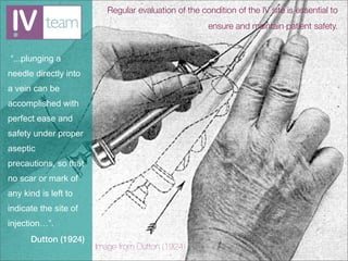
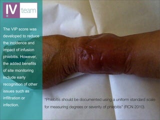
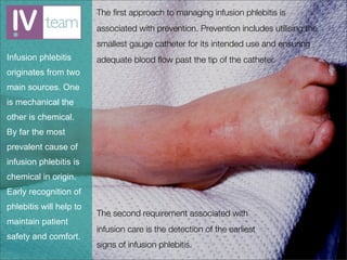
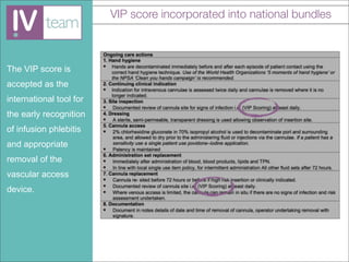
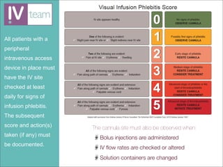
IV Care & Management: Site Monitoring
Implementing the VIP score
Regular evaluation of the condition of the IV site is essential to
                                                      ensure and maintain patient safety.


“...plunging a
needle directly into
a vein can be
accomplished with
perfect ease and
safety under proper
aseptic
precautions, so that
no scar or mark of
any kind is left to
indicate the site of
injection…”.
      Dutton (1924)
                       Image from Dutton (1924)
The VIP score was
developed to reduce
the incidence and
impact of infusion
phlebitis. However,
the added benefits
of site monitoring
include early
recognition of other
issues such as
infiltration or        “Phlebitis should be documented using a uniform standard scale
infection.             for measuring degrees or severity of phlebitis” (RCN 2010).
The first approach to managing infusion phlebitis is
                         associated with prevention. Prevention includes utilising the
                         smallest gauge catheter for its intended use and ensuring
Infusion phlebitis       adequate blood flow past the tip of the catheter.
originates from two
main sources. One
is mechanical the
other is chemical.
By far the most
prevalent cause of
infusion phlebitis is
chemical in origin.
Early recognition of
phlebitis will help to
                         The second requirement associated with
maintain patient
                         infusion care is the detection of the earliest
safety and comfort.
                         signs of infusion phlebitis.
Blood flow in the
cephalic and basilic
veins in the upper
arms is 40 to 95ml/
min compared to
the superior vena
cava with a blood
flow of 2000 ml/min
(Stranz and
Kastango 2002).

                       Image from:
                       NAVAN (1998) Tip location of peripherally inserted central
                       catheters. Journal of Vascular Access Devices. 3(2), p.8-10.
Table from:
                       Stranz, M. and Kastango, E.S. (2002) A review of pH and
                       osmolarity. International Journal of Pharmaceutical
                       Compounding. 6(3), p.216-220.
"...phlebitis caused
by infusates of
incorrect pH and
osmolarity occurs
frequently... The
degree of cellular
damage from either
low or high pH is
determined by the
type of tissue
exposed to the pH
and the duration of
exposure” (Stranz
and Kastango 2002).
Failure to monitor
and document the
condition of a
peripheral
intravenous catheter
site may result in a
claim due to a
breach in duty of
care.


                       http://www.kennedys-law.com/media/docs/KennedysMedicalLawBriefMarch2010_832010.htm
VIP score recommendations

                       Gallant and Schultz (2006) state that...

                       “The VIP scale, as evaluated in this study, was
In 2006 Paulette
                       considered to be a valid and reliable measure for
Gallant and Alyce      determining when a PIV catheter should be
Schultz completed      removed” Galant and Schultz (2006).
an evaluation of the
                       The VIP score is the tool recommended by the RCN (2010)
VIP score as a tool
                       and the Department of Health (2010) in the UK.
that determines the
appropriate            Also, the VIP score is recommended in the Infusion
                       Nursing Standards of Practice (INS 2011). Stating that...
discontinuation of
peripheral             “The Visual Infusion Phlebitis (VIP) scale has content
intravenous            validity, inter-rater reliability, and is clinically feasible.
catheters.             This scale includes suggested actions matched to
                       each scale score” Infusion Nurses Society (2011).
VIP score incorporated into national bundles


The VIP score is
accepted as the
international tool for
the early recognition
of infusion phlebitis
and appropriate
removal of the
vascular access
device.
All patients with a
peripheral
intravenous access
device in place must
have the IV site
checked at least
daily for signs of
infusion phlebitis.
The subsequent
score and action(s)    The cannula site must also be observed when:
taken (if any) must       Bolus injections are administered
be documented.
                          IV flow rates are checked or altered
                          Solution containers are changed
References
                        Danchaivijitr, S., Srihapol, N., Pakaworawuth, S., Vaithayapiches, S., Judang, T., Pumsuwan, V. and
                        Kachintorn, K. (1995) Infusion-related phlebitis. Journal of the Medical Association of Thailand. 78,
                        Suppl 2:S85-90.

                        Department of Health (2010) High impact intervention: Peripheral intravenous cannula care.
                        DH, London.
Danchaivijitr et al     Dutton, W.F. (1924) Intravenous Therapy: Its application in the modern practice of medicine. F.A.
                        Davis Company, Philadelphia.
(1995) states 34.1
                        Gallant P and Schultz AA (2006) Evaluation of a visual infusion phlebitis scale for determining
per cent of infusions   appropriate discontinuation of peripheral intravenous catheters. Journal of Infusion Nursing.
                        vol. 29, no. 6, p. 338-45.
are interrupted by
                        INS (2011) Infusion Nursing Standards of Practice. Journal of Infusion Nursing. Supplement. 34(1s).
complications of
                        Jackson A. (1998) A battle in vein: infusion phlebitis. Nursing Times. 94 (4), p.68-71.
which 6.2 per cent
                        Jackson A. (2003) Reflecting on the nursing contribution to vascular access. British Journal of
were infusion           Nursing. 12(11), p.657-665.

phlebitis.              Mowry, J.L. and Hartman, L.S. (2011) Intravascular thrombophlebitis related to the peripheral
                        infusion of amiodarone and vancomycin. Western Journal of Nursing Research.33(3), p.
                        457-471

                        NAVAN (1998) Tip location of peripherally inserted central catheters. Journal of Vascular Access
                        Devices. 3(2), p.8-10.

                        RCN (2010) Standards for infusion therapy. Royal College of Nursing, London.

                        Stranz, M. and Kastango, E.S. (2002) A review of pH and osmolarity. International Journal of
                        Pharmaceutical Compounding. 6(3), p.216-220.

Introduction to the Visual Infusion Phlebitis (VIP) score

  • 1.
    IV Care &Management: Site Monitoring Implementing the VIP score
  • 2.
    Regular evaluation ofthe condition of the IV site is essential to ensure and maintain patient safety. “...plunging a needle directly into a vein can be accomplished with perfect ease and safety under proper aseptic precautions, so that no scar or mark of any kind is left to indicate the site of injection…”. Dutton (1924) Image from Dutton (1924)
  • 3.
    The VIP scorewas developed to reduce the incidence and impact of infusion phlebitis. However, the added benefits of site monitoring include early recognition of other issues such as infiltration or “Phlebitis should be documented using a uniform standard scale infection. for measuring degrees or severity of phlebitis” (RCN 2010).
  • 4.
    The first approachto managing infusion phlebitis is associated with prevention. Prevention includes utilising the smallest gauge catheter for its intended use and ensuring Infusion phlebitis adequate blood flow past the tip of the catheter. originates from two main sources. One is mechanical the other is chemical. By far the most prevalent cause of infusion phlebitis is chemical in origin. Early recognition of phlebitis will help to The second requirement associated with maintain patient infusion care is the detection of the earliest safety and comfort. signs of infusion phlebitis.
  • 5.
    Blood flow inthe cephalic and basilic veins in the upper arms is 40 to 95ml/ min compared to the superior vena cava with a blood flow of 2000 ml/min (Stranz and Kastango 2002). Image from: NAVAN (1998) Tip location of peripherally inserted central catheters. Journal of Vascular Access Devices. 3(2), p.8-10.
  • 6.
    Table from: Stranz, M. and Kastango, E.S. (2002) A review of pH and osmolarity. International Journal of Pharmaceutical Compounding. 6(3), p.216-220. "...phlebitis caused by infusates of incorrect pH and osmolarity occurs frequently... The degree of cellular damage from either low or high pH is determined by the type of tissue exposed to the pH and the duration of exposure” (Stranz and Kastango 2002).
  • 7.
    Failure to monitor anddocument the condition of a peripheral intravenous catheter site may result in a claim due to a breach in duty of care. http://www.kennedys-law.com/media/docs/KennedysMedicalLawBriefMarch2010_832010.htm
  • 8.
    VIP score recommendations Gallant and Schultz (2006) state that... “The VIP scale, as evaluated in this study, was In 2006 Paulette considered to be a valid and reliable measure for Gallant and Alyce determining when a PIV catheter should be Schultz completed removed” Galant and Schultz (2006). an evaluation of the The VIP score is the tool recommended by the RCN (2010) VIP score as a tool and the Department of Health (2010) in the UK. that determines the appropriate Also, the VIP score is recommended in the Infusion Nursing Standards of Practice (INS 2011). Stating that... discontinuation of peripheral “The Visual Infusion Phlebitis (VIP) scale has content intravenous validity, inter-rater reliability, and is clinically feasible. catheters. This scale includes suggested actions matched to each scale score” Infusion Nurses Society (2011).
  • 9.
    VIP score incorporatedinto national bundles The VIP score is accepted as the international tool for the early recognition of infusion phlebitis and appropriate removal of the vascular access device.
  • 10.
    All patients witha peripheral intravenous access device in place must have the IV site checked at least daily for signs of infusion phlebitis. The subsequent score and action(s) The cannula site must also be observed when: taken (if any) must Bolus injections are administered be documented. IV flow rates are checked or altered Solution containers are changed
  • 11.
    References Danchaivijitr, S., Srihapol, N., Pakaworawuth, S., Vaithayapiches, S., Judang, T., Pumsuwan, V. and Kachintorn, K. (1995) Infusion-related phlebitis. Journal of the Medical Association of Thailand. 78, Suppl 2:S85-90. Department of Health (2010) High impact intervention: Peripheral intravenous cannula care. DH, London. Danchaivijitr et al Dutton, W.F. (1924) Intravenous Therapy: Its application in the modern practice of medicine. F.A. Davis Company, Philadelphia. (1995) states 34.1 Gallant P and Schultz AA (2006) Evaluation of a visual infusion phlebitis scale for determining per cent of infusions appropriate discontinuation of peripheral intravenous catheters. Journal of Infusion Nursing. vol. 29, no. 6, p. 338-45. are interrupted by INS (2011) Infusion Nursing Standards of Practice. Journal of Infusion Nursing. Supplement. 34(1s). complications of Jackson A. (1998) A battle in vein: infusion phlebitis. Nursing Times. 94 (4), p.68-71. which 6.2 per cent Jackson A. (2003) Reflecting on the nursing contribution to vascular access. British Journal of were infusion Nursing. 12(11), p.657-665. phlebitis. Mowry, J.L. and Hartman, L.S. (2011) Intravascular thrombophlebitis related to the peripheral infusion of amiodarone and vancomycin. Western Journal of Nursing Research.33(3), p. 457-471 NAVAN (1998) Tip location of peripherally inserted central catheters. Journal of Vascular Access Devices. 3(2), p.8-10. RCN (2010) Standards for infusion therapy. Royal College of Nursing, London. Stranz, M. and Kastango, E.S. (2002) A review of pH and osmolarity. International Journal of Pharmaceutical Compounding. 6(3), p.216-220.