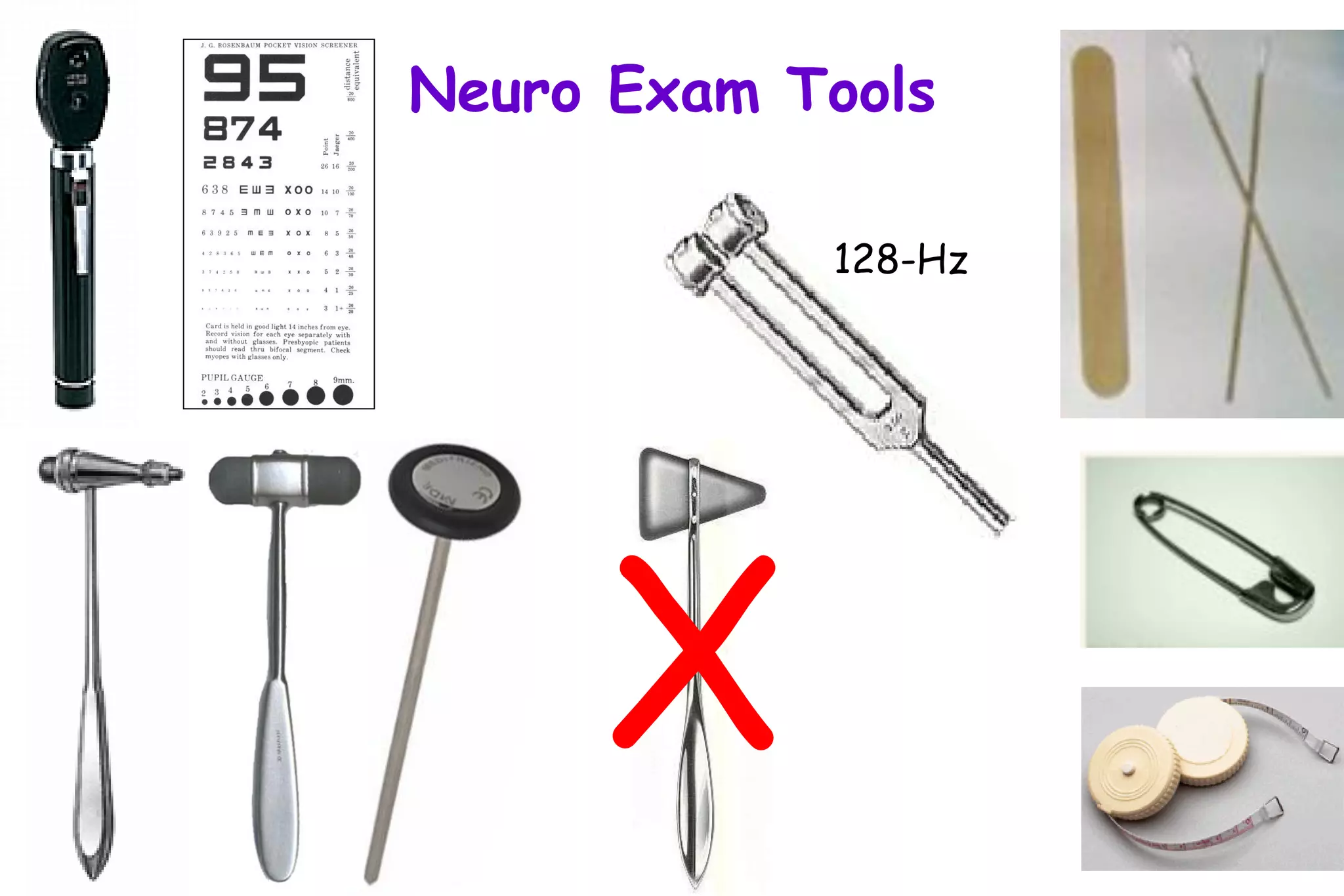
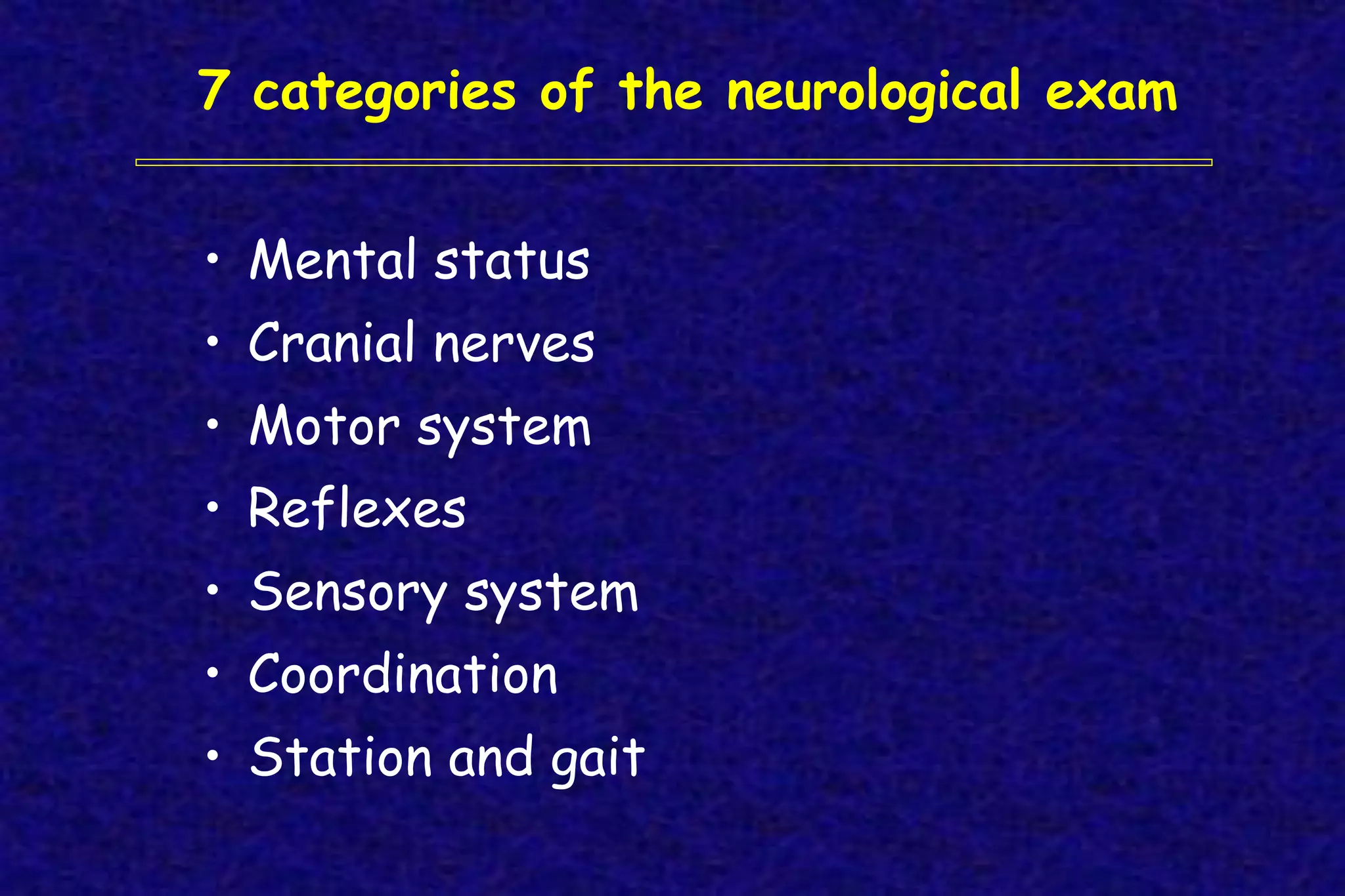
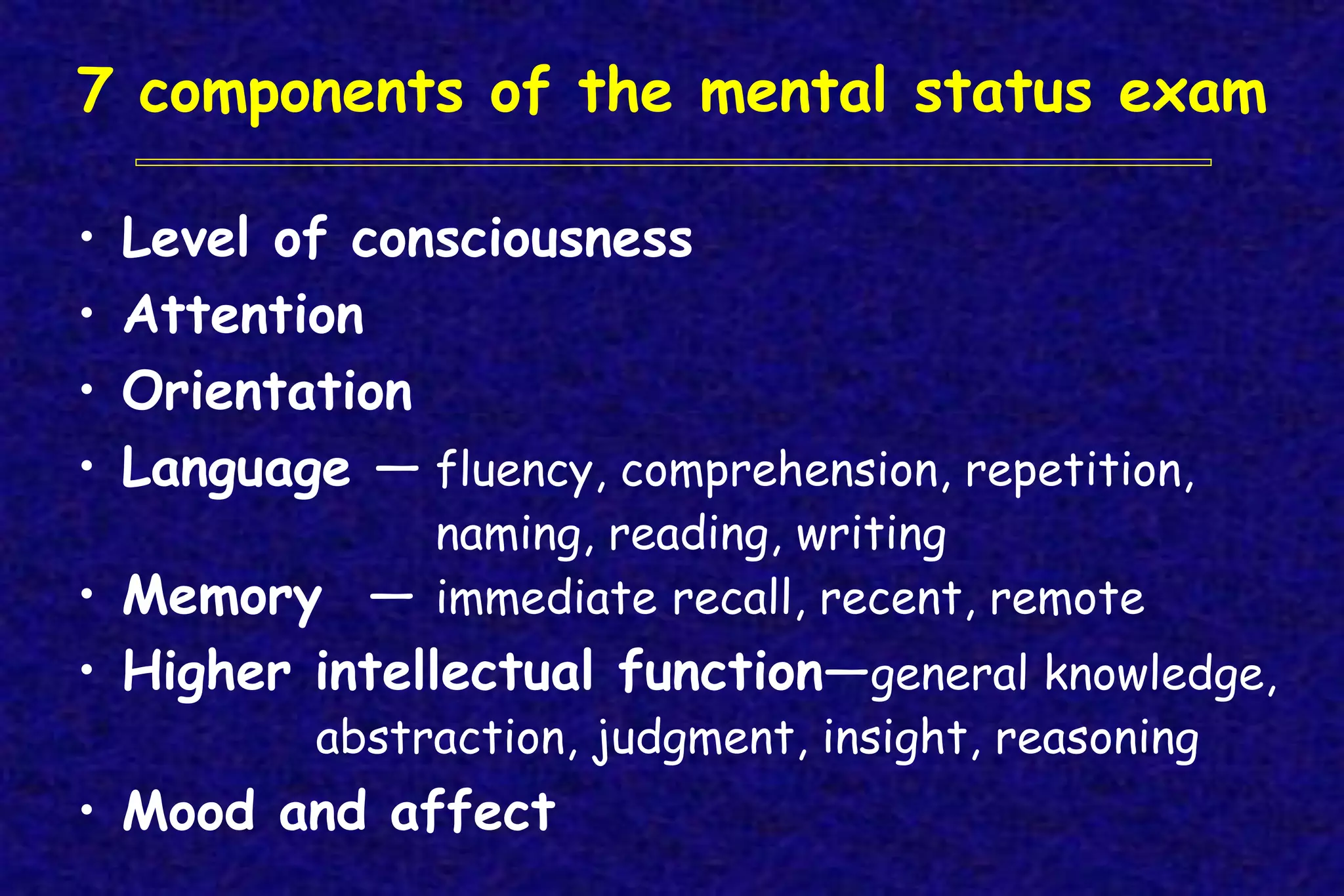
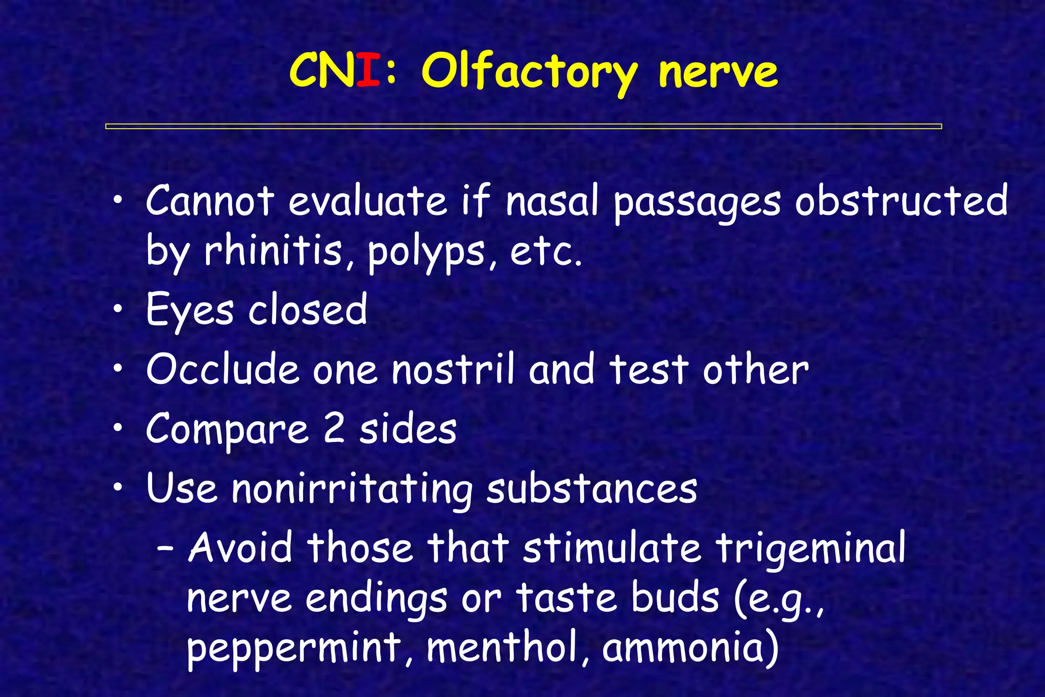
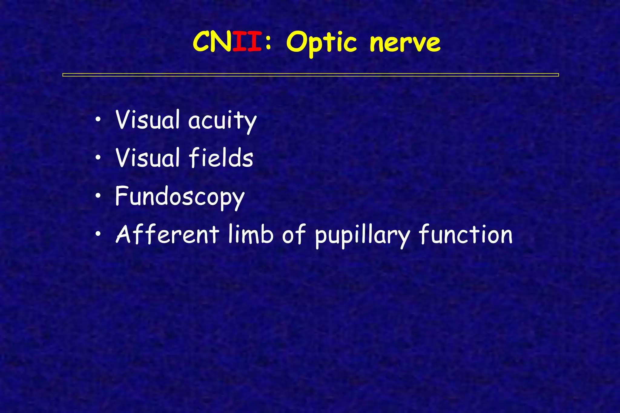
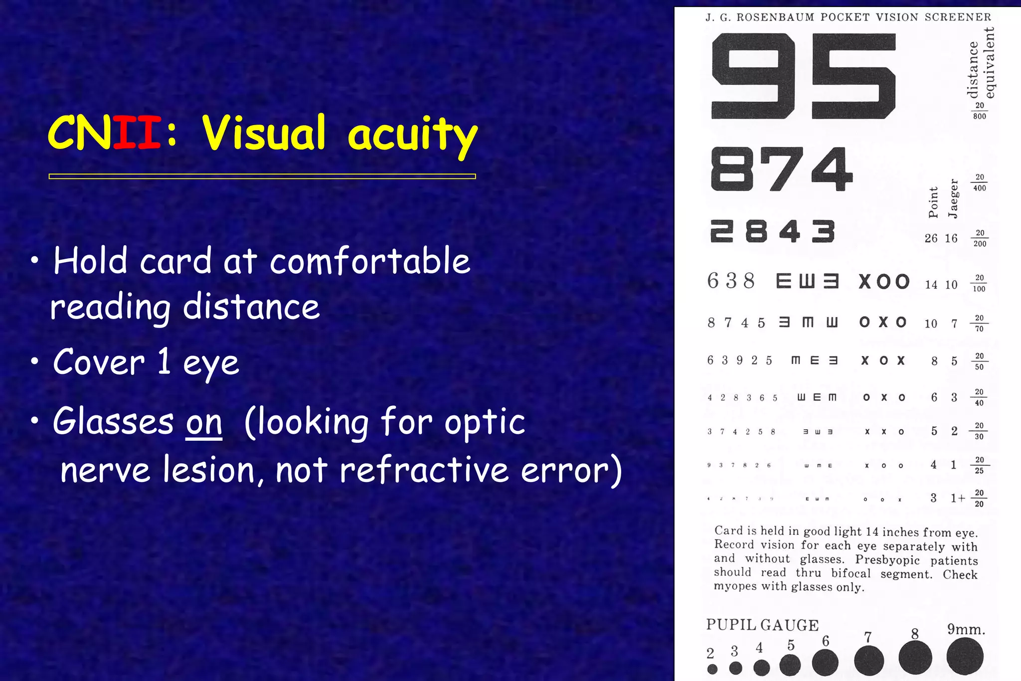
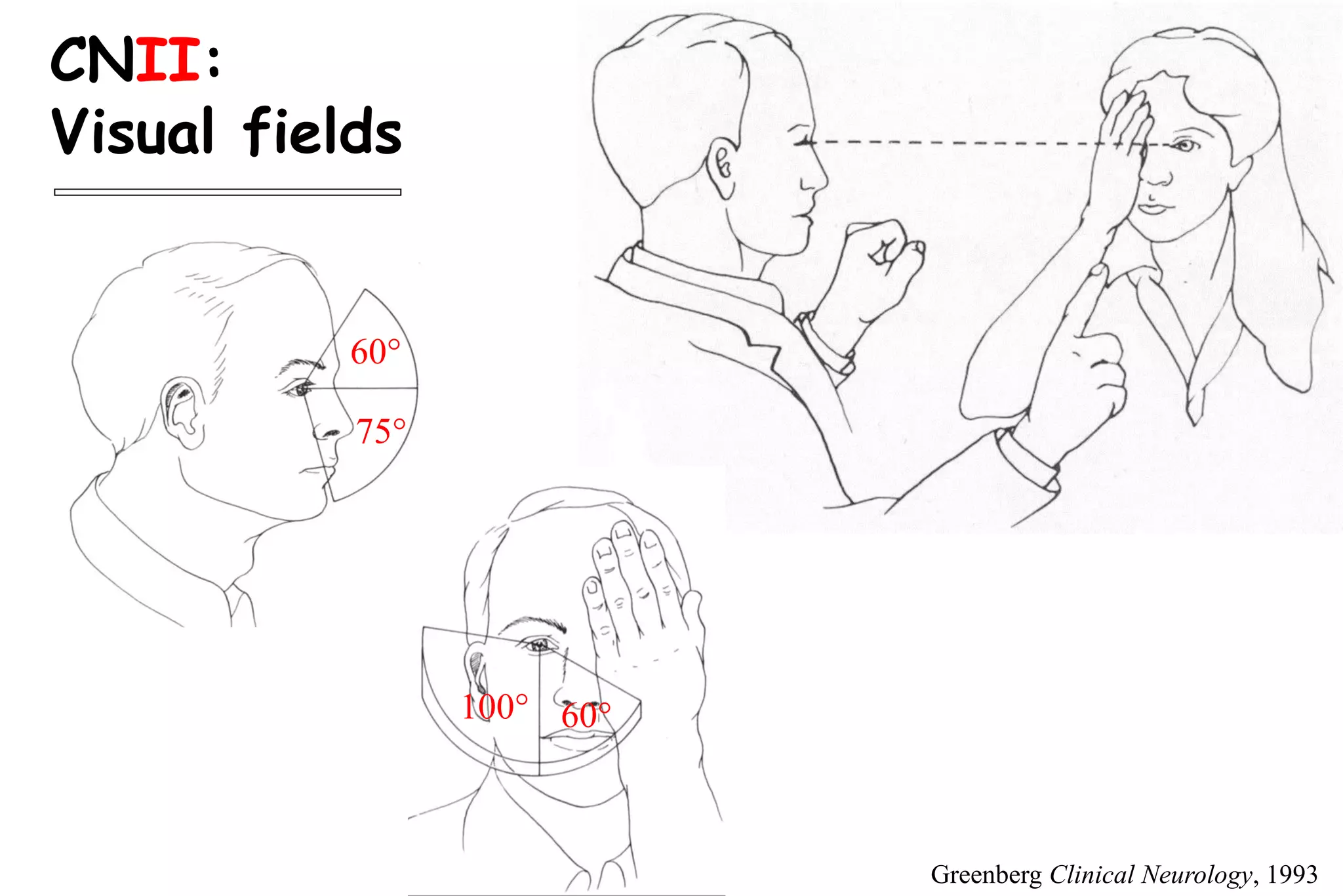
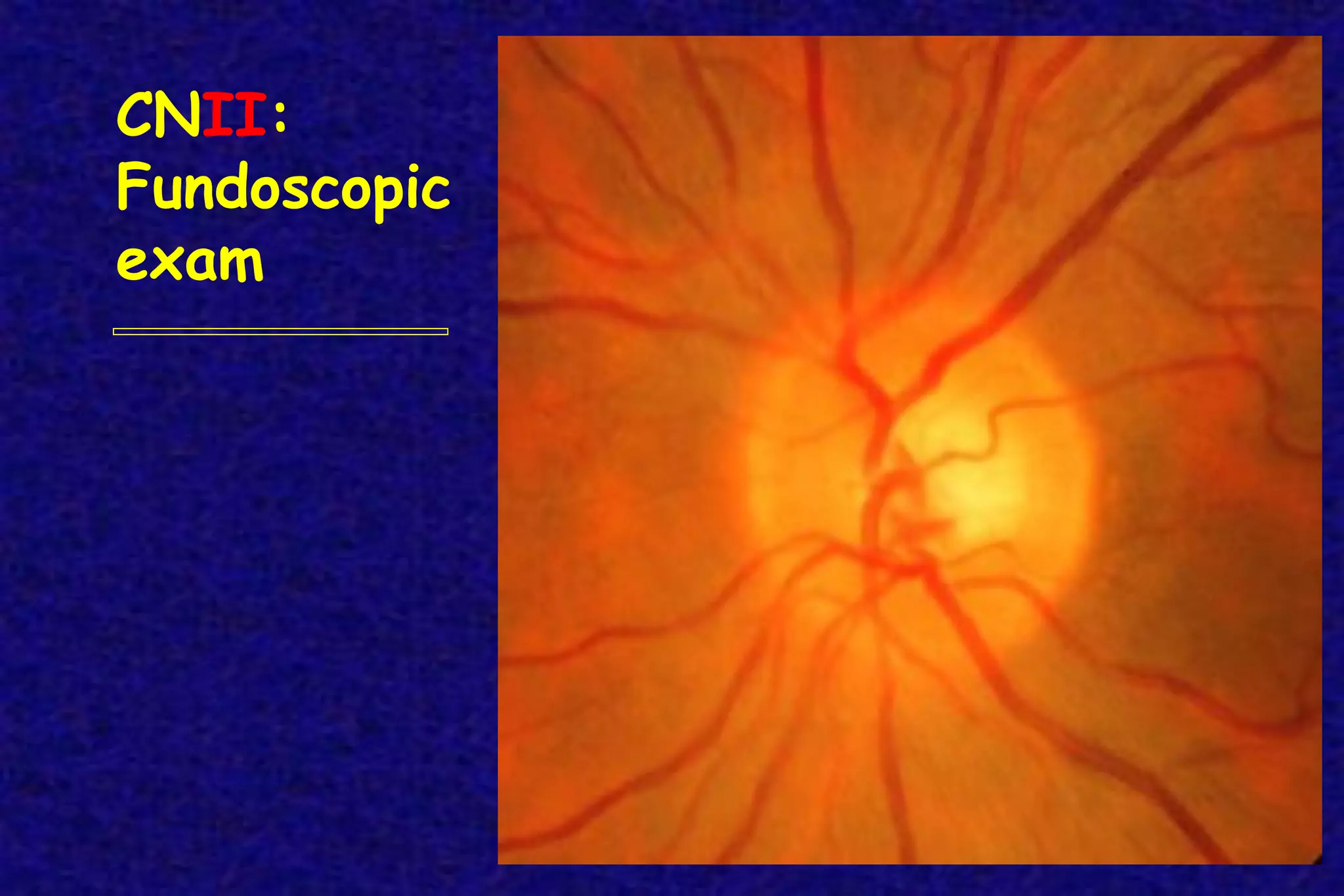
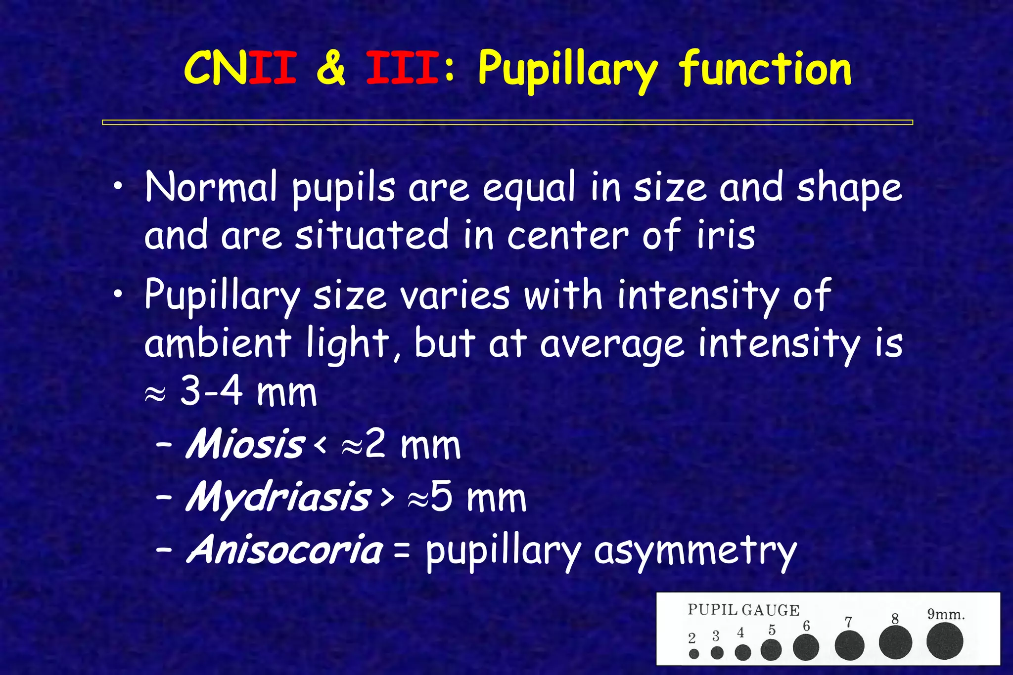
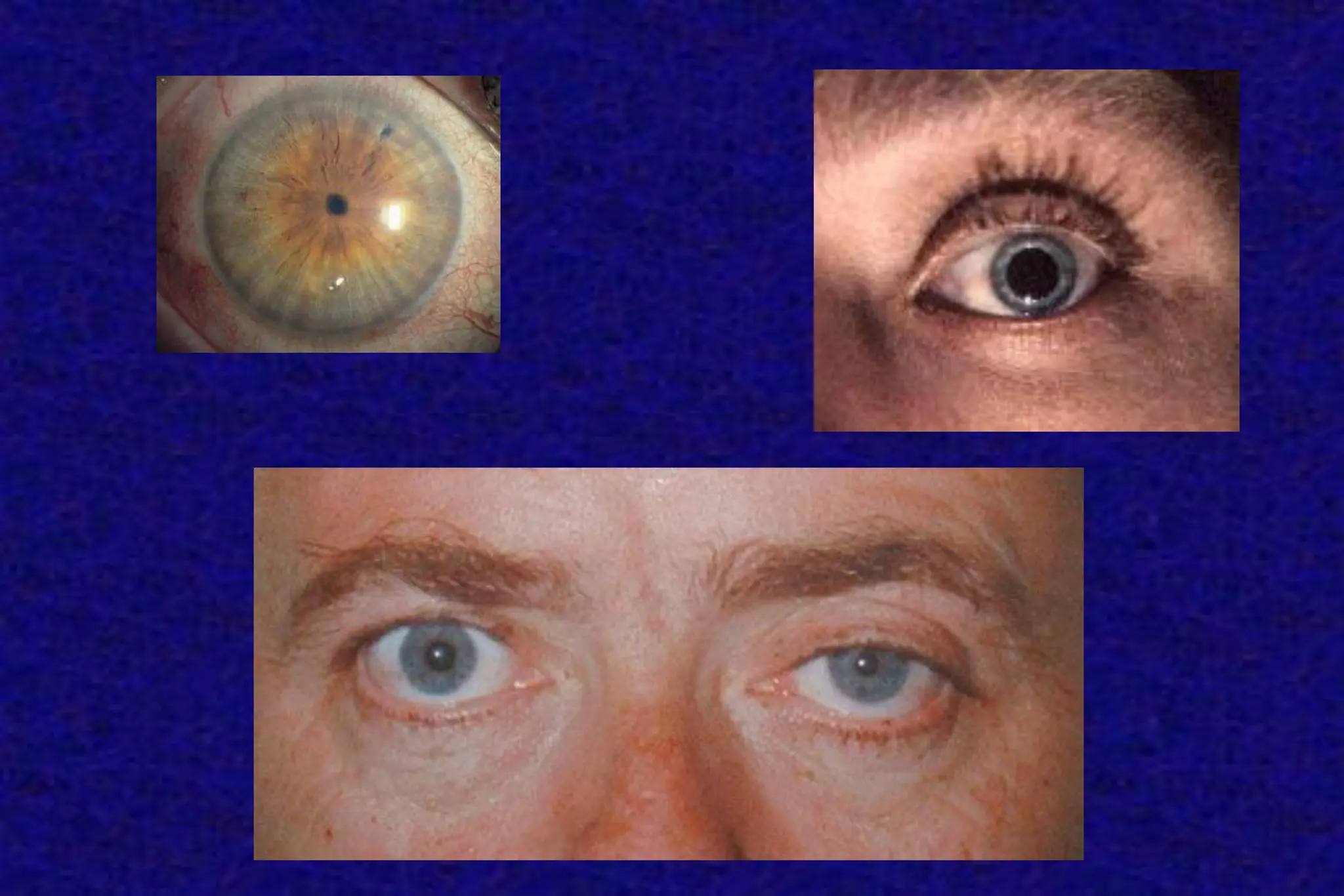
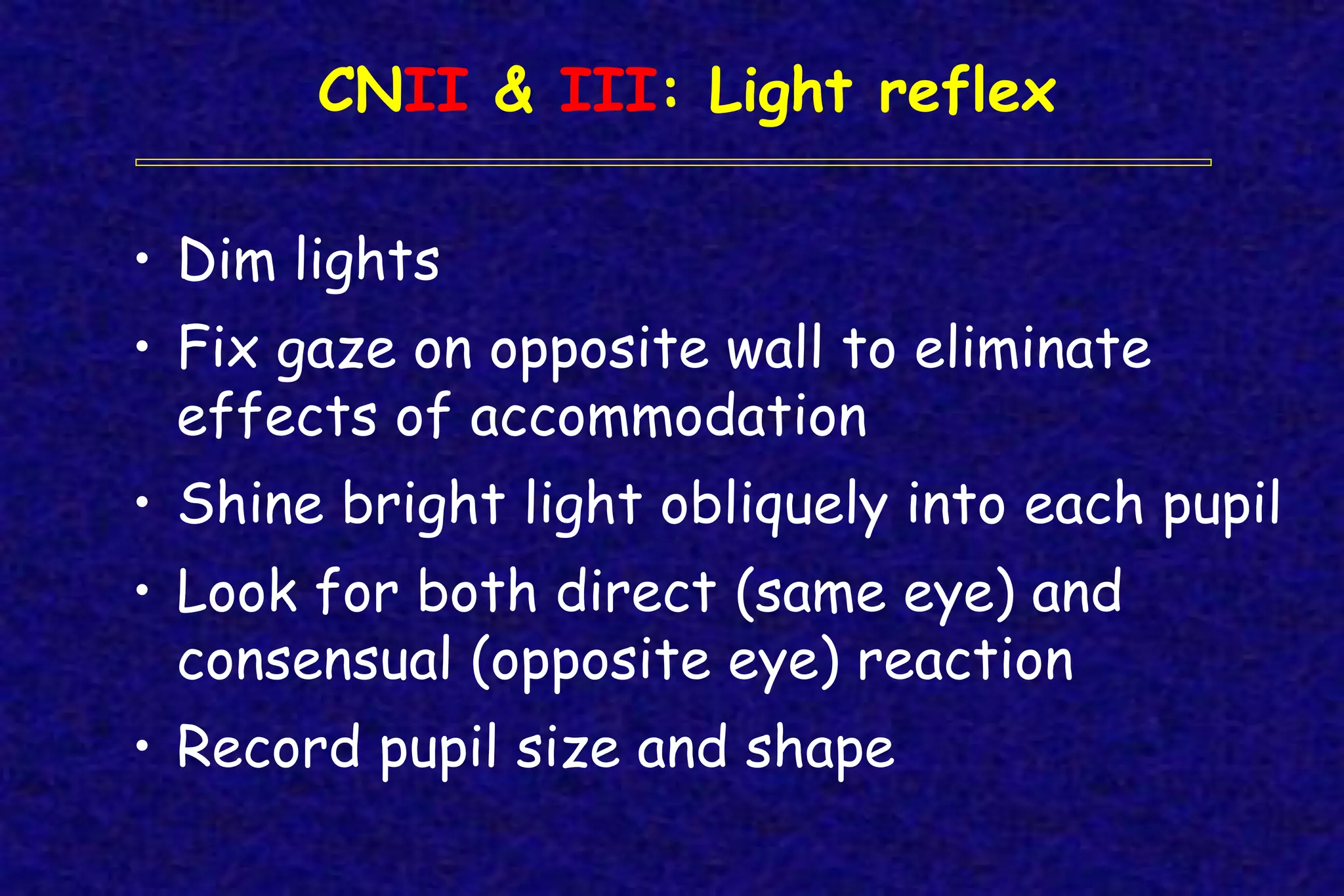
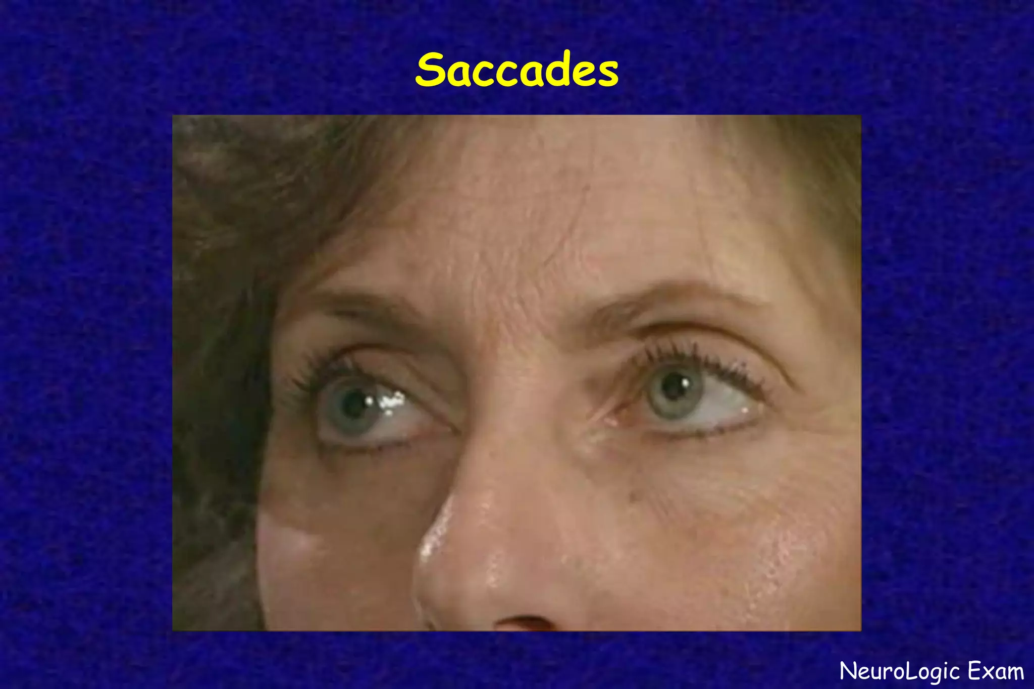
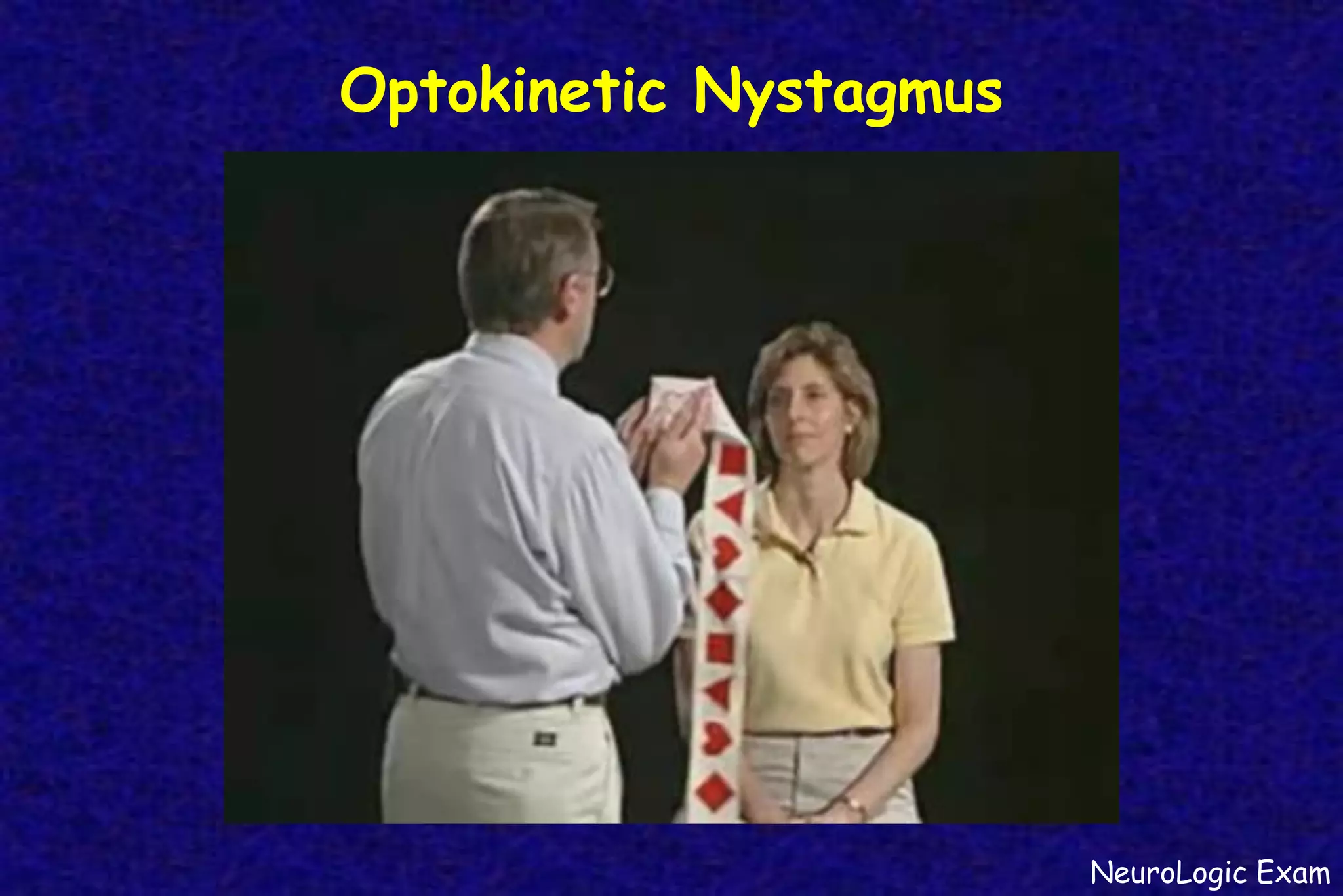
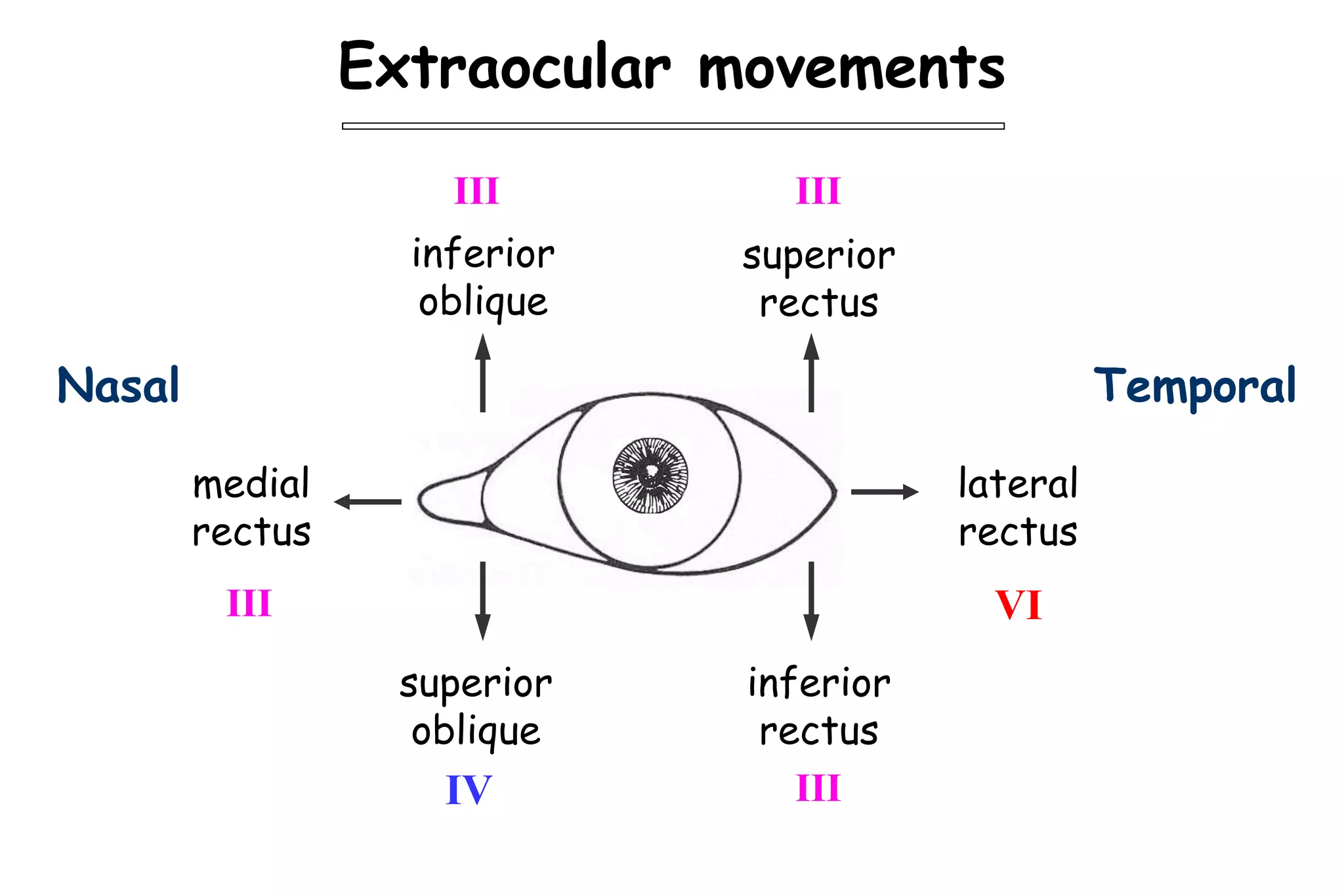
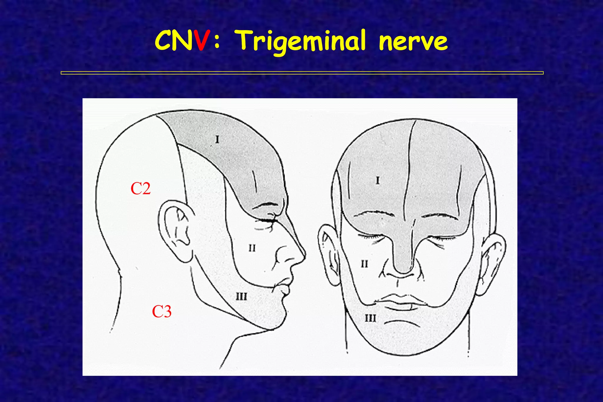
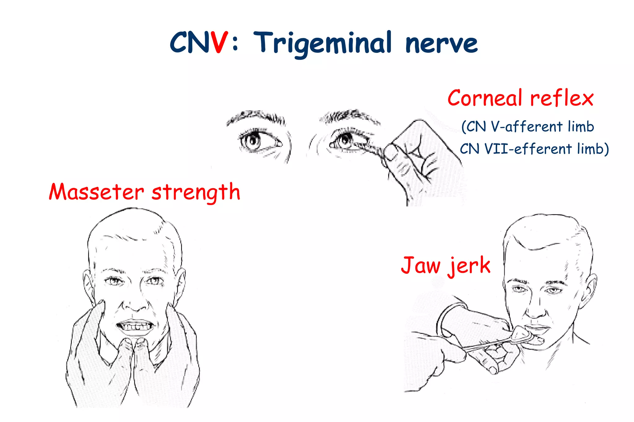
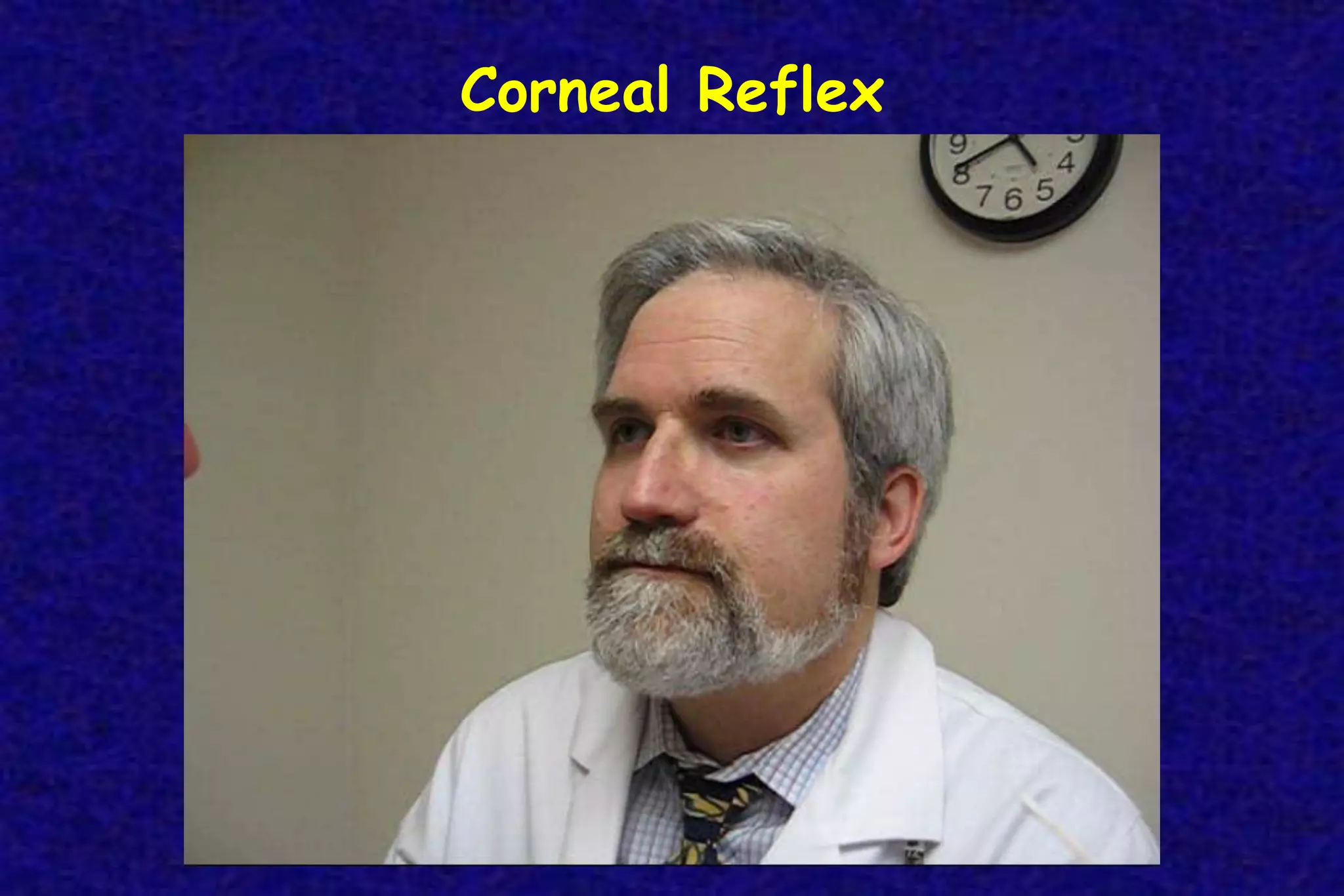
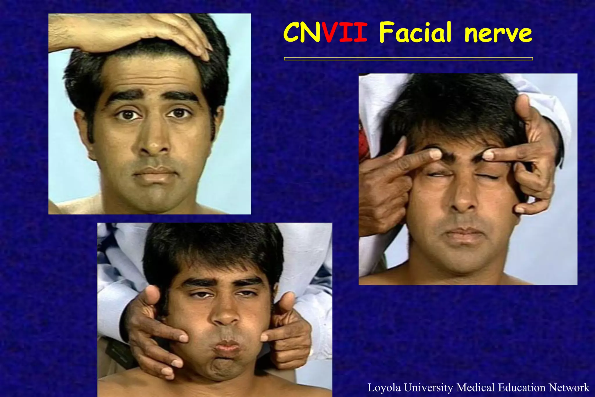
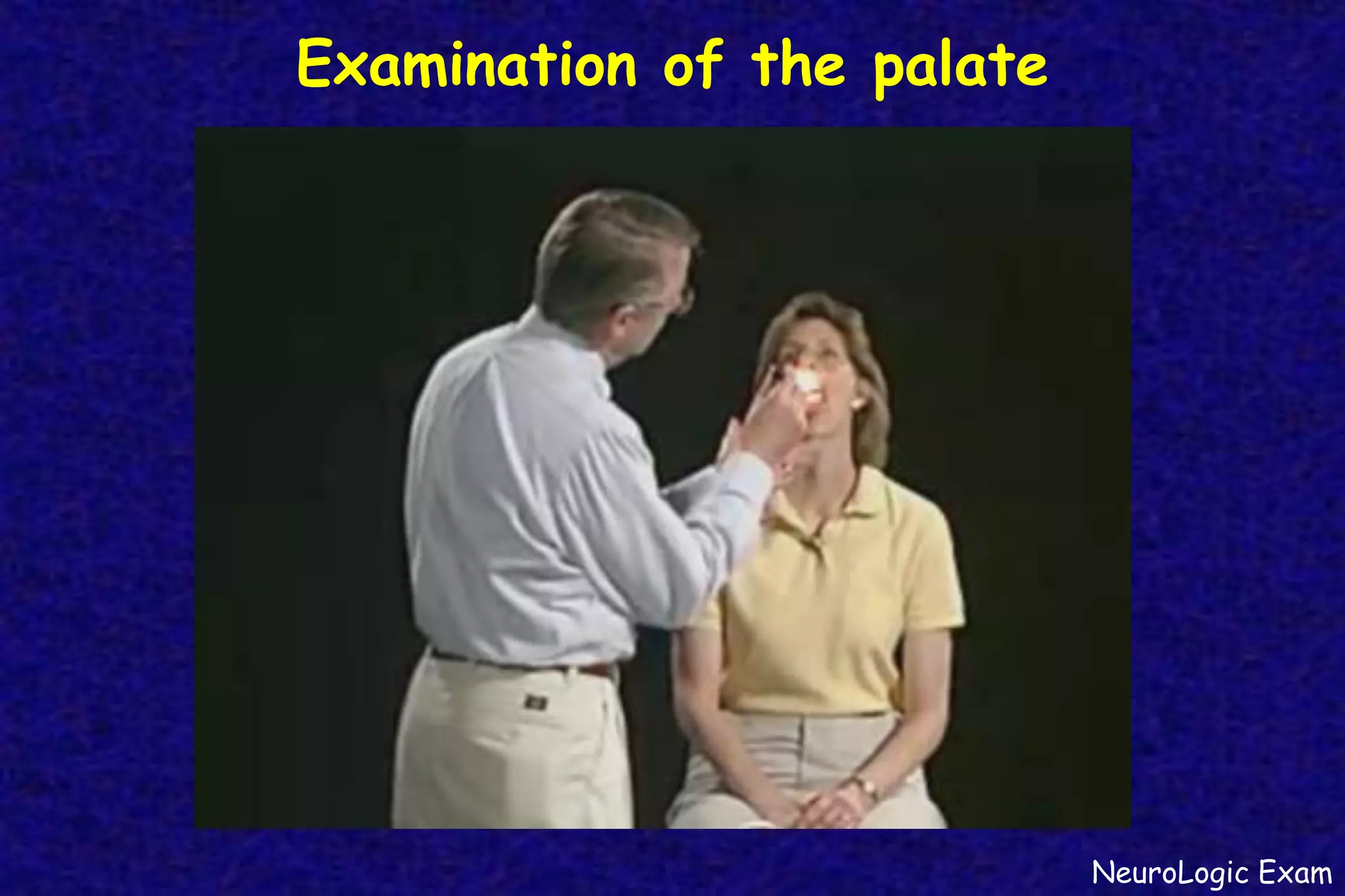
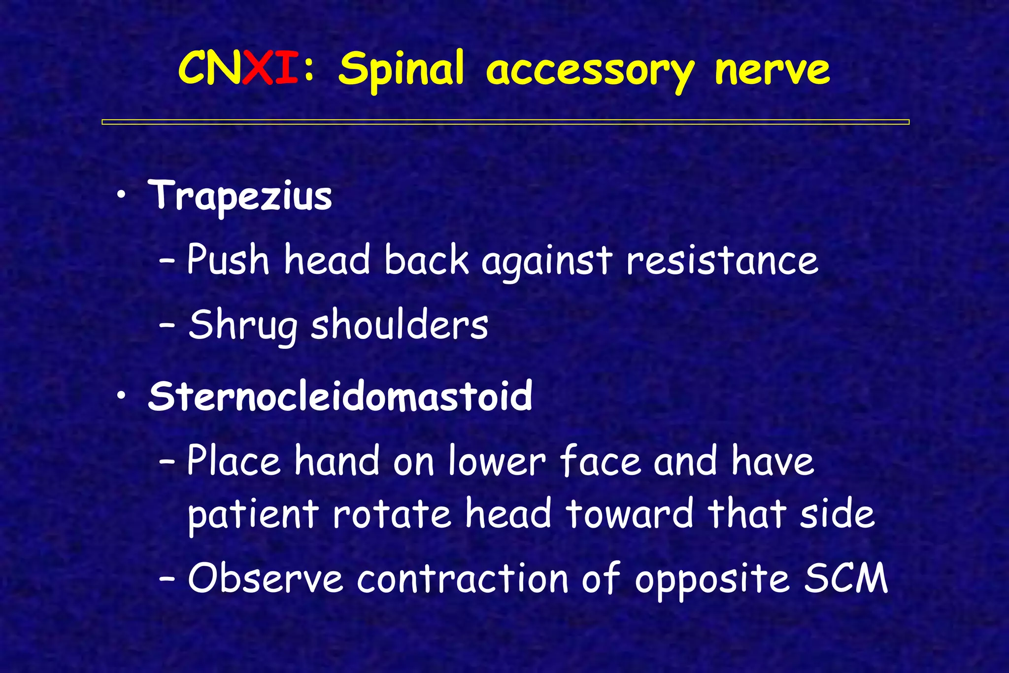
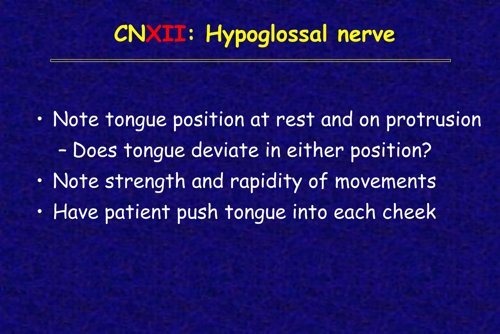
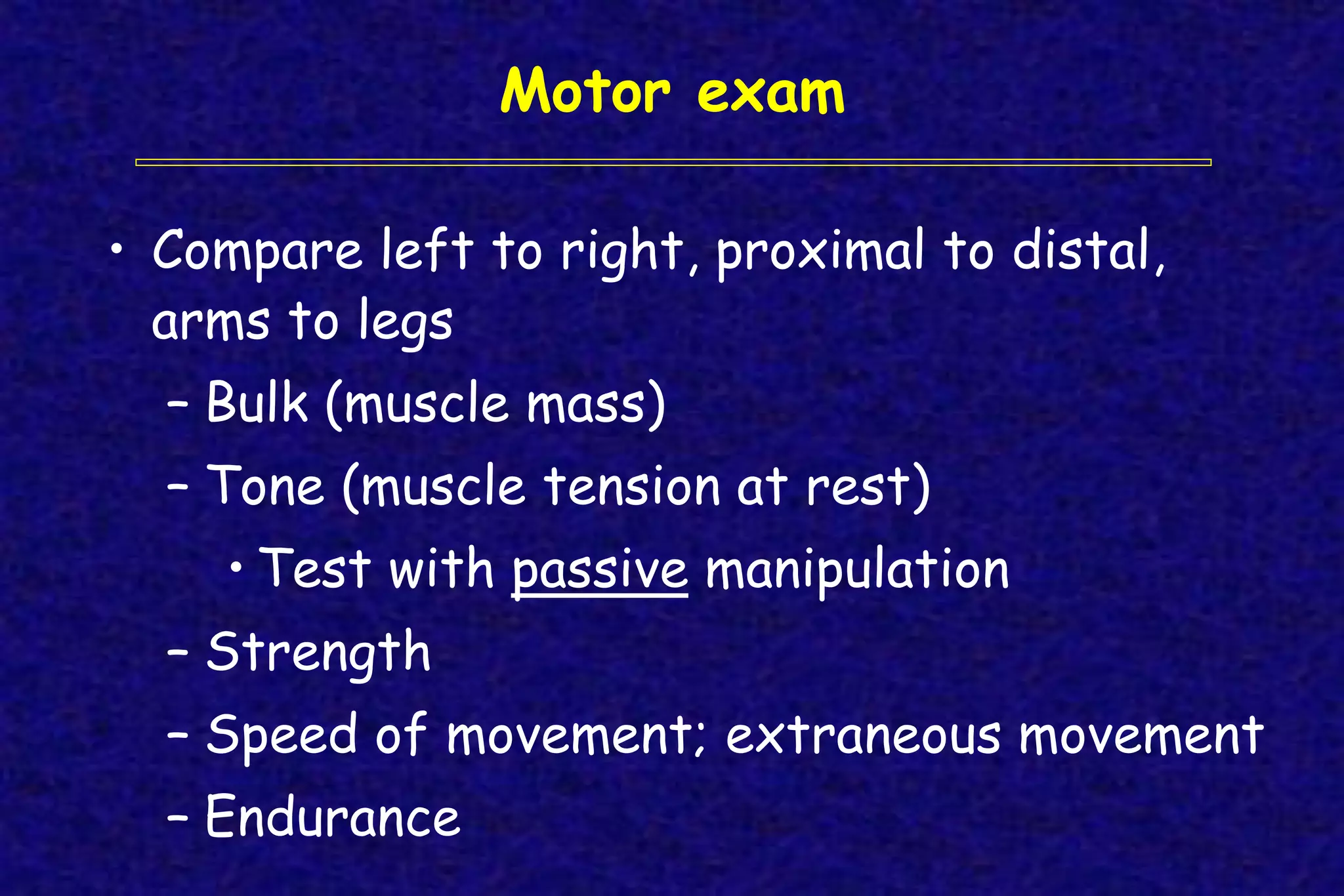
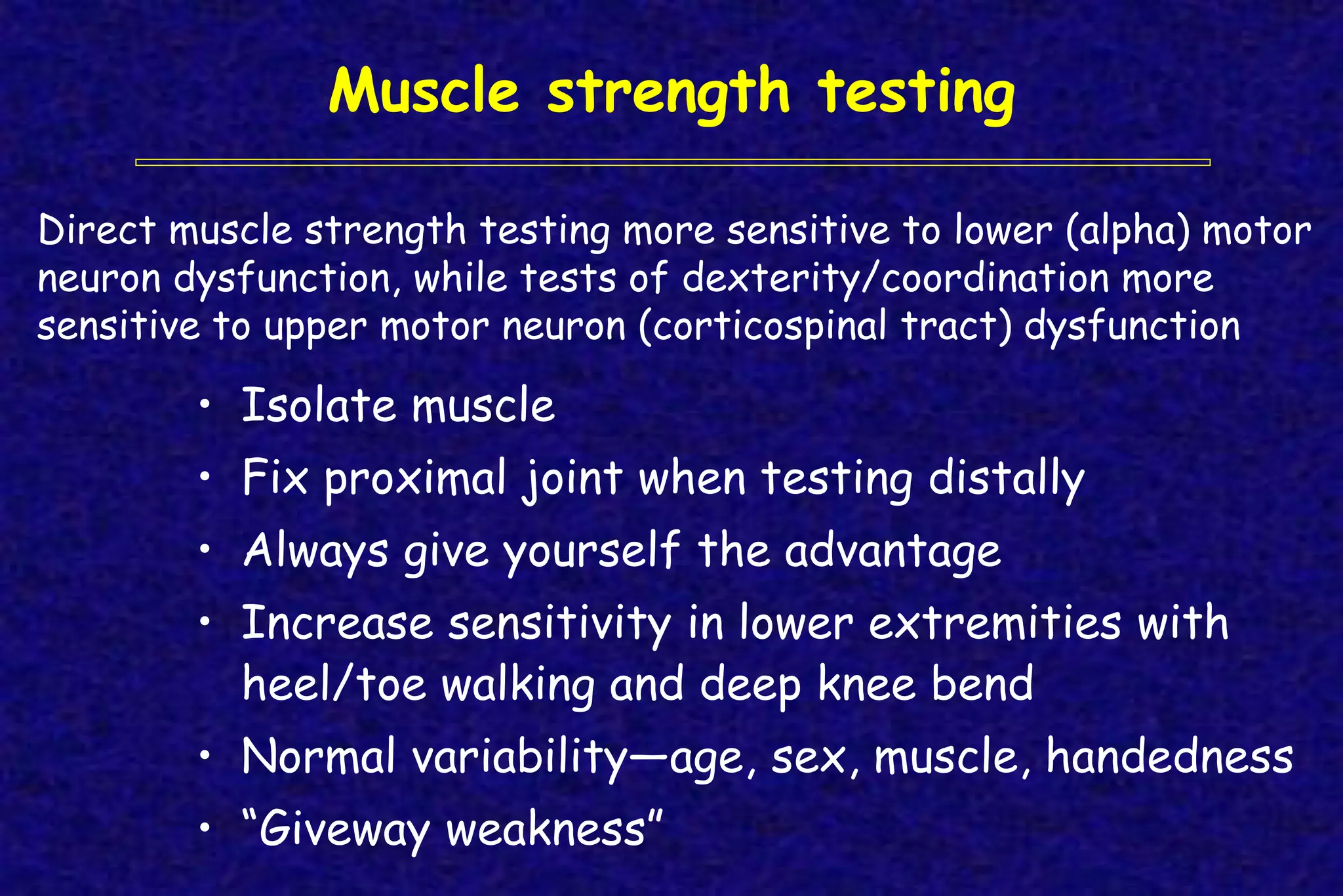
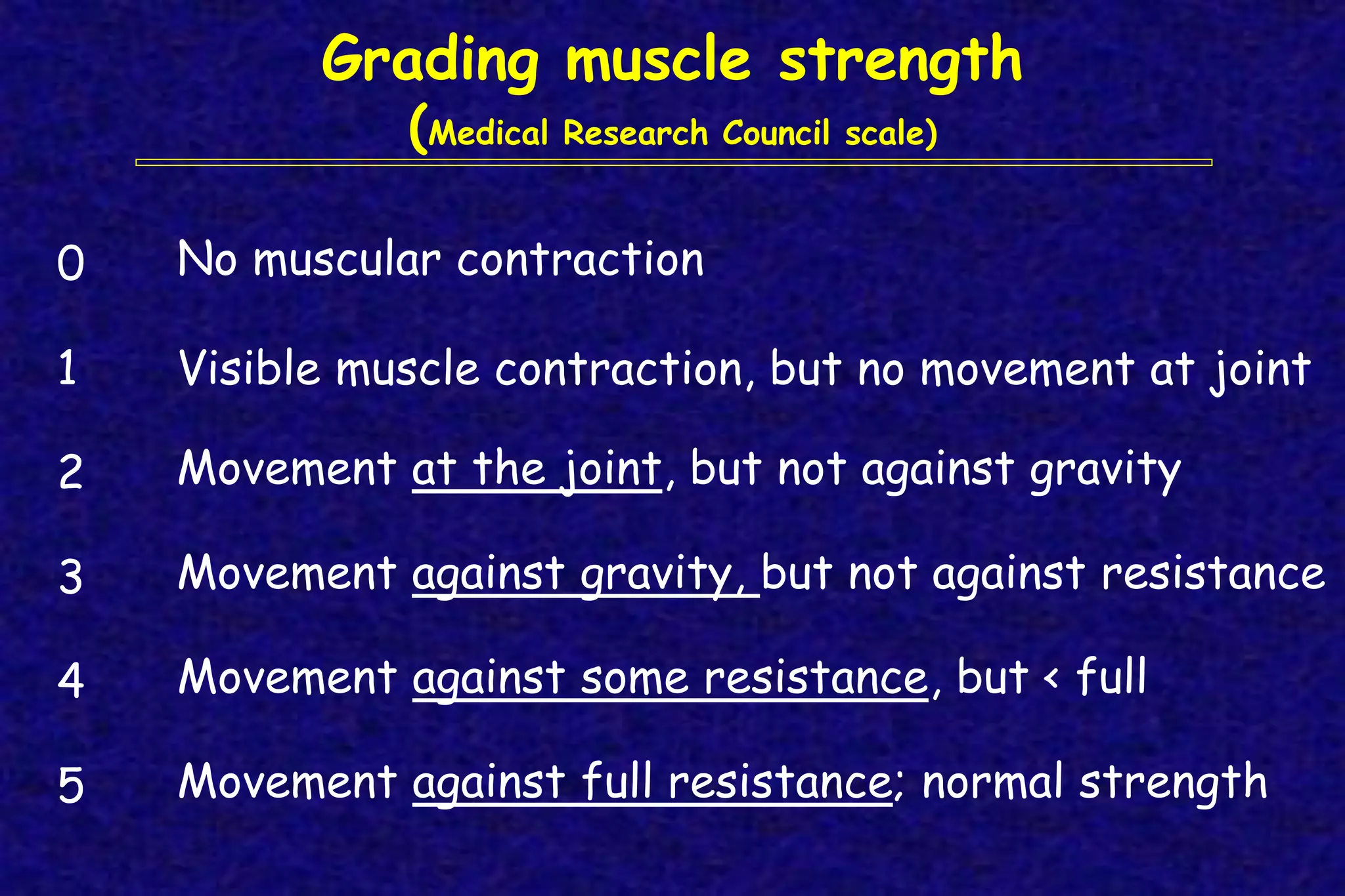
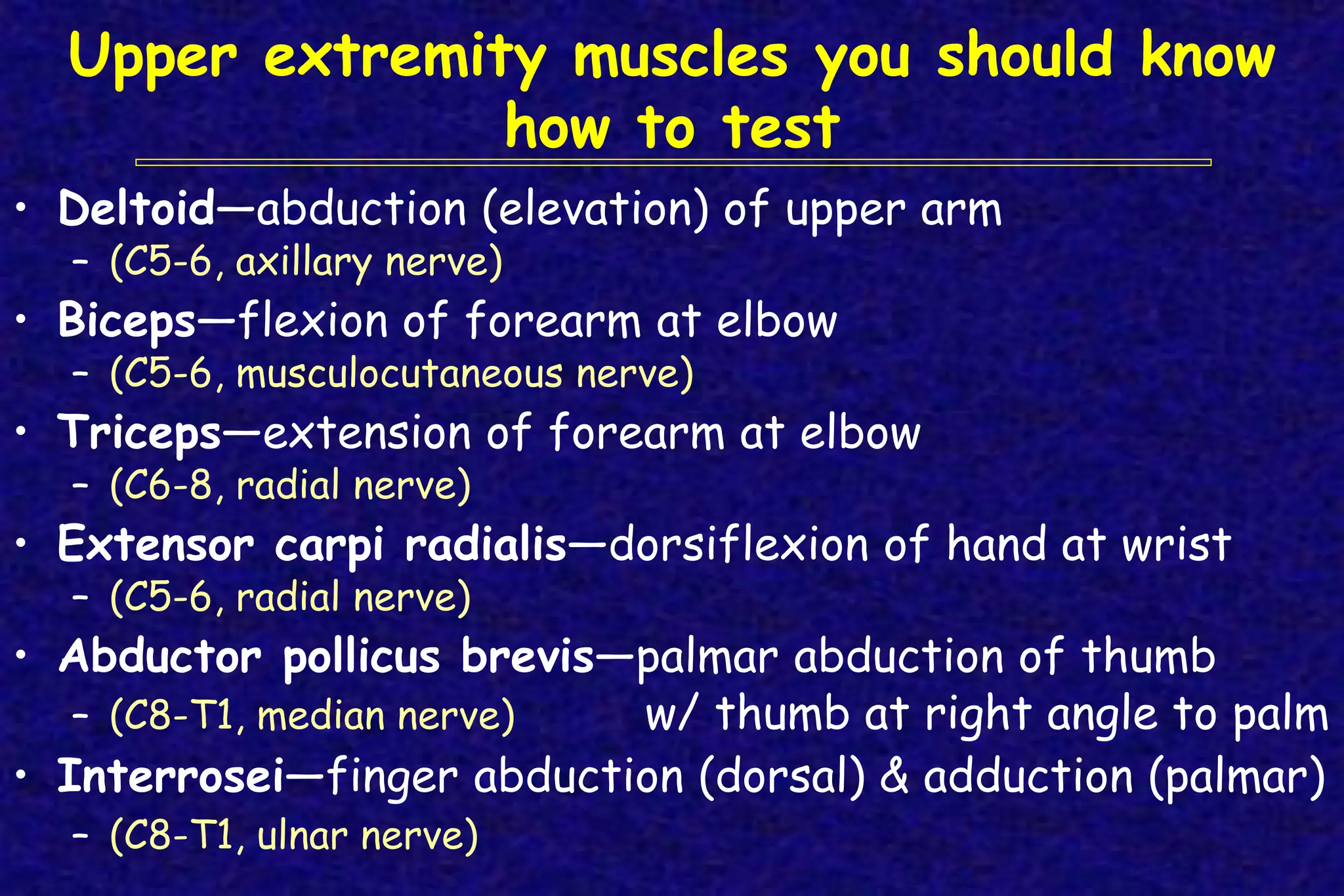
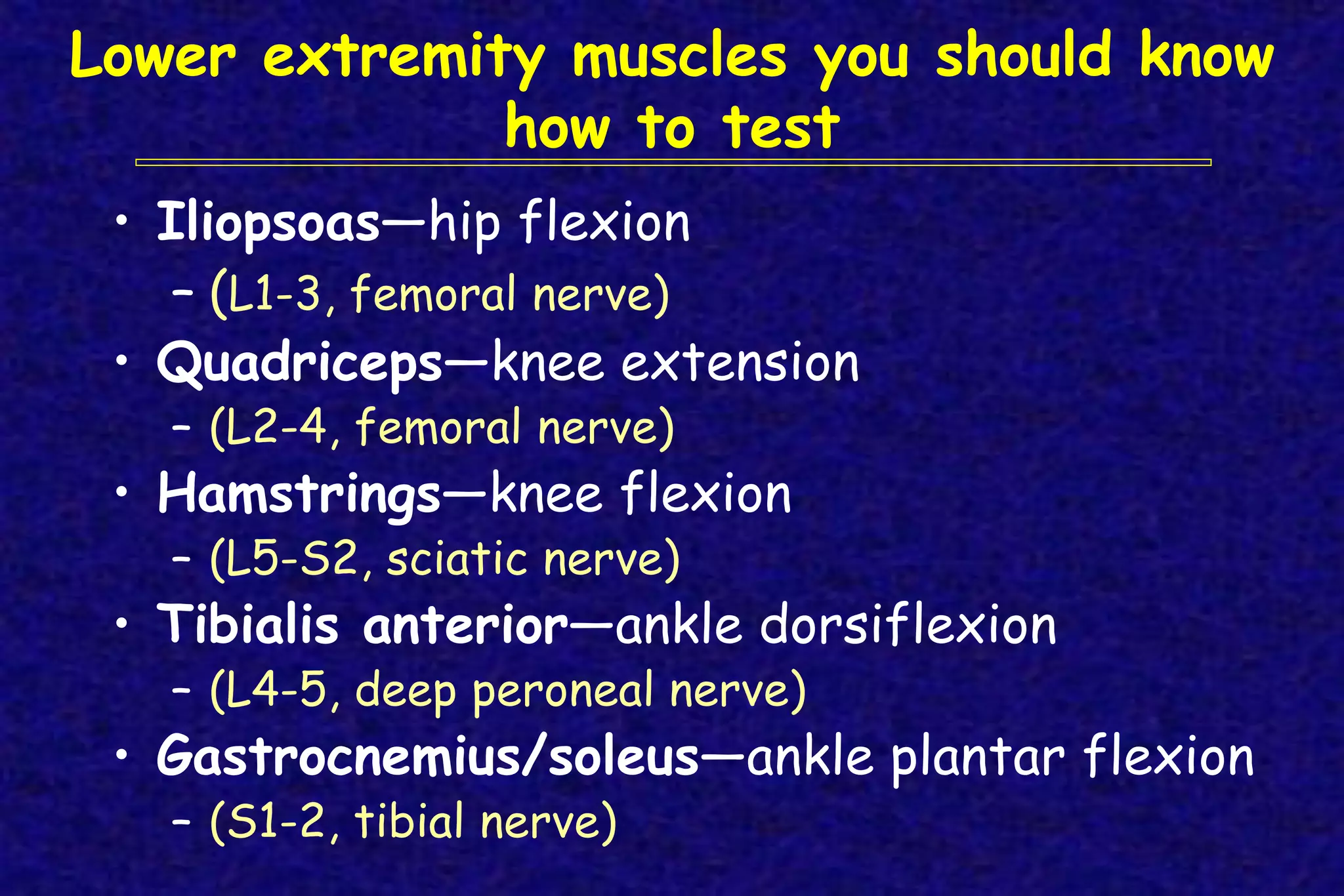
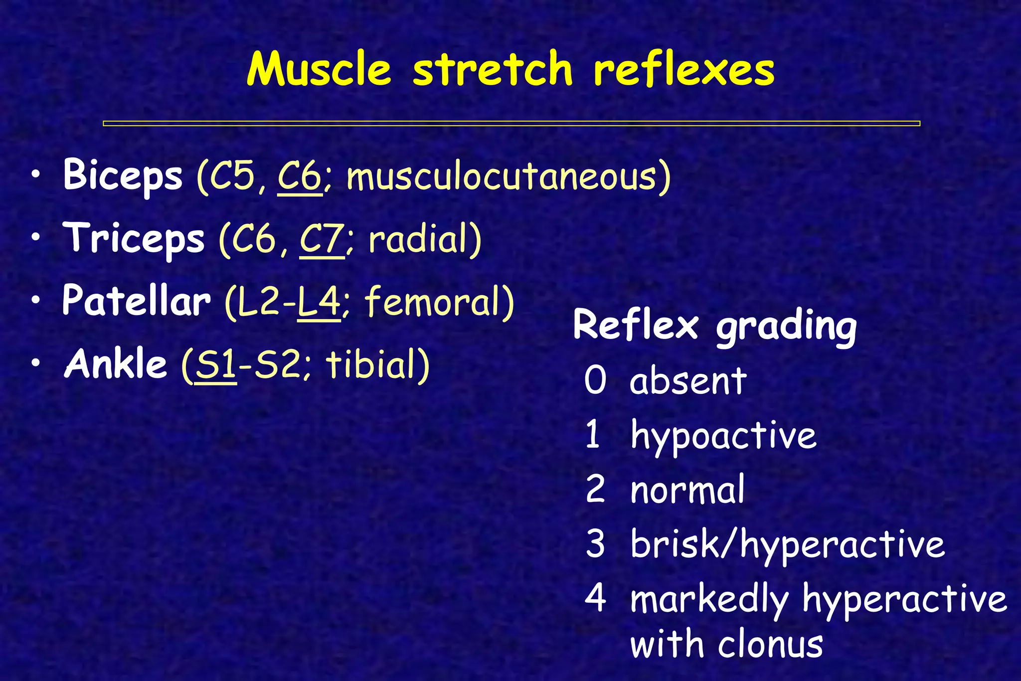
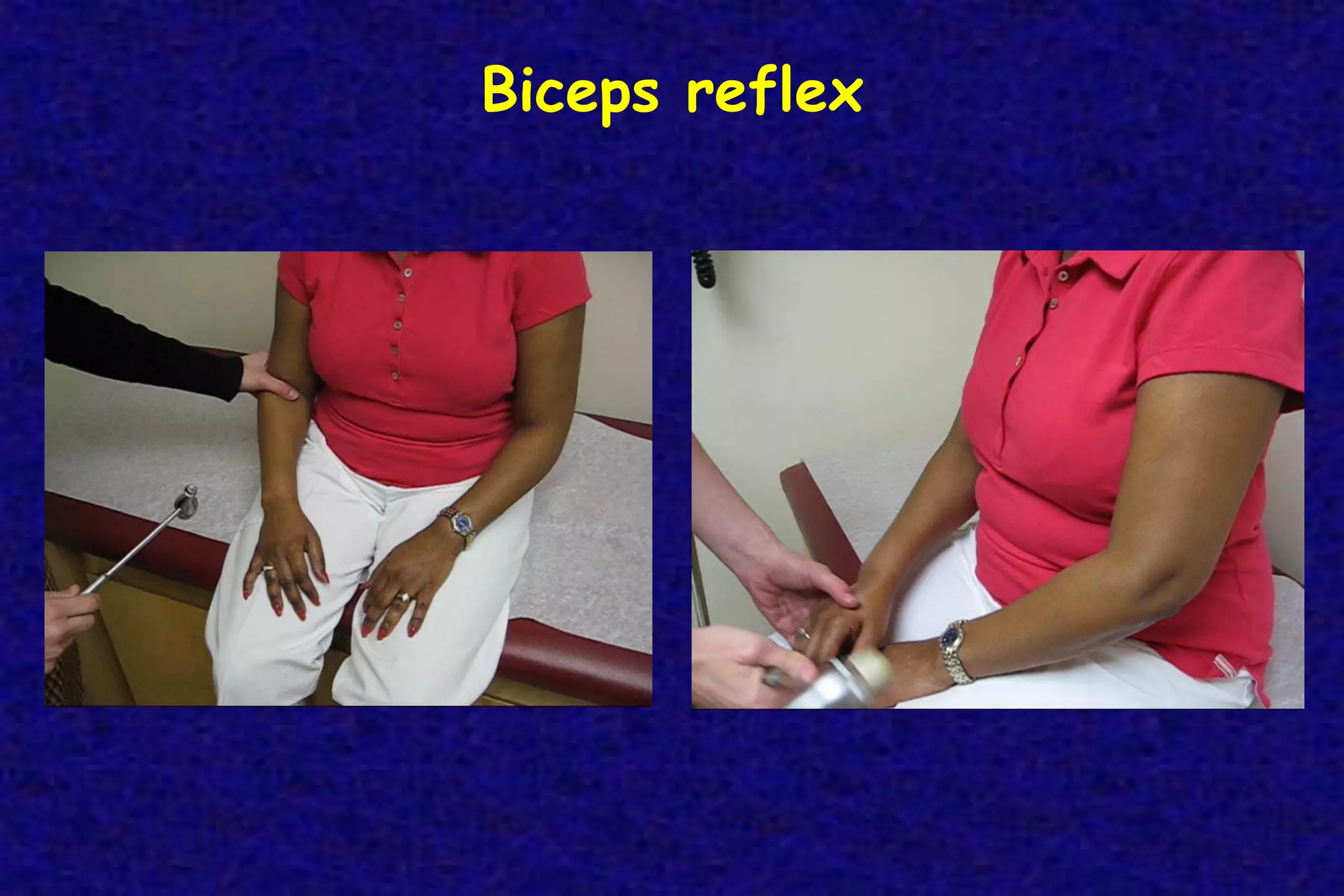
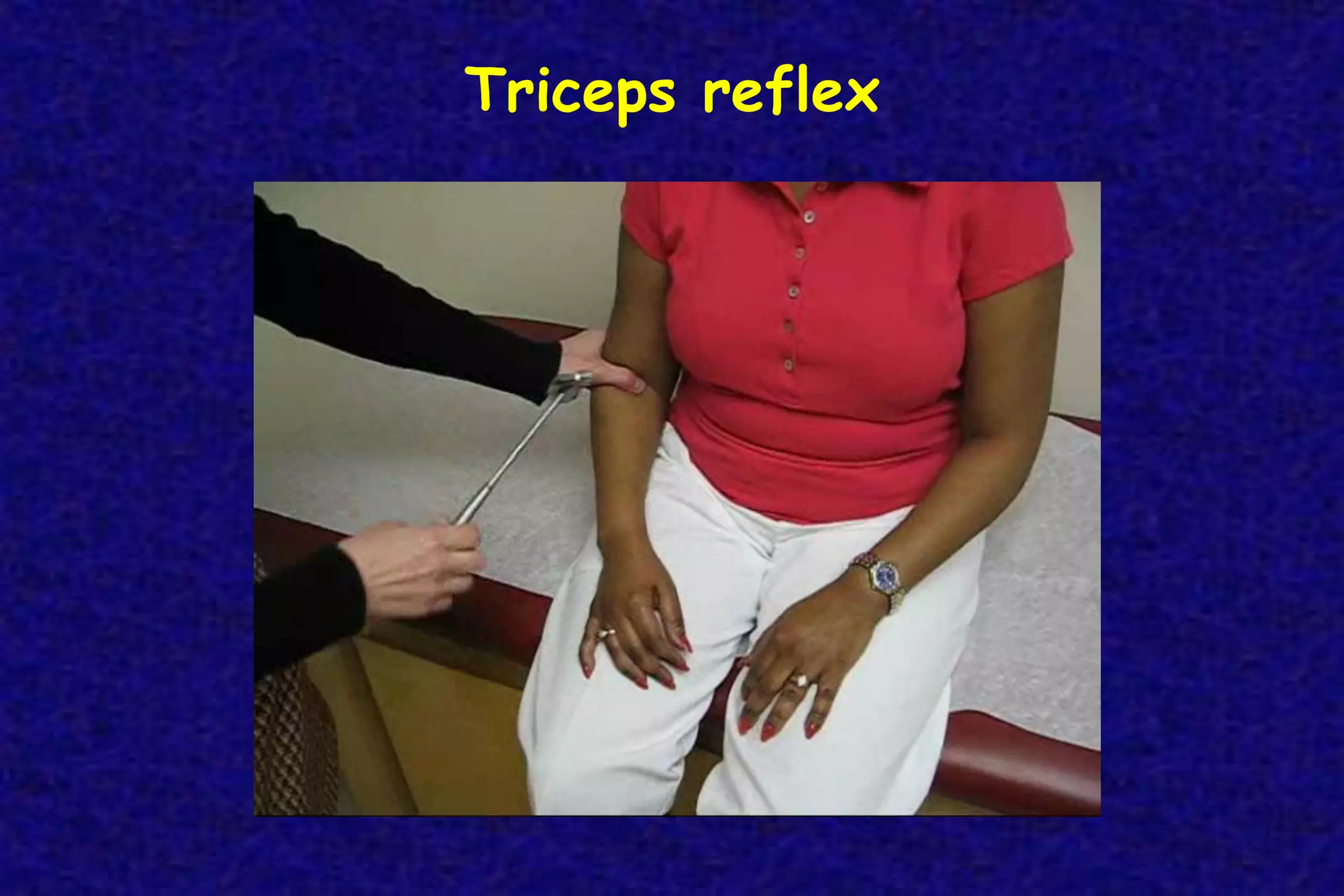
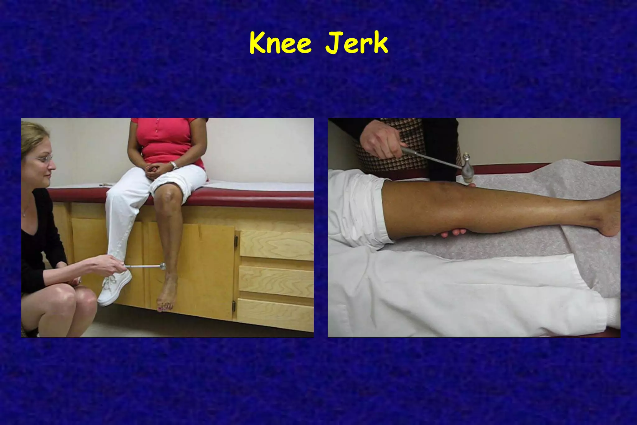
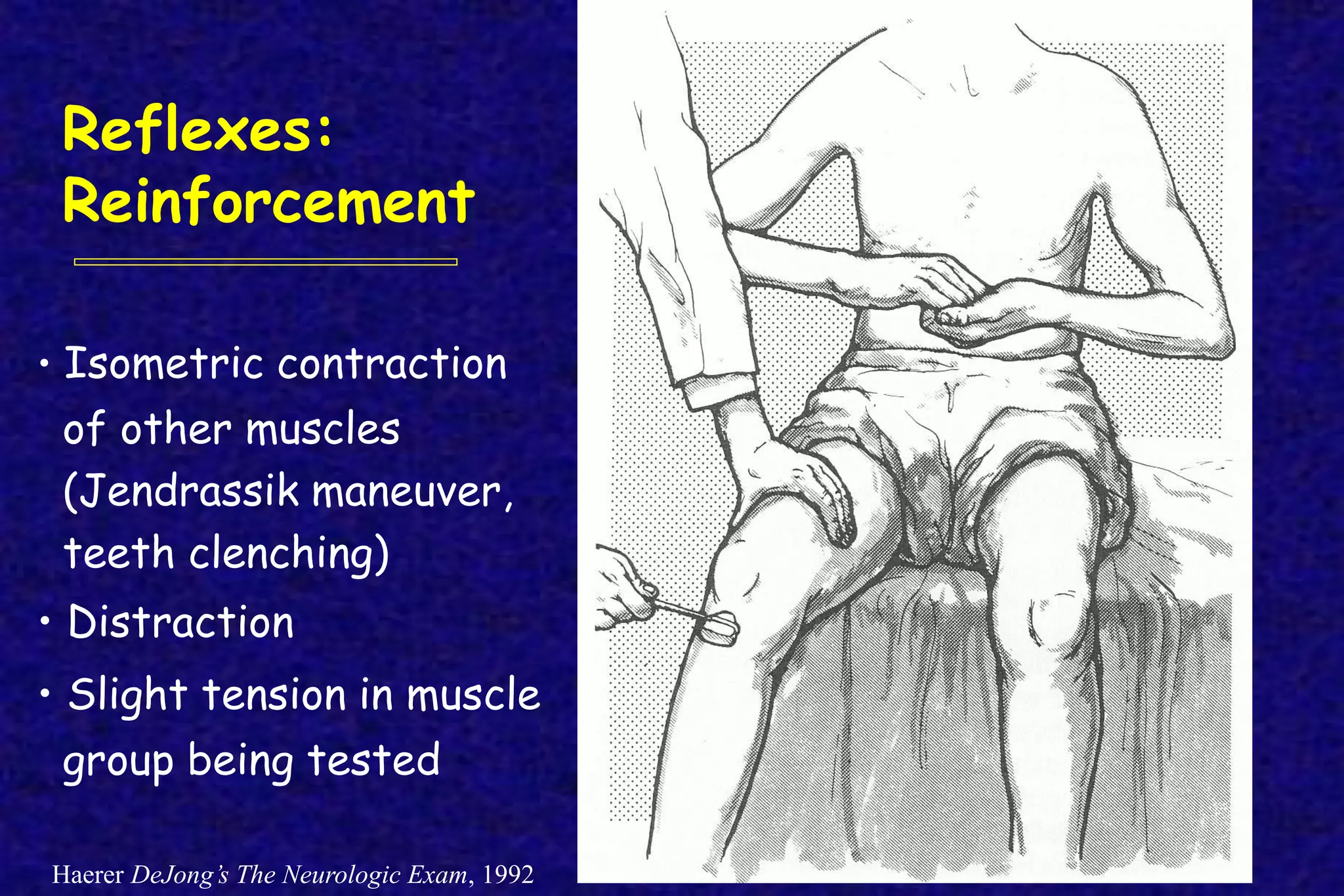
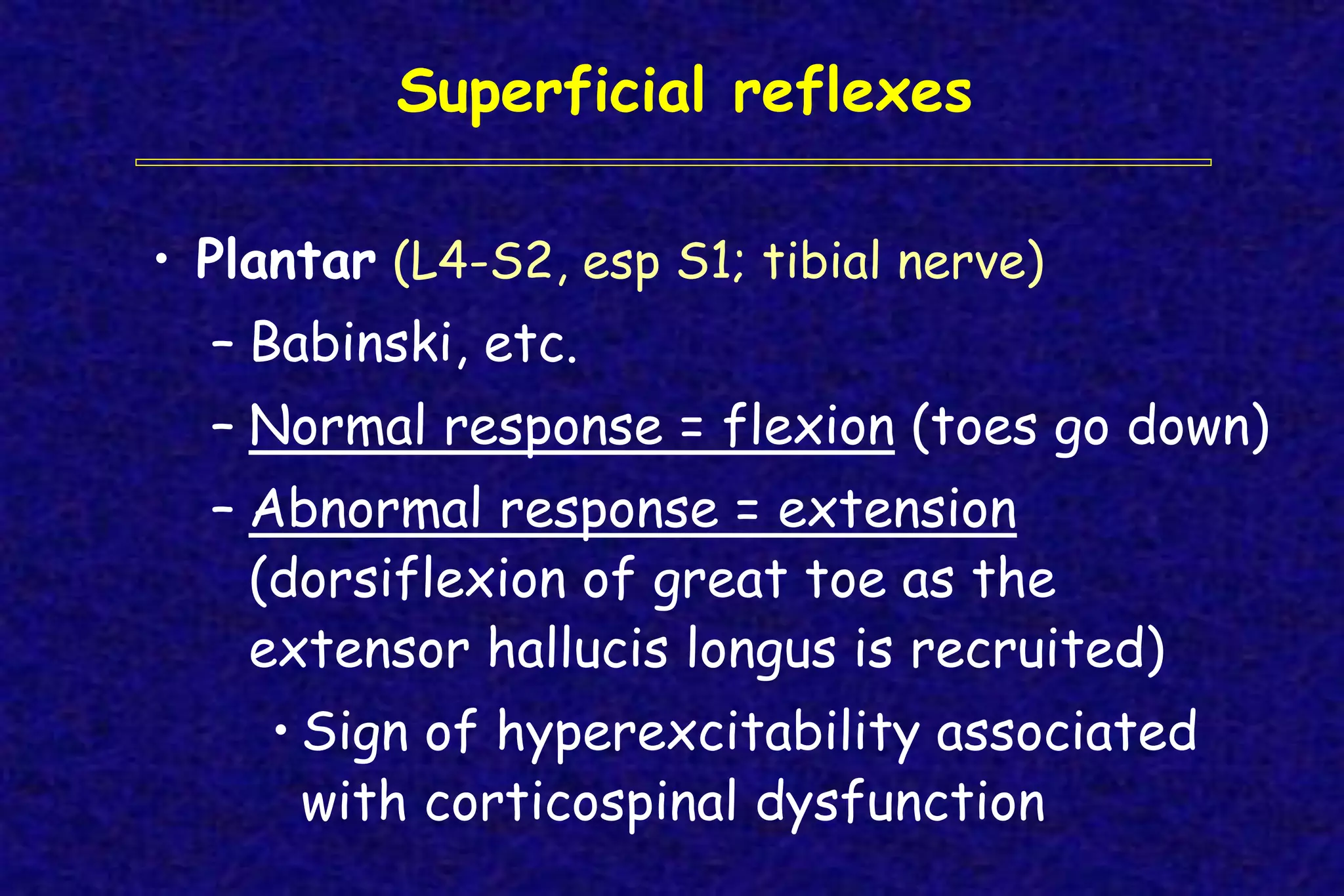
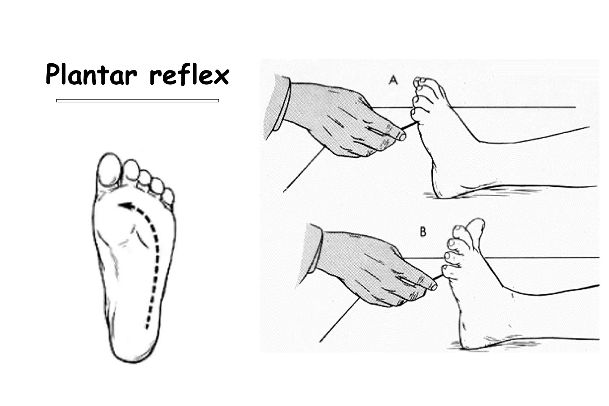
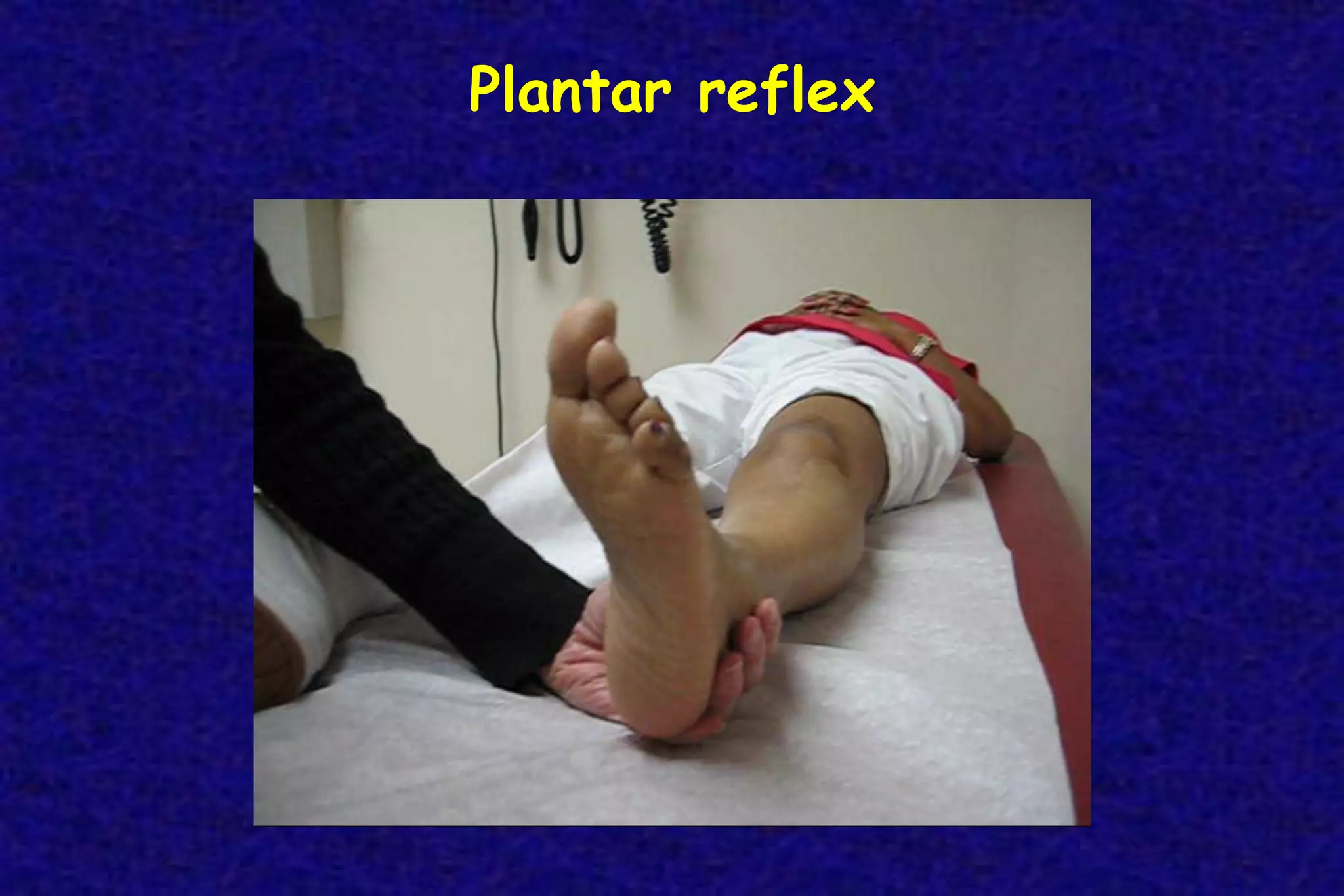
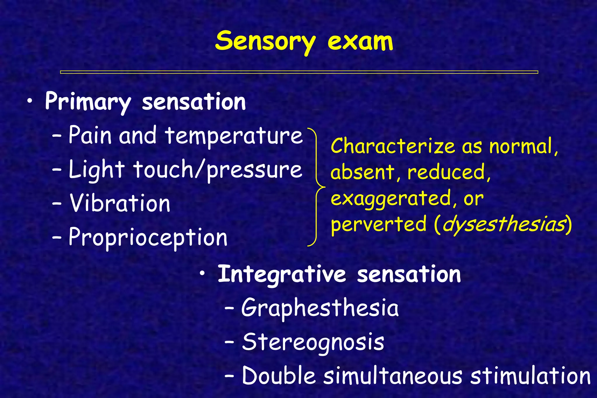
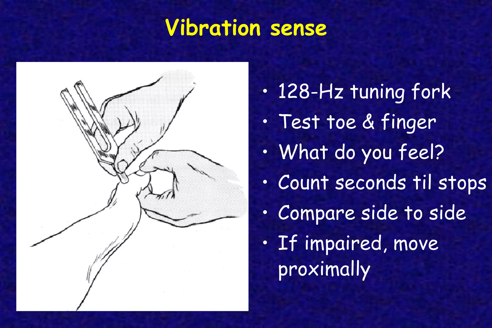
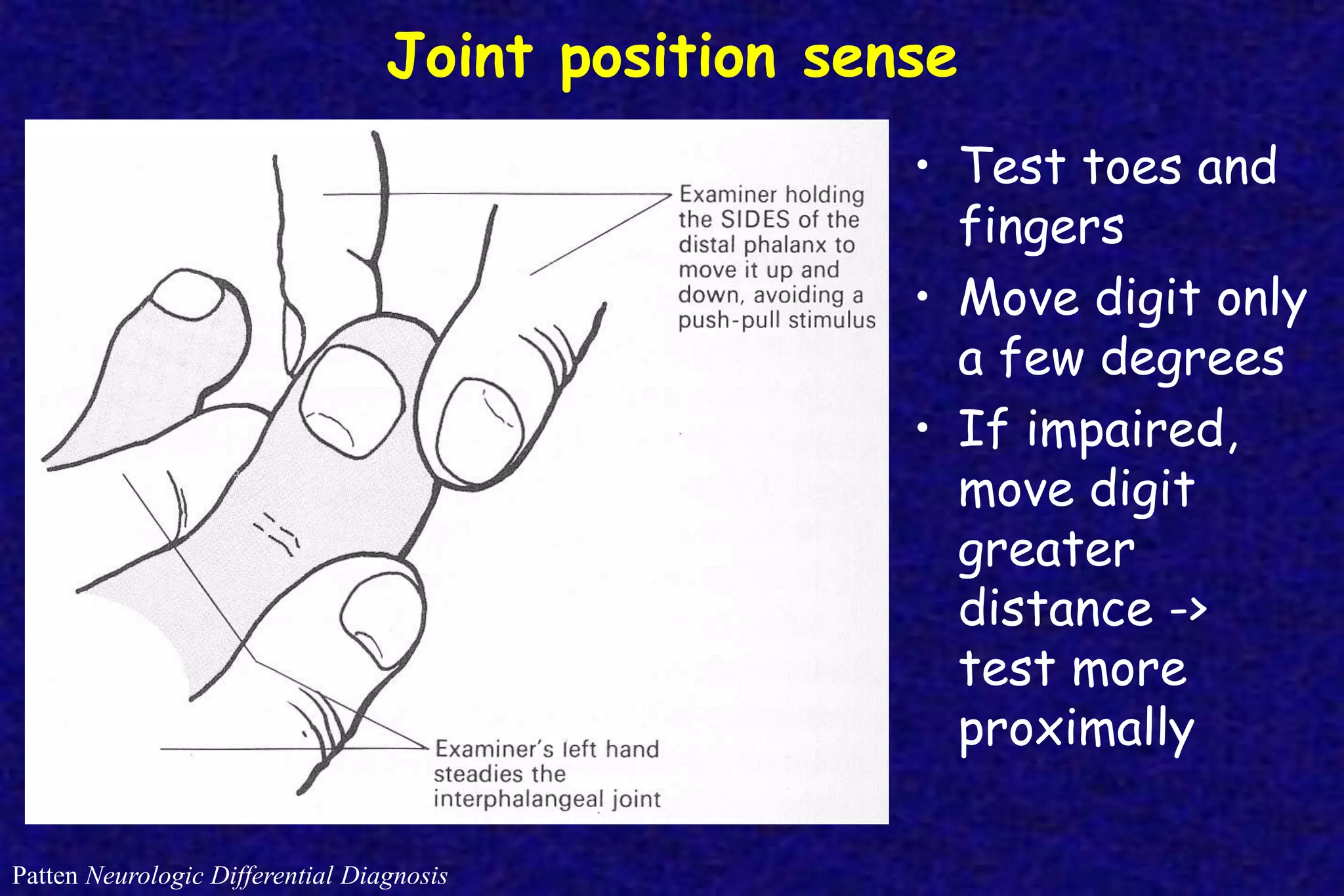
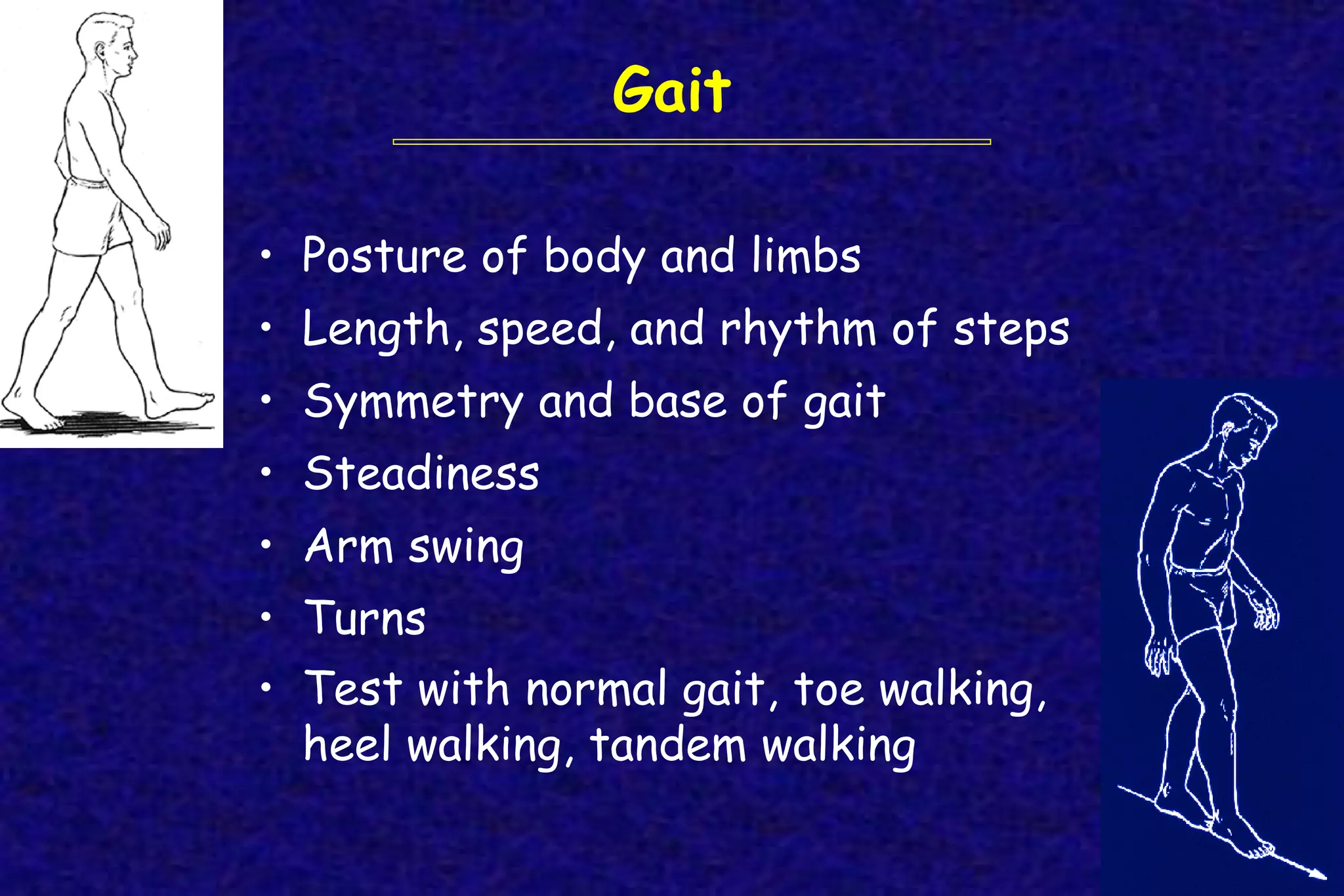
The Neurological Examination
Neuro Exam Tools
X
128-Hz
7 categories of the neurological exam
• Mental status
• Cranial nerves
• Motor system
• Reflexes
• Sensory system
• Coordination
• Station and gait
7 components of the mental status exam
• Level of consciousness
• Attention
• Orientation
• Language — fluency, comprehension, repetition,
naming, reading, writing
• Memory — immediate recall, recent, remote
• Higher intellectual function—general knowledge,
abstraction, judgment, insight, reasoning
• Mood and affect
CNI: Olfactory nerve
• Cannot evaluate if nasal passages obstructed
by rhinitis, polyps, etc.
• Eyes closed
• Occlude one nostril and test other
• Compare 2 sides
• Use nonirritating substances
– Avoid those that stimulate trigeminal
nerve endings or taste buds (e.g.,
peppermint, menthol, ammonia)
CNII: Optic nerve
• Visual acuity
• Visual fields
• Fundoscopy
• Afferent limb of pupillary function
• Hold card at comfortable
reading distance
• Cover 1 eye
• Glasses on (looking for optic
nerve lesion, not refractive error)
CNII: Visual acuity
CNII:
Visual fields
60
75
100 60
Greenberg Clinical Neurology, 1993
CNII:
Fundoscopic
exam
CNII & III: Pupillary function
• Normal pupils are equal in size and shape
and are situated in center of iris
• Pupillary size varies with intensity of
ambient light, but at average intensity is
 3-4 mm
– Miosis < 2 mm
– Mydriasis > 5 mm
– Anisocoria = pupillary asymmetry
CNII & III: Light reflex
• Dim lights
• Fix gaze on opposite wall to eliminate
effects of accommodation
• Shine bright light obliquely into each pupil
• Look for both direct (same eye) and
consensual (opposite eye) reaction
• Record pupil size and shape
CNII & III: Accommodation
• Hold finger 10 cm from patient’s nose
• Alternate looking into distance and at
finger
• Observe pupillary response
• CNIII Oculomotor nerve
• CNIV Trochlear nerve
• CNVI Abducens nerve
• Visual inspection: ocular alignment, lids
• Convergence
• Smooth pursuits
• Saccades
• Nystagmus
• 6 cardinal directions of gaze
CNIII, IV, VI: Ocular nerves
Smooth Pursuits
NeuroLogic Exam
Saccades
NeuroLogic Exam
Optokinetic Nystagmus
NeuroLogic Exam
Extraocular movements
IV III
VI
III
III
III
Nasal Temporal
inferior
oblique
superior
rectus
medial
rectus
superior
oblique
inferior
rectus
lateral
rectus
CNV: Trigeminal nerve
C3
C2
CNV: Trigeminal nerve
Corneal reflex
(CN V-afferent limb
CN VII-efferent limb)
Jaw jerk
Masseter strength
Corneal Reflex
Loyola University Medical Education Network
CNVII Facial nerve
CNVIII: Auditory nerve
• Hearing (cochlear nerve)
– Test with finger rubbing at arm’s length
– If can’t hear strong rubbing→impaired
– If can hear faint rubbing→normal
– Tuning fork tests (Weber, Rinne) have extremely poor sensitivity
CNIX & X: Glossopharyngeal & vagus nerves
• Testing centers on motor function
– Palate elevation
– Swallowing
– Voice
– Cough
– Gag reflex
Examination of the palate
NeuroLogic Exam
CNXI: Spinal accessory nerve
• Trapezius
– Push head back against resistance
– Shrug shoulders
• Sternocleidomastoid
– Place hand on lower face and have
patient rotate head toward that side
– Observe contraction of opposite SCM
CNXII: Hypoglossal nerve
• Note tongue position at rest and on protrusion
– Does tongue deviate in either position?
• Note strength and rapidity of movements
• Have patient push tongue into each cheek
Motor exam
• Compare left to right, proximal to distal,
arms to legs
– Bulk (muscle mass)
– Tone (muscle tension at rest)
• Test with passive manipulation
– Strength
– Speed of movement; extraneous movement
– Endurance
Muscle strength testing
• Isolate muscle
• Fix proximal joint when testing distally
• Always give yourself the advantage
• Increase sensitivity in lower extremities with
heel/toe walking and deep knee bend
• Normal variability—age, sex, muscle, handedness
• “Giveway weakness”
Direct muscle strength testing more sensitive to lower (alpha) motor
neuron dysfunction, while tests of dexterity/coordination more
sensitive to upper motor neuron (corticospinal tract) dysfunction
Grading muscle strength
(Medical Research Council scale)
No muscular contraction
Visible muscle contraction, but no movement at joint
Movement at the joint, but not against gravity
Movement against gravity, but not against resistance
Movement against some resistance, but < full
0
1
2
3
4
5 Movement against full resistance; normal strength
Upper extremity muscles you should know
how to test
• Deltoid—abduction (elevation) of upper arm
– (C5-6, axillary nerve)
• Biceps—flexion of forearm at elbow
– (C5-6, musculocutaneous nerve)
• Triceps—extension of forearm at elbow
– (C6-8, radial nerve)
• Extensor carpi radialis—dorsiflexion of hand at wrist
– (C5-6, radial nerve)
• Abductor pollicus brevis—palmar abduction of thumb
– (C8-T1, median nerve) w/ thumb at right angle to palm
• Interrosei—finger abduction (dorsal) & adduction (palmar)
– (C8-T1, ulnar nerve)
Lower extremity muscles you should know
how to test
• Iliopsoas—hip flexion
– (L1-3, femoral nerve)
• Quadriceps—knee extension
– (L2-4, femoral nerve)
• Hamstrings—knee flexion
– (L5-S2, sciatic nerve)
• Tibialis anterior—ankle dorsiflexion
– (L4-5, deep peroneal nerve)
• Gastrocnemius/soleus—ankle plantar flexion
– (S1-2, tibial nerve)
Muscle stretch reflexes
• Biceps (C5, C6; musculocutaneous)
• Triceps (C6, C7; radial)
• Patellar (L2-L4; femoral)
• Ankle (S1-S2; tibial)
Reflex grading
0 absent
1 hypoactive
2 normal
3 brisk/hyperactive
4 markedly hyperactive
with clonus
Biceps reflex
Triceps reflex
Knee Jerk
Ankle Jerk
Reflexes:
Reinforcement
Haerer DeJong’s The Neurologic Exam, 1992
• Isometric contraction
of other muscles
(Jendrassik maneuver,
teeth clenching)
• Distraction
• Slight tension in muscle
group being tested
Superficial reflexes
• Plantar (L4-S2, esp S1; tibial nerve)
– Babinski, etc.
– Normal response = flexion (toes go down)
– Abnormal response = extension
(dorsiflexion of great toe as the
extensor hallucis longus is recruited)
• Sign of hyperexcitability associated
with corticospinal dysfunction
Plantar reflex
Plantar reflex
• Explain each test before you do it
• Patient’s eyes should be closed during testing
• Test all 4 extremities
• Avoid leading questions like “Is this sharp?”
• Compare side-to-side and distal-to-proximal
asking if they are “about the same”
• When you detect an area of sensory loss, map
out its boundaries in detail
Sensory Exam: General Points
Sensory exam
• Primary sensation
– Pain and temperature
– Light touch/pressure
– Vibration
– Proprioception
Characterize as normal,
absent, reduced,
exaggerated, or
perverted (dysesthesias)
• Integrative sensation
– Graphesthesia
– Stereognosis
– Double simultaneous stimulation
Vibration sense
• 128-Hz tuning fork
• Test toe & finger
• What do you feel?
• Count seconds til stops
• Compare side to side
• If impaired, move
proximally
Joint position sense
Patten Neurologic Differential Diagnosis
• Test toes and
fingers
• Move digit only
a few degrees
• If impaired,
move digit
greater
distance ->
test more
proximally
Pain sensation
• Test for distal gradient of sensory loss in the leg
• Test for sensory loss in most commonly affected
nerve and nerve root distributions
– Palmar aspect of index finger (median nerve).
– Palmar aspect of 5th finger (ulnar nerve)
– Web space between thumb and index finger on
dorsal surface of hand (radial nerve).
– Lateral surface of foot (L5).
– Posterior aspect of leg (S1).
• Ask patient if the sensation is “about the same”
Light Touch & Double Simultaneous Stimulation
• Lightly touch face and extremities in
random order, asking patient to respond
whenever a touch is felt
• Touch both sides of face or body
simultaneously, asking patient to indicate
whether touch is felt on left, right, or both
sides
Stereognosis & Graphesthesia
Romberg sign
• Ability to maintain upright position with
feet together and eyes open
• Sway/fall when eyes closed
• Indicates impaired proprioception or
vestibular dysfunction
Coordination
• Control, precision, rhythm, synergy of movement
• Test at rest and with action in trunk and limbs
– Finger-nose-finger
– Rapid alternating movements
– Heel-knee-shin
– Finger or toe tapping
Gait
• Posture of body and limbs
• Length, speed, and rhythm of steps
• Symmetry and base of gait
• Steadiness
• Arm swing
• Turns
• Test with normal gait, toe walking,
heel walking, tandem walking
Meningeal signs
• Neck mobility—look for nuchal rigidity
(neck stiffness)
Movies marked as from NeuroLogic Exam and PediNeuroLogic Exam movies are used by
permission of Paul D. Larsen, M.D., University of Nebraska Medical Center and Suzanne S.
Stensaas, Ph.D., University of Utah School of Medicine. Additional materials were drawn from
resources provided by Alejandro Stern, Stern Foundation, Buenos Aires, Argentina; Kathleen
Digre, M.D., University of Utah; and Daniel Jacobson, M.D., Marshfield Clinic, Wisconsin.
Subsequent re-use of any materials outside of this program, presentation, or website requires
permission from the original producers. Contact SLICE@media.utah.edu.
Recommended resource:

neurologic-examination.pdf

  • 1.
  • 2.
  • 3.
    7 categories ofthe neurological exam • Mental status • Cranial nerves • Motor system • Reflexes • Sensory system • Coordination • Station and gait
  • 4.
    7 components ofthe mental status exam • Level of consciousness • Attention • Orientation • Language — fluency, comprehension, repetition, naming, reading, writing • Memory — immediate recall, recent, remote • Higher intellectual function—general knowledge, abstraction, judgment, insight, reasoning • Mood and affect
  • 5.
    CNI: Olfactory nerve •Cannot evaluate if nasal passages obstructed by rhinitis, polyps, etc. • Eyes closed • Occlude one nostril and test other • Compare 2 sides • Use nonirritating substances – Avoid those that stimulate trigeminal nerve endings or taste buds (e.g., peppermint, menthol, ammonia)
  • 6.
    CNII: Optic nerve •Visual acuity • Visual fields • Fundoscopy • Afferent limb of pupillary function
  • 7.
    • Hold cardat comfortable reading distance • Cover 1 eye • Glasses on (looking for optic nerve lesion, not refractive error) CNII: Visual acuity
  • 8.
  • 9.
  • 10.
    CNII & III:Pupillary function • Normal pupils are equal in size and shape and are situated in center of iris • Pupillary size varies with intensity of ambient light, but at average intensity is  3-4 mm – Miosis < 2 mm – Mydriasis > 5 mm – Anisocoria = pupillary asymmetry
  • 12.
    CNII & III:Light reflex • Dim lights • Fix gaze on opposite wall to eliminate effects of accommodation • Shine bright light obliquely into each pupil • Look for both direct (same eye) and consensual (opposite eye) reaction • Record pupil size and shape
  • 13.
    CNII & III:Accommodation • Hold finger 10 cm from patient’s nose • Alternate looking into distance and at finger • Observe pupillary response
  • 14.
    • CNIII Oculomotornerve • CNIV Trochlear nerve • CNVI Abducens nerve • Visual inspection: ocular alignment, lids • Convergence • Smooth pursuits • Saccades • Nystagmus • 6 cardinal directions of gaze CNIII, IV, VI: Ocular nerves
  • 15.
  • 16.
  • 17.
  • 18.
    Extraocular movements IV III VI III III III NasalTemporal inferior oblique superior rectus medial rectus superior oblique inferior rectus lateral rectus
  • 19.
  • 20.
    CNV: Trigeminal nerve Cornealreflex (CN V-afferent limb CN VII-efferent limb) Jaw jerk Masseter strength
  • 21.
  • 22.
    Loyola University MedicalEducation Network CNVII Facial nerve
  • 23.
    CNVIII: Auditory nerve •Hearing (cochlear nerve) – Test with finger rubbing at arm’s length – If can’t hear strong rubbing→impaired – If can hear faint rubbing→normal – Tuning fork tests (Weber, Rinne) have extremely poor sensitivity
  • 24.
    CNIX & X:Glossopharyngeal & vagus nerves • Testing centers on motor function – Palate elevation – Swallowing – Voice – Cough – Gag reflex
  • 25.
    Examination of thepalate NeuroLogic Exam
  • 26.
    CNXI: Spinal accessorynerve • Trapezius – Push head back against resistance – Shrug shoulders • Sternocleidomastoid – Place hand on lower face and have patient rotate head toward that side – Observe contraction of opposite SCM
  • 27.
    CNXII: Hypoglossal nerve •Note tongue position at rest and on protrusion – Does tongue deviate in either position? • Note strength and rapidity of movements • Have patient push tongue into each cheek
  • 28.
    Motor exam • Compareleft to right, proximal to distal, arms to legs – Bulk (muscle mass) – Tone (muscle tension at rest) • Test with passive manipulation – Strength – Speed of movement; extraneous movement – Endurance
  • 29.
    Muscle strength testing •Isolate muscle • Fix proximal joint when testing distally • Always give yourself the advantage • Increase sensitivity in lower extremities with heel/toe walking and deep knee bend • Normal variability—age, sex, muscle, handedness • “Giveway weakness” Direct muscle strength testing more sensitive to lower (alpha) motor neuron dysfunction, while tests of dexterity/coordination more sensitive to upper motor neuron (corticospinal tract) dysfunction
  • 30.
    Grading muscle strength (MedicalResearch Council scale) No muscular contraction Visible muscle contraction, but no movement at joint Movement at the joint, but not against gravity Movement against gravity, but not against resistance Movement against some resistance, but < full 0 1 2 3 4 5 Movement against full resistance; normal strength
  • 31.
    Upper extremity musclesyou should know how to test • Deltoid—abduction (elevation) of upper arm – (C5-6, axillary nerve) • Biceps—flexion of forearm at elbow – (C5-6, musculocutaneous nerve) • Triceps—extension of forearm at elbow – (C6-8, radial nerve) • Extensor carpi radialis—dorsiflexion of hand at wrist – (C5-6, radial nerve) • Abductor pollicus brevis—palmar abduction of thumb – (C8-T1, median nerve) w/ thumb at right angle to palm • Interrosei—finger abduction (dorsal) & adduction (palmar) – (C8-T1, ulnar nerve)
  • 32.
    Lower extremity musclesyou should know how to test • Iliopsoas—hip flexion – (L1-3, femoral nerve) • Quadriceps—knee extension – (L2-4, femoral nerve) • Hamstrings—knee flexion – (L5-S2, sciatic nerve) • Tibialis anterior—ankle dorsiflexion – (L4-5, deep peroneal nerve) • Gastrocnemius/soleus—ankle plantar flexion – (S1-2, tibial nerve)
  • 33.
    Muscle stretch reflexes •Biceps (C5, C6; musculocutaneous) • Triceps (C6, C7; radial) • Patellar (L2-L4; femoral) • Ankle (S1-S2; tibial) Reflex grading 0 absent 1 hypoactive 2 normal 3 brisk/hyperactive 4 markedly hyperactive with clonus
  • 34.
  • 35.
  • 36.
  • 37.
  • 38.
    Reflexes: Reinforcement Haerer DeJong’s TheNeurologic Exam, 1992 • Isometric contraction of other muscles (Jendrassik maneuver, teeth clenching) • Distraction • Slight tension in muscle group being tested
  • 39.
    Superficial reflexes • Plantar(L4-S2, esp S1; tibial nerve) – Babinski, etc. – Normal response = flexion (toes go down) – Abnormal response = extension (dorsiflexion of great toe as the extensor hallucis longus is recruited) • Sign of hyperexcitability associated with corticospinal dysfunction
  • 40.
  • 41.
  • 42.
    • Explain eachtest before you do it • Patient’s eyes should be closed during testing • Test all 4 extremities • Avoid leading questions like “Is this sharp?” • Compare side-to-side and distal-to-proximal asking if they are “about the same” • When you detect an area of sensory loss, map out its boundaries in detail Sensory Exam: General Points
  • 43.
    Sensory exam • Primarysensation – Pain and temperature – Light touch/pressure – Vibration – Proprioception Characterize as normal, absent, reduced, exaggerated, or perverted (dysesthesias) • Integrative sensation – Graphesthesia – Stereognosis – Double simultaneous stimulation
  • 44.
    Vibration sense • 128-Hztuning fork • Test toe & finger • What do you feel? • Count seconds til stops • Compare side to side • If impaired, move proximally
  • 45.
    Joint position sense PattenNeurologic Differential Diagnosis • Test toes and fingers • Move digit only a few degrees • If impaired, move digit greater distance -> test more proximally
  • 46.
    Pain sensation • Testfor distal gradient of sensory loss in the leg • Test for sensory loss in most commonly affected nerve and nerve root distributions – Palmar aspect of index finger (median nerve). – Palmar aspect of 5th finger (ulnar nerve) – Web space between thumb and index finger on dorsal surface of hand (radial nerve). – Lateral surface of foot (L5). – Posterior aspect of leg (S1). • Ask patient if the sensation is “about the same”
  • 47.
    Light Touch &Double Simultaneous Stimulation • Lightly touch face and extremities in random order, asking patient to respond whenever a touch is felt • Touch both sides of face or body simultaneously, asking patient to indicate whether touch is felt on left, right, or both sides
  • 48.
  • 49.
    Romberg sign • Abilityto maintain upright position with feet together and eyes open • Sway/fall when eyes closed • Indicates impaired proprioception or vestibular dysfunction
  • 50.
    Coordination • Control, precision,rhythm, synergy of movement • Test at rest and with action in trunk and limbs – Finger-nose-finger – Rapid alternating movements – Heel-knee-shin – Finger or toe tapping
  • 51.
    Gait • Posture ofbody and limbs • Length, speed, and rhythm of steps • Symmetry and base of gait • Steadiness • Arm swing • Turns • Test with normal gait, toe walking, heel walking, tandem walking
  • 52.
    Meningeal signs • Neckmobility—look for nuchal rigidity (neck stiffness)
  • 53.
    Movies marked asfrom NeuroLogic Exam and PediNeuroLogic Exam movies are used by permission of Paul D. Larsen, M.D., University of Nebraska Medical Center and Suzanne S. Stensaas, Ph.D., University of Utah School of Medicine. Additional materials were drawn from resources provided by Alejandro Stern, Stern Foundation, Buenos Aires, Argentina; Kathleen Digre, M.D., University of Utah; and Daniel Jacobson, M.D., Marshfield Clinic, Wisconsin. Subsequent re-use of any materials outside of this program, presentation, or website requires permission from the original producers. Contact SLICE@media.utah.edu. Recommended resource: