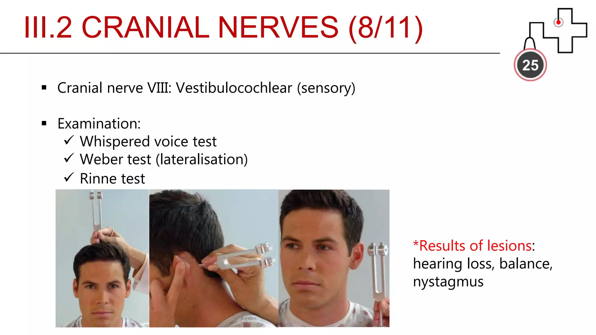
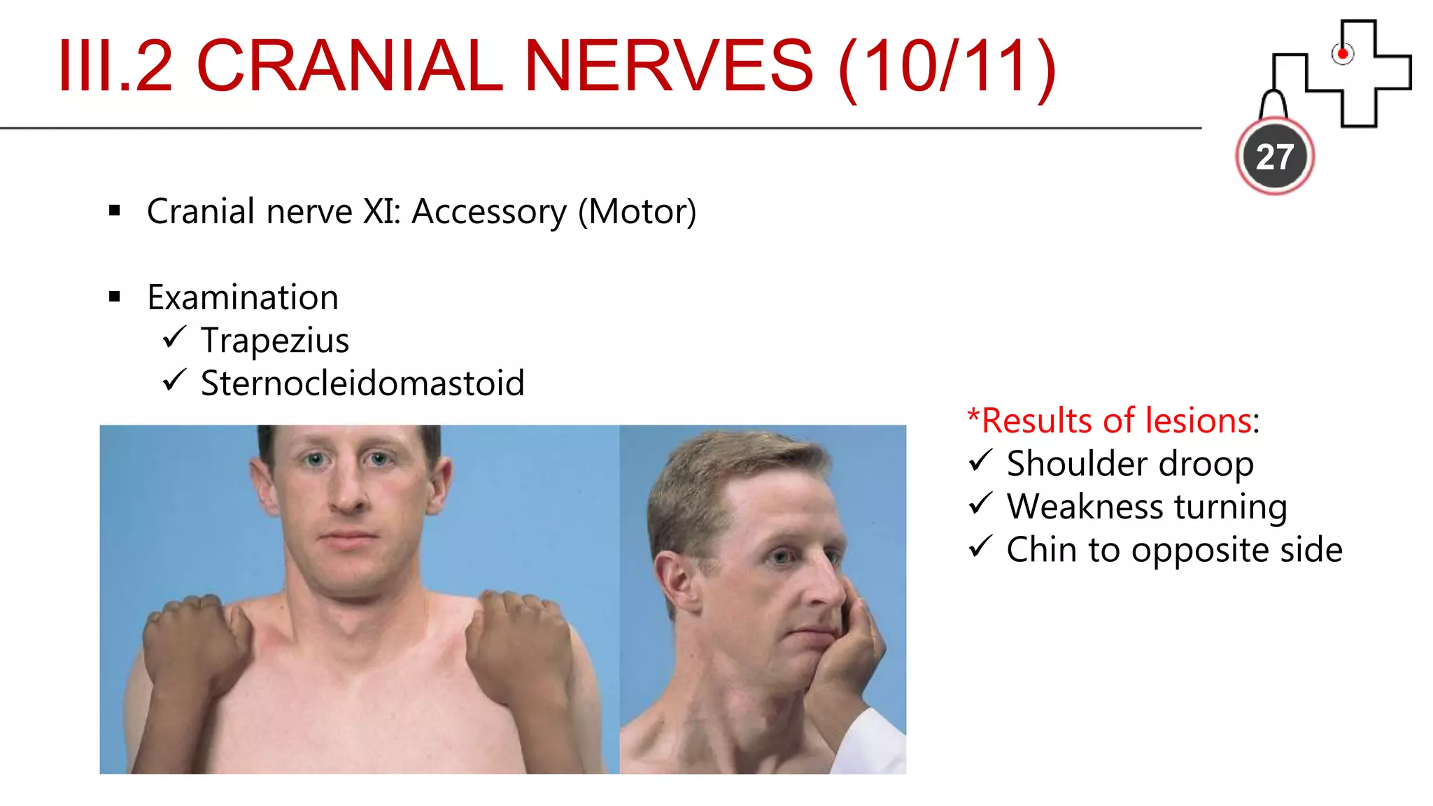
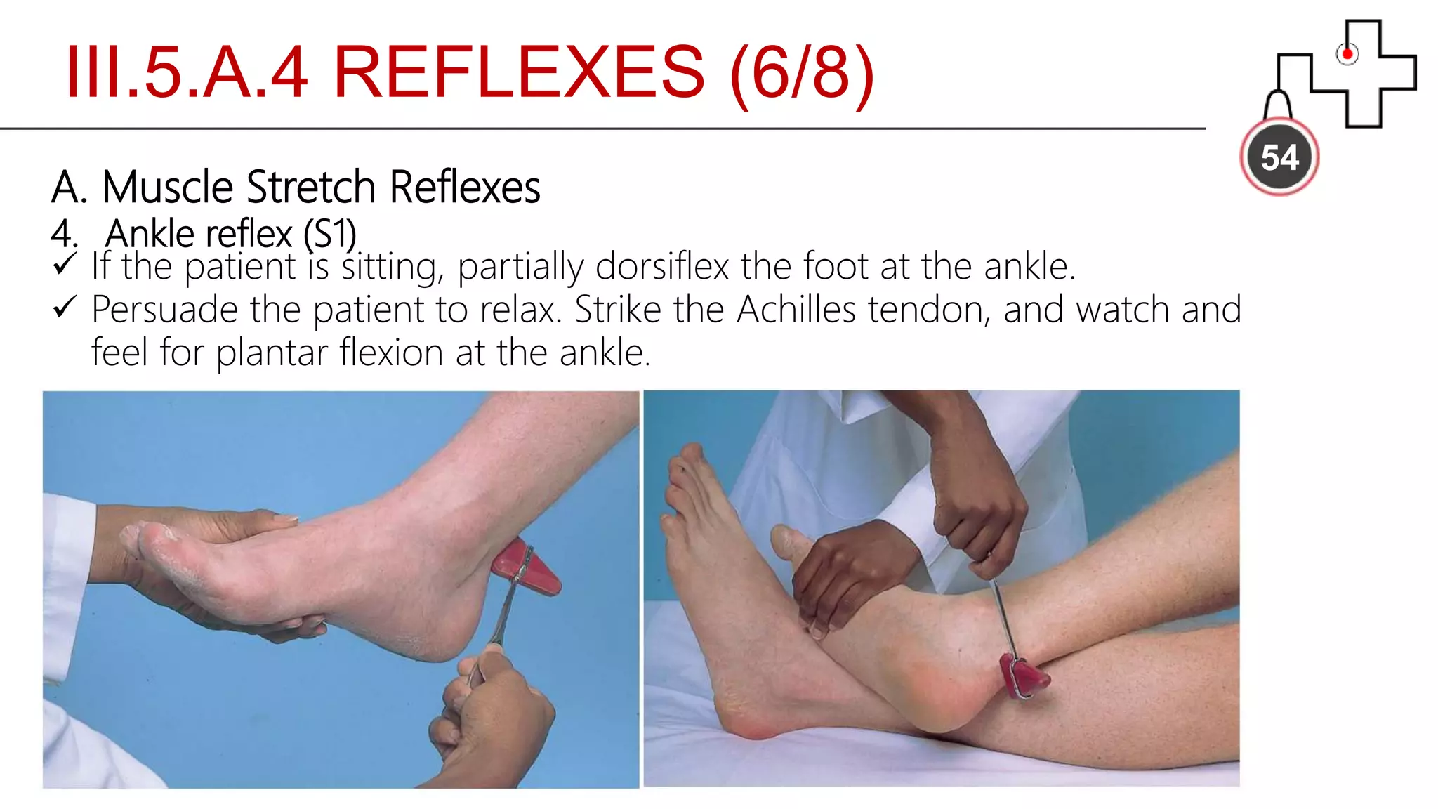
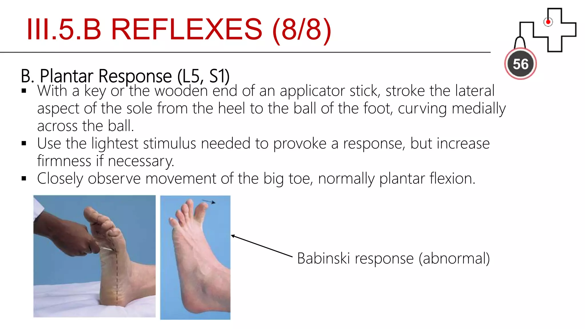
THE NEUROLOGIC
EXAMINATION
CelestinBilong Mbangtang JNR,MG4
Facultyof Medicine and BiomedicalSciences
Universityof Yaounde I
Cameroon
OBJECTIVES
02
L2, L3: Appreciate the burden of neuroscience in the neurologic
examination and familiarize with clinical terms in neurology.
M1: Understand and master the facets of the neurologic examination.
PLAN
03Introduction
I. History taking
II. Common Symptoms
III. Essential screening exam
IV. Some syndromes
Conclusion
References
INTRODUCTION
04
 Prerequisite: Neuroscience
 Adopt a fixed routine or examination sequence
 Dedication and Practice
I. HISTORY TAKING
05
 Proforma is same as other systems but for identification:
handedness
 Goal of history taking: site of lesion, nature of lesion
 Basic description:
 Site
 Severity
 Onset
 Duration
 Frequency
 Precipitating/Relieving factors
 Time of occurrence
II. COMMON SYMPTOMS
06
 Headaches
 Dizziness or Vertigo
 Weakness (generalized, proximal or distal)
 Numbness, abnormal or absent sensations
 Fainting and Blackout
 Seizures
 Tremors
 Cognitive function impairment
 Autonomic disorder
 …
III. ESSENTIAL SCREENING EXAM
07
1. Mental status
2. Cranial nerves
3. Motor system
4. Sensory system
5. Reflexes
6. Meningeal signs
III. ESSENTIAL EXAM
08
 Requirements
1. Reflex hammer
2. Tuning fork (128 Hz)
3. Opthalmoscope
4. Cotton bud
5. Tongue depressor
6. Olfactory stimulus e.g soap
III. 1 MENTAL STATUS
09
III.1. MENTAL STATUS (1/7)
10
 Evaluated throughout history taking
 Appearance and behavior (consciousness)
Glasgow Coma Scale (GCS)
 Cognitive function:
Attentiveness
Memory
Calculation
Abstract thinking
Speech and Language
III.1. MENTAL STATUS (2/7)
11
Glasgow Coma Scale
Peripheral pressure point (E):
 Nail bed
Central pressure points (M):
 Trapezius pinch
 Supraorbital notch pressure
 Check
 Observe
 Stimulate
 Rate
III.1. MENTAL STATUS (3/7)
12
 Cognitive function: Memory
Digit span (immediate recall)
Remote memory (e.g birthdays)
Recent memory (e.g events of the day)
New learning ability (long-term)
*Poor performance: dementia, delirium, Amnesia (anterograde, retrograde or both)
III.1. MENTAL STATUS (4/7)
13
 Cognitive function: Calculation
Serial 7s
Simple problem
Operation variety
*Poor performance: dementia, delirium, intellectual disability, level of education
III.1. MENTAL STATUS (5/7)
14
 Cognitive function: Speech and language
Fluency
Comprehension
Repetition
Naming
Reading
Writing
*Poor performance: Aphonia, Dysphonia, Dysarthria, Aphasia
III.1. MENTAL STATUS (6/7)
15
 Cognitive function: Speech and language
 Disorders of speech:
Phonation of voice (Aphonia, Dysphonia)
Articulation of words (Dysarthria)
Production and comprehension of language (Broca and Wernicke
Aphasia)
III.1. MENTAL STATUS (7/7)
16
 Cognitive function: Speech and language
III. 2 CRANIAL NERVES
17
III.2 CRANIAL NERVES (1/11)
18
 Cranial nerve I: Olfactory (sensory)
 Examination:
 Make sure each nasal passage is patent
 Ask the patient to close both eyes
 Occlude one nostril and test smell with the other. Repeat
procedure contralaterally
 Typical stimuli: Coffee, soap, lemon, vanilla; avoid menthol,
ammonia or peppermint
 Normal response: Bilateral perception and identification
*Results of lesions: Anosmia, Dysosmia, Hyposmia, CSF rhinorrhea
III.2 CRANIAL NERVES (2/11)
19
 Cranial nerve II: Optic (sensory)
 Examination:
 Visual acuity (Snellen chart)
 Visual field (confrontation)
 Fundoscopy
*Results of lesions:
 Visual field deficits
 Loss of pupillary light reflex (with III)
Static finger wiggle test
III.2 CRANIAL NERVES (3/11)
20
 Cranial nerve III: Oculomotor (Motor)
 Examination:
 Check pupillary light reflex
 Convergence
 Accommodation
*Results of lesions: Loss of pupillary light reflex, external strabismus,
ptosis, loss of accommodation, dilated pupil, loss of parallel gaze
III.2 CRANIAL NERVES (4/11)
21
 Cranial nerve III, IV and VI: Oculomotor, Trochlear and Abducens
(Motor)
 Examination:
 Visual inspection
 6 cardinal directions of gaze
 Smooth pursuits, saccades
 Convergence
 Pupillary light reflex
*Results of lesions: (In addition to III), weakness looking down in
adduction, internal strabismus
III.2 CRANIAL NERVES (5/11)
22
 Cranial nerve V: Trigeminal (Mixed)
 Examination:
 Motor (a & b)
 Sensory (c)
 Corneal reflex (d)
*Results of lesions: Loss of blink reflex
with VII, loss of general sensation in
corresponding areas.
(a) (b)
(c) (d)
III.2 CRANIAL NERVES (6/11)
23
 Cranial nerve VII: Facial (mixed)
 Examination:
 Raise both eye brows
 Frown
 Close eyes and test muscle strength while opening
 Smile
 Show upper and lower teeth
 Puff out both cheeks
*Results of lesions: Bell palsy (contralateral lower facial weakness if
central lesion), Loss of taste of anterior 2/3 of the tongue
III.2 CRANIAL NERVES (7/11)
24
Corticobulbar innervation of facial motor neurons
Cranial nerve VII:
Facial (Mixed)
III.2 CRANIAL NERVES (8/11)
25
 Cranial nerve VIII: Vestibulocochlear (sensory)
 Examination:
 Whispered voice test
 Weber test (lateralisation)
 Rinne test
*Results of lesions:
hearing loss, balance,
nystagmus
III.2 CRANIAL NERVES (9/11)
26
 Cranial nerve IX & X: Glossopharyngeal and Vagus (mixed)
 Examination
 Listen to patient’s voice: note any hoarseness, nasal quality
 Ask patient to swallow and cough
 Test gag reflex
*Results of lesions: Lost of gag reflex, dysphagia, nasal speech
III.2 CRANIAL NERVES (10/11)
27
 Cranial nerve XI: Accessory (Motor)
 Examination
 Trapezius
 Sternocleidomastoid
*Results of lesions:
 Shoulder droop
 Weakness turning
 Chin to opposite side
III.2 CRANIAL NERVES (11/11)
28
 Cranial nerve XII: Hypoglossal (Motor)
 Examination
 Note tongue position at rest in the mouth and on protrusion.
Is there deviation in any position?
 Ask patient to stick out tongue and move it from side to side.
Note strength and rapidity of movements
 Have patient push tongue into each cheek while you push
from the outside. Note strength.
*Results of lesion: Tongue deviation on protrusion towards side
of the lesion
III. 3 MOTOR SYSTEM
29
30
1. Visual inspection
2. Muscle bulk
3. Muscle tone
4. Muscle strength and endurance
5. Coordination
III.3 MOTOR SYSTEM (1/15)
311. Visual inspection
Observe the patient’s body position during movement and at rest
Watch for involuntary movements such as tremors, tics, chorea, or
fasciculations and characterize them.
2. Muscle bulk
Inspect the size and contours of muscles. Do the muscles look flat or
concave, suggesting loss of muscle bulk from atrophy or wasting? If
is the process unilateral or bilateral? . . . proximal or distal?
III.3.1-2 MOTOR SYSTEM (2/15)
32III.3.2 MOTOR SYSTEM (3/15)
III.3.3 MOTOR SYSTEM (4/15)
33
3. Muscle tone
 Ask patient to relax
 Flex and extend patient’s wrists, elbows, ankles and knees
 Look for increased (spasticity, rigidity) or decreased
resistance
34
Disorders of
muscle tone
III.3.4 MOTOR SYSTEM (6/15)
35
4. Muscle strength and endurance
 Isolate muscles when testing to avoid compensation
 Fix proximal joint when testing distally e.g fix humerus during
pronation
 Muscle strength gradation (0-5)
III.3.4 MOTOR SYSTEM (7/15)
36
4. Muscle strength and endurance (commonly tested)
 Biceps: flexion of fore arm at elbow
 Triceps: extension of forearm at elbow
 Extensor carpi radialis: dorsiflexion of hand at wrist
 Quadriceps femoris: knee extension
 Hamstrings: knee flexion
 Gastrocnemius/soleus: plantar flexion
37
III.3.4 MOTOR SYSTEM (8/15)
III.3.5 MOTOR SYSTEM (9/15)
38
5. Coordination
A. Rapid alternating movements
B. Point to point movements
C. Gait
D. Stance
III.3.5.A MOTOR SYSTEM (10/15)
39
5. Coordination
A. Rapid alternating movements
(a) Alternating arm movement
(b) Rapid finger tapping
Cerebellar disease: Dysmetria, dysdiadochokinesia, intention tremor
III.3.5.B MOTOR SYSTEM (11/15)
40
5. Coordination
B. Point to point movements
 Finger-nose test
 Heel-shin test
III.3.5.C MOTOR SYSTEM (12/15)
415. Coordination
C. Gait
 Observe the patient do the following:
 Rise from a seated position
 Walk across room, turn and come back
 Walk on toes
 Walk on heels
 Walk heel to toe (tandem gait) in a straight line
 Pay attention to posture, base of gait, arm swing, steadiness,
turning
 Be prepared to catch
III.3.5.D MOTOR SYSTEM (13/15)
425. Coordination
D. Stance
 Romberg test
 Pronator drift
43
Abnormalities of
Gait and Posture
(1/2)
44
Abnormalities of Gait and Posture (2/2)
III.3.5.D MOTOR SYSTEM (15/15)
III. 4 SENSORY SYSTEM
45
III. 4 SENSORY SYSTEM (1/2)
46
 Pain and temperature
 Position and vibration
 Light touch
 Discriminative sensations
NB: Could also be classified as superficial or
deep
III. 4 SENSORY SYSTEM (2/2)
47
 Discriminative sensations
 Stereognosia
 Graphesthesia
 Two-point discrimination
*Lesions to sensory cortex: Astereognosia, Graphanesthesia, loss of two-point discrimination
III. 5 REFLEXES
48
III. 5 REFLEXES (1/8)
49
A. Muscle Stretch Reflexes
B. Plantar Response
III.5.A REFLEXES (2/8)
50
A. Muscle Stretch Reflexes
 Encourage patient to relax and position limbs properly
 Quickly tap the tendon to which the muscle is attached
 Observe vigor and briskness of response and compare side-side
 If reflexes are diminished, try reinforcement.
III.5.A.1 REFLEXES (3/8)
51
A. Muscle Stretch Reflexes
 Test at least the following reflexes: biceps, triceps, patellar, ankle
1. Biceps reflex (C5, C6)
 The patient’s elbow should be partially flexed and the forearm
pronated with palm down.
 Place your thumb or finger firmly on the biceps tendon.
 Aim the strike with the reflex hammer directly through your digit toward
the biceps tendon
 Contraction of the biceps muscle
 Flexion of the elbow
III.5.A.2 REFLEXES (4/8)
52
A. Muscle Stretch Reflexes
2. Triceps reflex (C6, C7)
 The patient may be sitting or supine.
 Flex the patient’s arm at the elbow, with palm toward the body,
and pull it slightly across the chest.
 Strike the triceps tendon with a direct blow directly behind and
 just above the elbow
 Contraction of the triceps
 Extension of the elbow
III.5.A.3 REFLEXES (5/8)
53
A. Muscle Stretch Reflexes
3. Patellar reflex (L2, L3, L4)
The patient may be either sitting or lying down as long as the knee is
Briskly tap the patellar tendon just below the patella.
 Contraction of the quadriceps femoris
 Extension of the knee
 Placing your hand on the anterior thigh lets you feel this
III.5.A.4 REFLEXES (6/8)
54
A. Muscle Stretch Reflexes
4. Ankle reflex (S1)
 If the patient is sitting, partially dorsiflex the foot at the ankle.
 Persuade the patient to relax. Strike the Achilles tendon, and watch and
feel for plantar flexion at the ankle.
III.5.A REFLEXES (7/8)
55
A. Muscle Stretch Reflexes
 Test for ankle clonus if reflexes seem hyperactive.
 Support knee in a partly flexed position
 With patient relaxed, quickly dorsiflex the foot
 Observe for rhythmic oscillations
III.5.B REFLEXES (8/8)
56
B. Plantar Response (L5, S1)
 With a key or the wooden end of an applicator stick, stroke the lateral
aspect of the sole from the heel to the ball of the foot, curving medially
across the ball.
 Use the lightest stimulus needed to provoke a response, but increase
firmness if necessary.
 Closely observe movement of the big toe, normally plantar flexion.
Babinski response (abnormal)
III. 6 MENINGEAL SIGNS
57
III. 6 MENINGEAL SIGNS (1/4)
58
A. Neck mobility/Nuchal rigidity
B. Kernig sign
C. Brudzinski sign
III. 6 MENINGEAL SIGNS (2/4)
59
A. Neck mobility/Nuchal rigidity
 First, make sure there is no injury or fracture to the cervical vertebrae
or cervical cord.
(In trauma settings, this often requires radiologic evaluation)
 Then, with the patient supine, place your hands behind the patient’s
head and flex the neck forward, if possible until the chin touches the
chest.
 Normally the neck is supple, and the patient can easily bend the head
and neck forward.
*Neck stiffness with resistance to flexion is mostly seen in patients with
bacterial meningitis and subarachnoid hemorrhage.
III. 6 MENINGEAL SIGNS (3/4)
60
B. Kernig sign
Flex the patient’s leg at both the hip and the knee, and then
slowly extend the leg and straighten the knee.
Discomfort behind the knee during full extension is normal
should not produce pain.
III. 6 MENINGEAL SIGNS (4/4)
61
C. Brudzinski sign
As you flex the neck, watch the hips and knees in reaction to
your maneuver.
Normally they should remain relaxed and motionless.
IV. SOME SYNDROMES
62
IV. SOME SYNDROMES (1/5)
63
 Brainstem:
A. Lateral medullary syndrome (Wallenberg)
B. Medial midbrain syndrome (Weber)
C. Dorsal midbrain syndrome (Parinaud)
 Spinal cord: Brown-Sequard syndrome
IV. SOME SYNDROMES (2/5)
64
 Brainstem:
A. Lateral medullary syndrome (Wallenberg)
 Vertigo, nausea, nystagmus
 Ipsilateral limb ataxia
 Ipsilateral loss of pain and
temperature to the face
 Ipsilateral paralysis of larynx,
pharynx, soft palate (dysphagia,
dysarthria, loss of gag reflex)
 Contralateral loss of pain and
temperature
 Ipsilateral Horner’s syndrome
*PICA
IV. SOME SYNDROMES (3/5)
65
 Brainstem:
B. Medial midbrain syndrome (Weber)
 Contralateral hemispastic
paresis
 Contralateral spastic paresis
of lower face
 Ipsilateral oculomotor palsy
*PCA
IV. SOME SYNDROMES (4/5)
66
 Brainstem:
C. Dorsal midbrain syndrome (Parinaud)
 Paralysis of upward gaze
 Various pupillary abnormalities
 Non communicating hydrocephalus
IV. SOME SYNDROMES (5/5)
67 Spinal cord: Brown-Sequard syndrome
 At lesion:
Loss of all sensation and flaccid
weakness
 Below lesion:
UMN of corticospinal tract:
ipsilateral spastic paresis
Medial lemniscal: ipsilateral
impaired proprioception, vibration,
2-point discrimination
Spinothalamic: Contralateral
impaired pain and temperature
sensation 1-2 seg.b.
CONCLUSION
68
 Neuroscience basics are of utmost importance
 Use a routine clinical examination
 Dedication and practice
REFERENCES
69
 Bates Guide to Physical examination and History taking, 12th
edition, Lynn S. Bickley
 USMLE Kaplan Anatomy, 2016
 Clinical Skills: Neurological Examination, Lecture notes
Alan Glass,M.D
Allyson Zazulia, M.D
Neurologic exam

Neurologic exam

  • 1.
    THE NEUROLOGIC EXAMINATION CelestinBilong MbangtangJNR,MG4 Facultyof Medicine and BiomedicalSciences Universityof Yaounde I Cameroon
  • 2.
    OBJECTIVES 02 L2, L3: Appreciatethe burden of neuroscience in the neurologic examination and familiarize with clinical terms in neurology. M1: Understand and master the facets of the neurologic examination.
  • 3.
    PLAN 03Introduction I. History taking II.Common Symptoms III. Essential screening exam IV. Some syndromes Conclusion References
  • 4.
    INTRODUCTION 04  Prerequisite: Neuroscience Adopt a fixed routine or examination sequence  Dedication and Practice
  • 5.
    I. HISTORY TAKING 05 Proforma is same as other systems but for identification: handedness  Goal of history taking: site of lesion, nature of lesion  Basic description:  Site  Severity  Onset  Duration  Frequency  Precipitating/Relieving factors  Time of occurrence
  • 6.
    II. COMMON SYMPTOMS 06 Headaches  Dizziness or Vertigo  Weakness (generalized, proximal or distal)  Numbness, abnormal or absent sensations  Fainting and Blackout  Seizures  Tremors  Cognitive function impairment  Autonomic disorder  …
  • 7.
    III. ESSENTIAL SCREENINGEXAM 07 1. Mental status 2. Cranial nerves 3. Motor system 4. Sensory system 5. Reflexes 6. Meningeal signs
  • 8.
    III. ESSENTIAL EXAM 08 Requirements 1. Reflex hammer 2. Tuning fork (128 Hz) 3. Opthalmoscope 4. Cotton bud 5. Tongue depressor 6. Olfactory stimulus e.g soap
  • 9.
    III. 1 MENTALSTATUS 09
  • 10.
    III.1. MENTAL STATUS(1/7) 10  Evaluated throughout history taking  Appearance and behavior (consciousness) Glasgow Coma Scale (GCS)  Cognitive function: Attentiveness Memory Calculation Abstract thinking Speech and Language
  • 11.
    III.1. MENTAL STATUS(2/7) 11 Glasgow Coma Scale Peripheral pressure point (E):  Nail bed Central pressure points (M):  Trapezius pinch  Supraorbital notch pressure  Check  Observe  Stimulate  Rate
  • 12.
    III.1. MENTAL STATUS(3/7) 12  Cognitive function: Memory Digit span (immediate recall) Remote memory (e.g birthdays) Recent memory (e.g events of the day) New learning ability (long-term) *Poor performance: dementia, delirium, Amnesia (anterograde, retrograde or both)
  • 13.
    III.1. MENTAL STATUS(4/7) 13  Cognitive function: Calculation Serial 7s Simple problem Operation variety *Poor performance: dementia, delirium, intellectual disability, level of education
  • 14.
    III.1. MENTAL STATUS(5/7) 14  Cognitive function: Speech and language Fluency Comprehension Repetition Naming Reading Writing *Poor performance: Aphonia, Dysphonia, Dysarthria, Aphasia
  • 15.
    III.1. MENTAL STATUS(6/7) 15  Cognitive function: Speech and language  Disorders of speech: Phonation of voice (Aphonia, Dysphonia) Articulation of words (Dysarthria) Production and comprehension of language (Broca and Wernicke Aphasia)
  • 16.
    III.1. MENTAL STATUS(7/7) 16  Cognitive function: Speech and language
  • 17.
    III. 2 CRANIALNERVES 17
  • 18.
    III.2 CRANIAL NERVES(1/11) 18  Cranial nerve I: Olfactory (sensory)  Examination:  Make sure each nasal passage is patent  Ask the patient to close both eyes  Occlude one nostril and test smell with the other. Repeat procedure contralaterally  Typical stimuli: Coffee, soap, lemon, vanilla; avoid menthol, ammonia or peppermint  Normal response: Bilateral perception and identification *Results of lesions: Anosmia, Dysosmia, Hyposmia, CSF rhinorrhea
  • 19.
    III.2 CRANIAL NERVES(2/11) 19  Cranial nerve II: Optic (sensory)  Examination:  Visual acuity (Snellen chart)  Visual field (confrontation)  Fundoscopy *Results of lesions:  Visual field deficits  Loss of pupillary light reflex (with III) Static finger wiggle test
  • 20.
    III.2 CRANIAL NERVES(3/11) 20  Cranial nerve III: Oculomotor (Motor)  Examination:  Check pupillary light reflex  Convergence  Accommodation *Results of lesions: Loss of pupillary light reflex, external strabismus, ptosis, loss of accommodation, dilated pupil, loss of parallel gaze
  • 21.
    III.2 CRANIAL NERVES(4/11) 21  Cranial nerve III, IV and VI: Oculomotor, Trochlear and Abducens (Motor)  Examination:  Visual inspection  6 cardinal directions of gaze  Smooth pursuits, saccades  Convergence  Pupillary light reflex *Results of lesions: (In addition to III), weakness looking down in adduction, internal strabismus
  • 22.
    III.2 CRANIAL NERVES(5/11) 22  Cranial nerve V: Trigeminal (Mixed)  Examination:  Motor (a & b)  Sensory (c)  Corneal reflex (d) *Results of lesions: Loss of blink reflex with VII, loss of general sensation in corresponding areas. (a) (b) (c) (d)
  • 23.
    III.2 CRANIAL NERVES(6/11) 23  Cranial nerve VII: Facial (mixed)  Examination:  Raise both eye brows  Frown  Close eyes and test muscle strength while opening  Smile  Show upper and lower teeth  Puff out both cheeks *Results of lesions: Bell palsy (contralateral lower facial weakness if central lesion), Loss of taste of anterior 2/3 of the tongue
  • 24.
    III.2 CRANIAL NERVES(7/11) 24 Corticobulbar innervation of facial motor neurons Cranial nerve VII: Facial (Mixed)
  • 25.
    III.2 CRANIAL NERVES(8/11) 25  Cranial nerve VIII: Vestibulocochlear (sensory)  Examination:  Whispered voice test  Weber test (lateralisation)  Rinne test *Results of lesions: hearing loss, balance, nystagmus
  • 26.
    III.2 CRANIAL NERVES(9/11) 26  Cranial nerve IX & X: Glossopharyngeal and Vagus (mixed)  Examination  Listen to patient’s voice: note any hoarseness, nasal quality  Ask patient to swallow and cough  Test gag reflex *Results of lesions: Lost of gag reflex, dysphagia, nasal speech
  • 27.
    III.2 CRANIAL NERVES(10/11) 27  Cranial nerve XI: Accessory (Motor)  Examination  Trapezius  Sternocleidomastoid *Results of lesions:  Shoulder droop  Weakness turning  Chin to opposite side
  • 28.
    III.2 CRANIAL NERVES(11/11) 28  Cranial nerve XII: Hypoglossal (Motor)  Examination  Note tongue position at rest in the mouth and on protrusion. Is there deviation in any position?  Ask patient to stick out tongue and move it from side to side. Note strength and rapidity of movements  Have patient push tongue into each cheek while you push from the outside. Note strength. *Results of lesion: Tongue deviation on protrusion towards side of the lesion
  • 29.
    III. 3 MOTORSYSTEM 29
  • 30.
    30 1. Visual inspection 2.Muscle bulk 3. Muscle tone 4. Muscle strength and endurance 5. Coordination III.3 MOTOR SYSTEM (1/15)
  • 31.
    311. Visual inspection Observethe patient’s body position during movement and at rest Watch for involuntary movements such as tremors, tics, chorea, or fasciculations and characterize them. 2. Muscle bulk Inspect the size and contours of muscles. Do the muscles look flat or concave, suggesting loss of muscle bulk from atrophy or wasting? If is the process unilateral or bilateral? . . . proximal or distal? III.3.1-2 MOTOR SYSTEM (2/15)
  • 32.
  • 33.
    III.3.3 MOTOR SYSTEM(4/15) 33 3. Muscle tone  Ask patient to relax  Flex and extend patient’s wrists, elbows, ankles and knees  Look for increased (spasticity, rigidity) or decreased resistance
  • 34.
  • 35.
    III.3.4 MOTOR SYSTEM(6/15) 35 4. Muscle strength and endurance  Isolate muscles when testing to avoid compensation  Fix proximal joint when testing distally e.g fix humerus during pronation  Muscle strength gradation (0-5)
  • 36.
    III.3.4 MOTOR SYSTEM(7/15) 36 4. Muscle strength and endurance (commonly tested)  Biceps: flexion of fore arm at elbow  Triceps: extension of forearm at elbow  Extensor carpi radialis: dorsiflexion of hand at wrist  Quadriceps femoris: knee extension  Hamstrings: knee flexion  Gastrocnemius/soleus: plantar flexion
  • 37.
  • 38.
    III.3.5 MOTOR SYSTEM(9/15) 38 5. Coordination A. Rapid alternating movements B. Point to point movements C. Gait D. Stance
  • 39.
    III.3.5.A MOTOR SYSTEM(10/15) 39 5. Coordination A. Rapid alternating movements (a) Alternating arm movement (b) Rapid finger tapping Cerebellar disease: Dysmetria, dysdiadochokinesia, intention tremor
  • 40.
    III.3.5.B MOTOR SYSTEM(11/15) 40 5. Coordination B. Point to point movements  Finger-nose test  Heel-shin test
  • 41.
    III.3.5.C MOTOR SYSTEM(12/15) 415. Coordination C. Gait  Observe the patient do the following:  Rise from a seated position  Walk across room, turn and come back  Walk on toes  Walk on heels  Walk heel to toe (tandem gait) in a straight line  Pay attention to posture, base of gait, arm swing, steadiness, turning  Be prepared to catch
  • 42.
    III.3.5.D MOTOR SYSTEM(13/15) 425. Coordination D. Stance  Romberg test  Pronator drift
  • 43.
  • 44.
    44 Abnormalities of Gaitand Posture (2/2) III.3.5.D MOTOR SYSTEM (15/15)
  • 45.
    III. 4 SENSORYSYSTEM 45
  • 46.
    III. 4 SENSORYSYSTEM (1/2) 46  Pain and temperature  Position and vibration  Light touch  Discriminative sensations NB: Could also be classified as superficial or deep
  • 47.
    III. 4 SENSORYSYSTEM (2/2) 47  Discriminative sensations  Stereognosia  Graphesthesia  Two-point discrimination *Lesions to sensory cortex: Astereognosia, Graphanesthesia, loss of two-point discrimination
  • 48.
  • 49.
    III. 5 REFLEXES(1/8) 49 A. Muscle Stretch Reflexes B. Plantar Response
  • 50.
    III.5.A REFLEXES (2/8) 50 A.Muscle Stretch Reflexes  Encourage patient to relax and position limbs properly  Quickly tap the tendon to which the muscle is attached  Observe vigor and briskness of response and compare side-side  If reflexes are diminished, try reinforcement.
  • 51.
    III.5.A.1 REFLEXES (3/8) 51 A.Muscle Stretch Reflexes  Test at least the following reflexes: biceps, triceps, patellar, ankle 1. Biceps reflex (C5, C6)  The patient’s elbow should be partially flexed and the forearm pronated with palm down.  Place your thumb or finger firmly on the biceps tendon.  Aim the strike with the reflex hammer directly through your digit toward the biceps tendon  Contraction of the biceps muscle  Flexion of the elbow
  • 52.
    III.5.A.2 REFLEXES (4/8) 52 A.Muscle Stretch Reflexes 2. Triceps reflex (C6, C7)  The patient may be sitting or supine.  Flex the patient’s arm at the elbow, with palm toward the body, and pull it slightly across the chest.  Strike the triceps tendon with a direct blow directly behind and  just above the elbow  Contraction of the triceps  Extension of the elbow
  • 53.
    III.5.A.3 REFLEXES (5/8) 53 A.Muscle Stretch Reflexes 3. Patellar reflex (L2, L3, L4) The patient may be either sitting or lying down as long as the knee is Briskly tap the patellar tendon just below the patella.  Contraction of the quadriceps femoris  Extension of the knee  Placing your hand on the anterior thigh lets you feel this
  • 54.
    III.5.A.4 REFLEXES (6/8) 54 A.Muscle Stretch Reflexes 4. Ankle reflex (S1)  If the patient is sitting, partially dorsiflex the foot at the ankle.  Persuade the patient to relax. Strike the Achilles tendon, and watch and feel for plantar flexion at the ankle.
  • 55.
    III.5.A REFLEXES (7/8) 55 A.Muscle Stretch Reflexes  Test for ankle clonus if reflexes seem hyperactive.  Support knee in a partly flexed position  With patient relaxed, quickly dorsiflex the foot  Observe for rhythmic oscillations
  • 56.
    III.5.B REFLEXES (8/8) 56 B.Plantar Response (L5, S1)  With a key or the wooden end of an applicator stick, stroke the lateral aspect of the sole from the heel to the ball of the foot, curving medially across the ball.  Use the lightest stimulus needed to provoke a response, but increase firmness if necessary.  Closely observe movement of the big toe, normally plantar flexion. Babinski response (abnormal)
  • 57.
  • 58.
    III. 6 MENINGEALSIGNS (1/4) 58 A. Neck mobility/Nuchal rigidity B. Kernig sign C. Brudzinski sign
  • 59.
    III. 6 MENINGEALSIGNS (2/4) 59 A. Neck mobility/Nuchal rigidity  First, make sure there is no injury or fracture to the cervical vertebrae or cervical cord. (In trauma settings, this often requires radiologic evaluation)  Then, with the patient supine, place your hands behind the patient’s head and flex the neck forward, if possible until the chin touches the chest.  Normally the neck is supple, and the patient can easily bend the head and neck forward. *Neck stiffness with resistance to flexion is mostly seen in patients with bacterial meningitis and subarachnoid hemorrhage.
  • 60.
    III. 6 MENINGEALSIGNS (3/4) 60 B. Kernig sign Flex the patient’s leg at both the hip and the knee, and then slowly extend the leg and straighten the knee. Discomfort behind the knee during full extension is normal should not produce pain.
  • 61.
    III. 6 MENINGEALSIGNS (4/4) 61 C. Brudzinski sign As you flex the neck, watch the hips and knees in reaction to your maneuver. Normally they should remain relaxed and motionless.
  • 62.
  • 63.
    IV. SOME SYNDROMES(1/5) 63  Brainstem: A. Lateral medullary syndrome (Wallenberg) B. Medial midbrain syndrome (Weber) C. Dorsal midbrain syndrome (Parinaud)  Spinal cord: Brown-Sequard syndrome
  • 64.
    IV. SOME SYNDROMES(2/5) 64  Brainstem: A. Lateral medullary syndrome (Wallenberg)  Vertigo, nausea, nystagmus  Ipsilateral limb ataxia  Ipsilateral loss of pain and temperature to the face  Ipsilateral paralysis of larynx, pharynx, soft palate (dysphagia, dysarthria, loss of gag reflex)  Contralateral loss of pain and temperature  Ipsilateral Horner’s syndrome *PICA
  • 65.
    IV. SOME SYNDROMES(3/5) 65  Brainstem: B. Medial midbrain syndrome (Weber)  Contralateral hemispastic paresis  Contralateral spastic paresis of lower face  Ipsilateral oculomotor palsy *PCA
  • 66.
    IV. SOME SYNDROMES(4/5) 66  Brainstem: C. Dorsal midbrain syndrome (Parinaud)  Paralysis of upward gaze  Various pupillary abnormalities  Non communicating hydrocephalus
  • 67.
    IV. SOME SYNDROMES(5/5) 67 Spinal cord: Brown-Sequard syndrome  At lesion: Loss of all sensation and flaccid weakness  Below lesion: UMN of corticospinal tract: ipsilateral spastic paresis Medial lemniscal: ipsilateral impaired proprioception, vibration, 2-point discrimination Spinothalamic: Contralateral impaired pain and temperature sensation 1-2 seg.b.
  • 68.
    CONCLUSION 68  Neuroscience basicsare of utmost importance  Use a routine clinical examination  Dedication and practice
  • 69.
    REFERENCES 69  Bates Guideto Physical examination and History taking, 12th edition, Lynn S. Bickley  USMLE Kaplan Anatomy, 2016  Clinical Skills: Neurological Examination, Lecture notes Alan Glass,M.D Allyson Zazulia, M.D