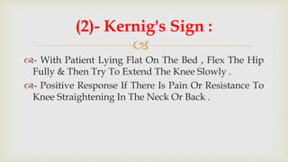
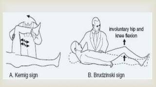
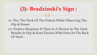
PREPARED BY THOBIAS B MSOMI

History Of Common Cases
1) Epilepsy :
2) Guillain-Barre Syndrome ( GBS ) :
3) Stroke :

- Introduce Yourself .
- Take Permission .
- Explain What You Are Going To Do .
- Maintain Privacy .
- Wash Your Hand .
Neurological Examination

A- Important Points In General
Examination

- General Look & Environment .
- Signs Of Respiratory Distress .
- Vital Signs : Pulse , BP , RR , Temp .
- Pallor , Jaundice .
- Skin Changes , Any Deformities .
- Any Dysmorphic Features .
A- Important Points In General
Examination

- Skull Shape , Fontanel Size & Closure .
- Abnormalities In The Spines ( Scoliosis , Kyphosis ,
Lordosis , Spina Pifida ) .
- Growth Parameters .

B- Signs Of Meningeal
Irritation

- Start By Active Movement , Ask The Patient To Flex
His Neck As Full As He Can & Then Proceed To
Passive Flexion.
(1)- Neck Stiffness :

- With Patient Lying Flat On The Bed , Flex The Hip
Fully & Then Try To Extend The Knee Slowly .
- Positive Response If There Is Pain Or Resistance To
Knee Straightening In The Neck Or Back .
(2)- Kernig's Sign :

- Flex The Neck Of The Patient While Observing The
Hip & Knees .
- Positive Response If There Is A Flexion In The Neck
Results In Hip & Knee Flexion With Pain On The Back
Of Neck .
(3)- Brudzinski's Sign :

D- Higher Function
Examination

- By Glasgow Coma Scale ( GCS ) :
The Glasgow Coma Scale provides a practical method
for assessment of impairment of conscious level in
response to defined stimuli.
Shown on the table below
1. Loss Of Consciousness(LOC):
By Glasgow Coma Scale ( GCS ) :

Interpretation Of GCS :
3 - 8 : Severe Brain Injury .
9 - 12 : Moderate Brain Injury .
13 - 15 : Mild Brain Injury

Ask the patient the following questions
 - What Is Your Name ?
 - What Is Your Age ? What Is Your Address ?
 - Where Are You Now ? What Is The Time , Day , Date
, And Month ?
# Disorientation May Be Acute ( Delirium ) Or
Chronic ( Dementia ) .
2. Orientation
( Frontal Lobe Function ) :

 - Short Term Memory : Ask About Any Recent News .
 - Long Term Memory : Ask About Event That Occur
More Than 5 Years Ago ( Marriage , Graduation , … ) .
3. Memory
( Temporal Lobe Function ) :

 - Shake The Patient Hand And Ask Is He Right Or Left
Handed .
 - To Identify The Dominant Hemisphere .
4. Handedness :

- Let The Patient Talks Freely , This Comes From
History Taking Already .
- Ask Patient To Describe The Room Or His Job To
Promote Flowing Speech .
- Then Test Comprehension By Applying Commands
" Touch Your Nose Then Ear Then , …" , Then By Yes
& No Questions .
5. Speech :

- Then Test Repetition , For Example : Repeat "
Alhamdullah " .
# If No Abnormality Detected There Is No Need To
Further Examination .

A- Dysphasia :
 - Receptive ( Posterior ) Dysphasia :
The Patient Can't Understand The Spoken Or Written Words.
Speech Is Fluent But Disorganized , Occur With Lesions In
Dominant Hemisphere : Wernicke's Area Specifically.
 - Expressive ( Anterior ) Dysphasia :
The Patient Understand But Can't Answer Appropriately.
Speech Isn't Fluent , Occur With Lesions In Dominant
Hemisphere : Broca's Area Specifically.
NOTE; Further Examination in
speech abnormality

 - Nominal Dysphasia :
The Patient Can't Names The Objects Rather That Is Normal ,
Occur With Lesions In Dominant Hemisphere : Posterior
Temporo-Parietal Area .
 - Conductive Dysphasia :
The Patient Repeats Statements Poorly But Can Follow
Commands , Lesion In Arcuate Fasciculus Or Other Connecting
Fibers .

B- Dysarthria :
Difficulty In Articulation Without Any Problems In
Content Of Speech , Occurs In UMNL Of Cranial
Nerves And Cerebellum Lesion .
C- Dysphonia :
Alteration Of The Sound Of The Voice , May Due To
Laryngeal Diseases Or RLN Palsy .

6. Cortical Sensation :
 - Astereognosis :
Ability To Recognize Familiar Objects When Put On
Hand While Closed Eyes .
 - Graphesthesia :
Ability Recognize Numbers & Letters When Written In
The Palm While The Eyes Closed .

 - Tactile Localization :
Ability To Recognize The Point Of Touch With Closed Eyes .
898
 - Two Points Discrimination :
Ability To Identify 2 Points Applied Simultaneously To The
Dorsum Of The Foot Or Pulp Of The Finger. Approximate
The Two Points Together Until He Perceives Them As One
Point , Minimal Separation 3 Cm .

E- Cranial Nerves Examination
( CNs )

" Not Tested Routinely "
- Ask The Patient To Close His Eyes & Say Yes If He
Smells Anything New ( Use Familiar Odour ) .
- Each Nostril Should Be Examined Separately
1- Olfactory Nerve ( I ) :

A- Visual Acuity : ( Pic.1 )
- Examine Near Vision By Reading & Far Vision By
Snellen Chart .
- Each Eye Should Examined Separately .
2- Optic Nerve ( II ) :
Pic.1

B- Color Vision : ( Pic.2 )
- By Ishihara Chart .
Pic.2

C- Visual Field : ( Pic.3 )
- By Confrontation Test , The Examiner Head Should
Be At Level Of Patient Head And Test Each Eye
Separately .
- Patient Looks Directly To Examiner Eye .
- By Finger Or Colored Pin Brought In From Outside
Until The Patient Can See It .
Visual Field

D- Papillary Reactions :
a) Reaction To Light :
Ask The Patient To Look Away From The Major
Source Of Light In The Room , Then Shine Pen
Torch On The Pupil From The Side Of The Eye .

Both Pupils On The Same Side ( Direct Light Reflex )
As Well As The Opposite Side ( Consensual Light
Reflex ) Well Constrict (Repeat From The Other Side) .
Remember That The Afferent Limb Is 2nd CN & The
Efferent Is 3rd CN .

b) Reaction To Accommodation Reflex :
Ask The Patient To Look At A Distant Object , Then
Ask Him To Focus On A Finger Held Close To His
Nose .
The Eyes Converge & The Pupils Constrict Attempting
To Look At A Close Object .

E- Fundoscopy :
To Examine The Optic Disc & Retina ( Not Routinely
Performed ) .

-All Muscles Are Supplied By Oculomotor Except
Superior Oblique ( Trochlear ) & Lateral Rectus
(Abducent) .
- Ask The Patient To Follow An Object ( E.g : Your
Finger ) In Different Direction .
-Move It In Vertical, Horizontal & Oblique Directions.
3- Oculomotor, Trochlear &
Abducent nerves ( III,IV, VI):

( Not Routinely Examined )
a) Motor Component :
Ask The Patient To Open & Close His Mouth , The
Jaw Will Be Deviated To The Paralyzed Side .
Ask The Patient To Clench His Teeth , Then Palpate
For Masseter & Temporalis Muscles.
4- Trigeminal nerve ( V ) :

b) Jaw Reflex Test ( Pic.4 ) :
Ask The Patient To Open His Mouth A Little , Place
Your Index Over The Chin & Then Tap Your Index In A
Downward Direction With A Tendon Hammer.
Normally There Is A Weak Or Absent Jerks Of The Jaw .
Pic.4

c) Sensory Component :
Touch The Face In 3 Different Regions With A Cotton
Over The Forehead ( Ophthalmic ) , Cheeks (
Maxillary ) , And Chin ( Mandibular ) .
Ask The Patient To Close His Eyes & Say Yes If He
Feels The Touch .

Corneal Reflex Test :
Ask The Patient To Look In One Direction & Approach
The Cornea From The Opposite Side With A Cotton ,
Observe For The Blink .

- Motor :
Facial Asymmetry ( Absence Of Frontal Wrinkles ) .
Flattening Of Naso-labial Folds .
Open Eye ( Widened Palpebral ) .
Deviation Of The Angle Of Mouth ( Drooping ) .
5- Facial nerve ( VII ) :
- Ask The Patient To :
Raise Eyebrows ( Occipito-Frontalis Muscle ) .
Close Eyes Tightly While You Are Trying To Open
Them ( Orbicularis Oculi ) .
Smile Or To Show His Teeth ( Levator Labii Muscle ) .
Whistle ( Orbicularis Oris ).
Puff Out Cheeks & To Keep Them Out While You Are
Tapping With Your Finger Over Both
Cheeks(Buccinators).

- Sensory :
Chorda Tympani Supplies Taste To The Anterior
Two-Thirds Of The Tongue .
Examination Of Taste Is Hard To Perform & Not
Usually Recommended .

- Cochlear Division :
Audiometry , Rinne's Test , Weber's Tests . ( Pic.5 )
- Vestibular Division :
It's Usually Tested Along With Cerebellar Function In
Order To Assess Balance & Gait .
6- Vestibulo-cochlear Nerve (VIII )
Pic.5

7- Glossopharyngeal & Vagus
Nerves ( IX, X ) :
- Talk To The Patient To Comment On His Voice
(Hoarseness Of Voice ) .
- Palatal Reflex : Touch The Patient's Soft Palate With
Tongue Depressor , It Will Lead To Elevation Of Soft
Palate & Retraction Of Uvula .
- Gag Reflex : Touch The Patient's Pharyngeal Wall
With Spatula , It Will Stimulate Gagging .

- Trapezius :
Ask The Patient To Shrug His Shoulder , Any Weakness
Result In Ipsilateral Dropping Of The Shoulder .
- Sternocleidomastoid :
Put Your Hand On The Medial Side Of The Patient's Jaw
, Ask Him To Push Against Your Hand While You Are
Palpating The Opposite Sternocleidomastoid Muscles .
8- Spinal Accessory Nerve ( XI ):
( Pic.6 )
Pic.6

9- Hypoglossal Nerve ( XII ) :
- Look At The Patient's Tongue , While Inside The
Mouth Observe For Size , Position , Wasting ,
Fasciculation .
- Ask The Patient To Stick His Tongue Out , Observe
For Any Deviation To Any Side .
- Ask The Patient To Push By His Tongue Against
Tongue Blade ( On Each Side ) .

F- Motor System Examination

- Posture .
- Gait .
- Muscle Bulk : Atrophy - Hypertrophy .
- Involuntary movements : Chorea , Tics , Tremor ,
Fasciculation .
- Inspection :

A- Upper Limb :
- Shaking Hands And Do Supination & Pronation .
- Flexion And Extension In The Elbow .
B- Lower Limb :
- Rolling .
- Flexion And Extension In Knee Joint .
- Raise Legs Up And Release It To Fall Down .
- Examination Of The Tone :
Notes :
- Grades Of Power :
0/5 : No Contraction .
1/5 : Just Flickering .
2/5 : Horizontal Movement With Gravity Only .
3/5 : Against Gravity , But WITHOUT Resistant .
4/5 : Against Gravity , With Minimal Resistant .
5/5 : Normal Strength .
- Movement Against Gravity = Grade 3 Or Above .

A- Upper Limb :
- Hand Grip .
- Flexion And Extension In Elbow With & Without
Resistant .
- Abduction & Adduction Of Both Arms With &
Without Resistant .
- Examination Of The Power :

B- Upper Limb :
- Abduction , Adduction , Flexion , Extension Of Hip
Joint With & Without Resistant .
- Flexion And Extension In Knee Joint With &
Without Resistant .
- Eversion , Inversion , Planter Flexion , Plantar
Extension Of Feet With & Without Resistant .

A- Deep Tendon Reflex : ( Pic.7 )
- Examination Of The Reflexes :

- Rating Of Deep Tendon Reflexes :
 0 : Absent = LMNL OR Early UMNL ( Shock Stage ) .
 +1 : Trace OR Seen Only With Reinforcement .
 +2 : Normal .
 +3 : Brisk .
 +4 : Non-Sustained Clonus .
 +5 : Sustained Clonus .
LMNL/UMNL = Lower/Upper Motor Neuron Lesion
Pic.7

B- Planter Reflex : ( Pic.8 )
- Scratch From Lateral Surface Of Dorsum Of Foot
And Goes Medially .
- Plantar Extension ( Big Toe Upward ) Called
Babinski Sign And It Indicates Upper Motor Neuron
Lesion(UMNL) .
Pic.8

C- Superficial Reflexes :
1- Abdominal Reflexes ( Epigastric T6-T9 , Abdominal
T9-T11 , Lower Abdomen T12-L1 ) : ( Pic.9 )
- By Lightly Stroking The Abdominal Wall Diagonally
Towards Umbilicus In Each Quadrant Of The
Abdomen .
- Normally : Contraction Of Abdominal Wall .
- Absent Of Contractions Indicates UMNL .

2- Cremasteric Reflex ( L1-L2 ) :
- Stroke Inner Part Of Thigh In A Downward
Direction .
- Cremasteric Muscle Contraction Causes Pull Up Of
Scrotum At Same Side .
Pic.9

G- Sensory System
Examination:

" Carried On Spino-Thalamic Tract “
1- Pain :
A Similar Approach Is Used With Stimulus Being The
Tip Of A Pin , The Patient Should Be Respond By
Saying Sharp OR Dull .
1- Superficial Sensation :

2- Temperature :
Use Two Tubes Of Cold & Warm Water , Touch The
Patient With One Of The Tubes And Ask Him To Tell
You If It's Cold Or Warm .
3- Fine Touch :
Touch The Skin Lightly With A Wisp Of Cotton ,
Then Ask Him To Say Yes If He Feel It With Closed
Eyes .

" Carried On (The Dorsal Column–Medial Lemniscus)
DCML Tract "
1- Proprioception :
Hold The Middle Phalanx Of The Patient's Index
Finger By Side, Then Flex & Extend The Distal
Phalanx Telling The Patient Which Direction You
Mean By Up & Down, Then Repeat It With His Eyes
Closed .
B- Deep Sensation :

2- Vibration :
By Tuning Fork On Bony Prominence While Eyes Are
Closed .
3- Deep Touch .

H- Cerebellar & Coordination
Examination :
( Not Routinely Examined )

A- Finger-Nose Test :
- Ask The Patient To Place The Tip Of The Index On
His Own Nose Starting From Full Abduction Of The
Arm With Eyes Open , Then Eyes Closed .
- Observe For Intention Tremor & Dysmetria (
Overshooting ) .
1- Upper Limbs :

B- Dysdiadochokinesia :
- Ask The Patient To Put One Hand On The Back Of
The Other Quickly & Regularly , Then Ask Him To
Tap The Back Of His Right Hand Alternately With
The Palm Of His Left Hand & Vice Versa , Also Ask
Him To Rapidly Supinate & Pronate His Hands
Together To Observe Symmetry & Regularity .

A- Heel-Shin Test : ( Pic.10 )
- Ask The Patient To Place His Heel On The Opposite
Knee & Then Run It Downwards Over The Shin Of
The Tibia To The Foot .
B- Toe-Finger Test :
- Ask The Patient To Left His Big Toe & Touch Your
Finger With It .
C- Gait : - Ask The Patient To Walk .
2- Lower Limbs :
Pic.10

## Romberg's Test : ( Pic.11 )
- Ask The Patient To Stand Erect With Eyes Open ,
Then Ask Him To Close His Eyes .
- It Is Positive When Unsteadiness Increase With
Closed Eyes .
- Positive Result Indicates Proprioception Loss .
Pic.11

Neurology examination

  • 1.
  • 2.
     History Of CommonCases 1) Epilepsy : 2) Guillain-Barre Syndrome ( GBS ) : 3) Stroke :
  • 3.
     - Introduce Yourself. - Take Permission . - Explain What You Are Going To Do . - Maintain Privacy . - Wash Your Hand . Neurological Examination
  • 4.
     A- Important PointsIn General Examination
  • 5.
     - General Look& Environment . - Signs Of Respiratory Distress . - Vital Signs : Pulse , BP , RR , Temp . - Pallor , Jaundice . - Skin Changes , Any Deformities . - Any Dysmorphic Features . A- Important Points In General Examination
  • 6.
     - Skull Shape, Fontanel Size & Closure . - Abnormalities In The Spines ( Scoliosis , Kyphosis , Lordosis , Spina Pifida ) . - Growth Parameters .
  • 7.
     B- Signs OfMeningeal Irritation
  • 8.
     - Start ByActive Movement , Ask The Patient To Flex His Neck As Full As He Can & Then Proceed To Passive Flexion. (1)- Neck Stiffness :
  • 9.
     - With PatientLying Flat On The Bed , Flex The Hip Fully & Then Try To Extend The Knee Slowly . - Positive Response If There Is Pain Or Resistance To Knee Straightening In The Neck Or Back . (2)- Kernig's Sign :
  • 11.
     - Flex TheNeck Of The Patient While Observing The Hip & Knees . - Positive Response If There Is A Flexion In The Neck Results In Hip & Knee Flexion With Pain On The Back Of Neck . (3)- Brudzinski's Sign :
  • 12.
  • 13.
     - By GlasgowComa Scale ( GCS ) : The Glasgow Coma Scale provides a practical method for assessment of impairment of conscious level in response to defined stimuli. Shown on the table below 1. Loss Of Consciousness(LOC):
  • 14.
    By Glasgow ComaScale ( GCS ) :
  • 15.
     Interpretation Of GCS: 3 - 8 : Severe Brain Injury . 9 - 12 : Moderate Brain Injury . 13 - 15 : Mild Brain Injury
  • 17.
     Ask the patientthe following questions  - What Is Your Name ?  - What Is Your Age ? What Is Your Address ?  - Where Are You Now ? What Is The Time , Day , Date , And Month ? # Disorientation May Be Acute ( Delirium ) Or Chronic ( Dementia ) . 2. Orientation ( Frontal Lobe Function ) :
  • 18.
      - ShortTerm Memory : Ask About Any Recent News .  - Long Term Memory : Ask About Event That Occur More Than 5 Years Ago ( Marriage , Graduation , … ) . 3. Memory ( Temporal Lobe Function ) :
  • 19.
      - ShakeThe Patient Hand And Ask Is He Right Or Left Handed .  - To Identify The Dominant Hemisphere . 4. Handedness :
  • 20.
     - Let ThePatient Talks Freely , This Comes From History Taking Already . - Ask Patient To Describe The Room Or His Job To Promote Flowing Speech . - Then Test Comprehension By Applying Commands " Touch Your Nose Then Ear Then , …" , Then By Yes & No Questions . 5. Speech :
  • 21.
     - Then TestRepetition , For Example : Repeat " Alhamdullah " . # If No Abnormality Detected There Is No Need To Further Examination .
  • 22.
     A- Dysphasia : - Receptive ( Posterior ) Dysphasia : The Patient Can't Understand The Spoken Or Written Words. Speech Is Fluent But Disorganized , Occur With Lesions In Dominant Hemisphere : Wernicke's Area Specifically.  - Expressive ( Anterior ) Dysphasia : The Patient Understand But Can't Answer Appropriately. Speech Isn't Fluent , Occur With Lesions In Dominant Hemisphere : Broca's Area Specifically. NOTE; Further Examination in speech abnormality
  • 23.
      - NominalDysphasia : The Patient Can't Names The Objects Rather That Is Normal , Occur With Lesions In Dominant Hemisphere : Posterior Temporo-Parietal Area .  - Conductive Dysphasia : The Patient Repeats Statements Poorly But Can Follow Commands , Lesion In Arcuate Fasciculus Or Other Connecting Fibers .
  • 24.
     B- Dysarthria : DifficultyIn Articulation Without Any Problems In Content Of Speech , Occurs In UMNL Of Cranial Nerves And Cerebellum Lesion . C- Dysphonia : Alteration Of The Sound Of The Voice , May Due To Laryngeal Diseases Or RLN Palsy .
  • 25.
     6. Cortical Sensation:  - Astereognosis : Ability To Recognize Familiar Objects When Put On Hand While Closed Eyes .  - Graphesthesia : Ability Recognize Numbers & Letters When Written In The Palm While The Eyes Closed .
  • 26.
      - TactileLocalization : Ability To Recognize The Point Of Touch With Closed Eyes . 898  - Two Points Discrimination : Ability To Identify 2 Points Applied Simultaneously To The Dorsum Of The Foot Or Pulp Of The Finger. Approximate The Two Points Together Until He Perceives Them As One Point , Minimal Separation 3 Cm .
  • 27.
     E- Cranial NervesExamination ( CNs )
  • 28.
     " Not TestedRoutinely " - Ask The Patient To Close His Eyes & Say Yes If He Smells Anything New ( Use Familiar Odour ) . - Each Nostril Should Be Examined Separately 1- Olfactory Nerve ( I ) :
  • 29.
     A- Visual Acuity: ( Pic.1 ) - Examine Near Vision By Reading & Far Vision By Snellen Chart . - Each Eye Should Examined Separately . 2- Optic Nerve ( II ) :
  • 30.
  • 31.
     B- Color Vision: ( Pic.2 ) - By Ishihara Chart . Pic.2
  • 32.
     C- Visual Field: ( Pic.3 ) - By Confrontation Test , The Examiner Head Should Be At Level Of Patient Head And Test Each Eye Separately . - Patient Looks Directly To Examiner Eye . - By Finger Or Colored Pin Brought In From Outside Until The Patient Can See It .
  • 33.
  • 34.
     D- Papillary Reactions: a) Reaction To Light : Ask The Patient To Look Away From The Major Source Of Light In The Room , Then Shine Pen Torch On The Pupil From The Side Of The Eye .
  • 35.
     Both Pupils OnThe Same Side ( Direct Light Reflex ) As Well As The Opposite Side ( Consensual Light Reflex ) Well Constrict (Repeat From The Other Side) . Remember That The Afferent Limb Is 2nd CN & The Efferent Is 3rd CN .
  • 36.
     b) Reaction ToAccommodation Reflex : Ask The Patient To Look At A Distant Object , Then Ask Him To Focus On A Finger Held Close To His Nose . The Eyes Converge & The Pupils Constrict Attempting To Look At A Close Object .
  • 37.
     E- Fundoscopy : ToExamine The Optic Disc & Retina ( Not Routinely Performed ) .
  • 38.
     -All Muscles AreSupplied By Oculomotor Except Superior Oblique ( Trochlear ) & Lateral Rectus (Abducent) . - Ask The Patient To Follow An Object ( E.g : Your Finger ) In Different Direction . -Move It In Vertical, Horizontal & Oblique Directions. 3- Oculomotor, Trochlear & Abducent nerves ( III,IV, VI):
  • 39.
     ( Not RoutinelyExamined ) a) Motor Component : Ask The Patient To Open & Close His Mouth , The Jaw Will Be Deviated To The Paralyzed Side . Ask The Patient To Clench His Teeth , Then Palpate For Masseter & Temporalis Muscles. 4- Trigeminal nerve ( V ) :
  • 40.
     b) Jaw ReflexTest ( Pic.4 ) : Ask The Patient To Open His Mouth A Little , Place Your Index Over The Chin & Then Tap Your Index In A Downward Direction With A Tendon Hammer. Normally There Is A Weak Or Absent Jerks Of The Jaw .
  • 41.
  • 42.
     c) Sensory Component: Touch The Face In 3 Different Regions With A Cotton Over The Forehead ( Ophthalmic ) , Cheeks ( Maxillary ) , And Chin ( Mandibular ) . Ask The Patient To Close His Eyes & Say Yes If He Feels The Touch .
  • 43.
     Corneal Reflex Test: Ask The Patient To Look In One Direction & Approach The Cornea From The Opposite Side With A Cotton , Observe For The Blink .
  • 44.
     - Motor : FacialAsymmetry ( Absence Of Frontal Wrinkles ) . Flattening Of Naso-labial Folds . Open Eye ( Widened Palpebral ) . Deviation Of The Angle Of Mouth ( Drooping ) . 5- Facial nerve ( VII ) :
  • 45.
    - Ask ThePatient To : Raise Eyebrows ( Occipito-Frontalis Muscle ) . Close Eyes Tightly While You Are Trying To Open Them ( Orbicularis Oculi ) . Smile Or To Show His Teeth ( Levator Labii Muscle ) . Whistle ( Orbicularis Oris ). Puff Out Cheeks & To Keep Them Out While You Are Tapping With Your Finger Over Both Cheeks(Buccinators).
  • 46.
     - Sensory : ChordaTympani Supplies Taste To The Anterior Two-Thirds Of The Tongue . Examination Of Taste Is Hard To Perform & Not Usually Recommended .
  • 47.
     - Cochlear Division: Audiometry , Rinne's Test , Weber's Tests . ( Pic.5 ) - Vestibular Division : It's Usually Tested Along With Cerebellar Function In Order To Assess Balance & Gait . 6- Vestibulo-cochlear Nerve (VIII )
  • 48.
  • 49.
     7- Glossopharyngeal &Vagus Nerves ( IX, X ) : - Talk To The Patient To Comment On His Voice (Hoarseness Of Voice ) . - Palatal Reflex : Touch The Patient's Soft Palate With Tongue Depressor , It Will Lead To Elevation Of Soft Palate & Retraction Of Uvula . - Gag Reflex : Touch The Patient's Pharyngeal Wall With Spatula , It Will Stimulate Gagging .
  • 50.
     - Trapezius : AskThe Patient To Shrug His Shoulder , Any Weakness Result In Ipsilateral Dropping Of The Shoulder . - Sternocleidomastoid : Put Your Hand On The Medial Side Of The Patient's Jaw , Ask Him To Push Against Your Hand While You Are Palpating The Opposite Sternocleidomastoid Muscles . 8- Spinal Accessory Nerve ( XI ): ( Pic.6 )
  • 51.
  • 52.
     9- Hypoglossal Nerve( XII ) : - Look At The Patient's Tongue , While Inside The Mouth Observe For Size , Position , Wasting , Fasciculation . - Ask The Patient To Stick His Tongue Out , Observe For Any Deviation To Any Side . - Ask The Patient To Push By His Tongue Against Tongue Blade ( On Each Side ) .
  • 53.
  • 54.
     - Posture . -Gait . - Muscle Bulk : Atrophy - Hypertrophy . - Involuntary movements : Chorea , Tics , Tremor , Fasciculation . - Inspection :
  • 55.
     A- Upper Limb: - Shaking Hands And Do Supination & Pronation . - Flexion And Extension In The Elbow . B- Lower Limb : - Rolling . - Flexion And Extension In Knee Joint . - Raise Legs Up And Release It To Fall Down . - Examination Of The Tone :
  • 56.
    Notes : - GradesOf Power : 0/5 : No Contraction . 1/5 : Just Flickering . 2/5 : Horizontal Movement With Gravity Only . 3/5 : Against Gravity , But WITHOUT Resistant . 4/5 : Against Gravity , With Minimal Resistant . 5/5 : Normal Strength . - Movement Against Gravity = Grade 3 Or Above .
  • 57.
     A- Upper Limb: - Hand Grip . - Flexion And Extension In Elbow With & Without Resistant . - Abduction & Adduction Of Both Arms With & Without Resistant . - Examination Of The Power :
  • 58.
     B- Upper Limb: - Abduction , Adduction , Flexion , Extension Of Hip Joint With & Without Resistant . - Flexion And Extension In Knee Joint With & Without Resistant . - Eversion , Inversion , Planter Flexion , Plantar Extension Of Feet With & Without Resistant .
  • 59.
     A- Deep TendonReflex : ( Pic.7 ) - Examination Of The Reflexes :
  • 60.
     - Rating OfDeep Tendon Reflexes :  0 : Absent = LMNL OR Early UMNL ( Shock Stage ) .  +1 : Trace OR Seen Only With Reinforcement .  +2 : Normal .  +3 : Brisk .  +4 : Non-Sustained Clonus .  +5 : Sustained Clonus . LMNL/UMNL = Lower/Upper Motor Neuron Lesion
  • 61.
  • 62.
     B- Planter Reflex: ( Pic.8 ) - Scratch From Lateral Surface Of Dorsum Of Foot And Goes Medially . - Plantar Extension ( Big Toe Upward ) Called Babinski Sign And It Indicates Upper Motor Neuron Lesion(UMNL) .
  • 63.
  • 64.
     C- Superficial Reflexes: 1- Abdominal Reflexes ( Epigastric T6-T9 , Abdominal T9-T11 , Lower Abdomen T12-L1 ) : ( Pic.9 ) - By Lightly Stroking The Abdominal Wall Diagonally Towards Umbilicus In Each Quadrant Of The Abdomen . - Normally : Contraction Of Abdominal Wall . - Absent Of Contractions Indicates UMNL .
  • 66.
     2- Cremasteric Reflex( L1-L2 ) : - Stroke Inner Part Of Thigh In A Downward Direction . - Cremasteric Muscle Contraction Causes Pull Up Of Scrotum At Same Side .
  • 67.
  • 68.
  • 69.
     " Carried OnSpino-Thalamic Tract “ 1- Pain : A Similar Approach Is Used With Stimulus Being The Tip Of A Pin , The Patient Should Be Respond By Saying Sharp OR Dull . 1- Superficial Sensation :
  • 70.
     2- Temperature : UseTwo Tubes Of Cold & Warm Water , Touch The Patient With One Of The Tubes And Ask Him To Tell You If It's Cold Or Warm . 3- Fine Touch : Touch The Skin Lightly With A Wisp Of Cotton , Then Ask Him To Say Yes If He Feel It With Closed Eyes .
  • 71.
     " Carried On(The Dorsal Column–Medial Lemniscus) DCML Tract " 1- Proprioception : Hold The Middle Phalanx Of The Patient's Index Finger By Side, Then Flex & Extend The Distal Phalanx Telling The Patient Which Direction You Mean By Up & Down, Then Repeat It With His Eyes Closed . B- Deep Sensation :
  • 72.
     2- Vibration : ByTuning Fork On Bony Prominence While Eyes Are Closed . 3- Deep Touch .
  • 73.
     H- Cerebellar &Coordination Examination : ( Not Routinely Examined )
  • 74.
     A- Finger-Nose Test: - Ask The Patient To Place The Tip Of The Index On His Own Nose Starting From Full Abduction Of The Arm With Eyes Open , Then Eyes Closed . - Observe For Intention Tremor & Dysmetria ( Overshooting ) . 1- Upper Limbs :
  • 75.
     B- Dysdiadochokinesia : -Ask The Patient To Put One Hand On The Back Of The Other Quickly & Regularly , Then Ask Him To Tap The Back Of His Right Hand Alternately With The Palm Of His Left Hand & Vice Versa , Also Ask Him To Rapidly Supinate & Pronate His Hands Together To Observe Symmetry & Regularity .
  • 76.
     A- Heel-Shin Test: ( Pic.10 ) - Ask The Patient To Place His Heel On The Opposite Knee & Then Run It Downwards Over The Shin Of The Tibia To The Foot . B- Toe-Finger Test : - Ask The Patient To Left His Big Toe & Touch Your Finger With It . C- Gait : - Ask The Patient To Walk . 2- Lower Limbs :
  • 77.
  • 78.
     ## Romberg's Test: ( Pic.11 ) - Ask The Patient To Stand Erect With Eyes Open , Then Ask Him To Close His Eyes . - It Is Positive When Unsteadiness Increase With Closed Eyes . - Positive Result Indicates Proprioception Loss .
  • 79.