
Recommended
More Related Content
Similar to centralnervoussystem-190923062339.pdf
Similar to centralnervoussystem-190923062339.pdf (20)
More from kiranpalepu5
More from kiranpalepu5 (20)
Recently uploaded
Recently uploaded (20)
centralnervoussystem-190923062339.pdf
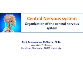
- 1. Central Nervous system Organization of the central nervous system Dr. S. Parasuraman, M.Pharm., Ph.D., Associate Professor, Faculty of Pharmacy, AIMST University.
- 2. 2 Divisions of the central nervous system
- 3. Divisions of the central nervous system • The central nervous system is made up of the brain and spinal cord. • The brain constitutes about one- fiftieth of the body weight and lies within the cranial cavity. • The brain parts are – Cerebrum – The brain stem (midbrain, pons, medulla oblongata) – Cerebellum 3 The average brain weight of the adult male was 1336 gr; for the adult female 1198 gr. With increasing age, brain weight decreases by 2.7 gr in males, and by 2.2 gr in females per year.
- 4. Divisions of the central nervous system • Cerebrum: – This is the largest part of the brain and it occupies the anterior and middle cranial fossae. – Cerebrum is made up of with two cerebral hemispheres and their cortices (outer layer of grey matter) and underlying regions of white matter. – The cerebral cortex is the outer covering of gray matter over the hemispheres. 4 Cerebral cortex
- 5. Divisions of the central nervous system • Cerebrum: – Each hemisphere of the cerebrum is divided into lobes which take the names of the bones of the cranium under which they lie: • frontal • parietal • temporal • Occipital 5 The boundaries of the lobes are marked by deep sulci (fissures). These are the central, lateral and parieto- occipital sulci.
- 6. Divisions of the central nervous system • Cerebrum: Functions of the cerebrum – Frontal Lobe- associated with reasoning, planning, parts of speech, movement, emotions, and problem solving – Parietal Lobe- associated with movement, orientation, recognition, perception of stimuli – Occipital Lobe- associated with visual processing – Temporal Lobe- associated with perception and recognition of auditory stimuli, memory, and speech 6
- 7. Divisions of the central nervous system • The brain stem (midbrain, pons, medulla oblongata) – Brain stem is the stem-like part of the base of the brain that is connected to the spinal cord. – This structure is responsible for basic vital life functions such as breathing, heartbeat, and blood pressure. – Medulla oblongata directly controls breathing, blood flow, and other essential functions. 7
- 8. Divisions of the central nervous system • Cerebellum – The cerebellum is situated behind the pons and immediately below the posterior portion of the cerebrum – It is ovoid in shape and has two hemispheres, separated by a narrow median strip called the vermis. 8
- 9. Divisions of the central nervous system • Cerebellum Functions of cerebellum – The cerebellum is concerned with the coordination of voluntary muscular movement, posture and balance. It coordinates activities associated with the maintenance of the balance and equilibrium of the body. – It is also involved in certain cognitive functions, such as language. – The cerebellum plays a major role in adapting and fine- tuning motor programs to make accurate movements through a trial-and-error process. – Damage to the cerebellum results in clumsy uncoordinated muscular movement, staggering gait and inability to carry out smooth, steady, precise movements. 9
- 10. Divisions of the central nervous system • Spinal cord – Spinal cord is continuous above with the medulla oblongata. – It is a long, thin bundle of nervous tissue. – It is approximately 45 cm long in an adult Caucasian male, and is about the thickness of the little finger. – The spinal cord receives and transmits electric signals throughout the enter body. 10
- 11. Divisions of the central nervous system • Spinal cord • The white matter tracts in the spinal cord are highways for nerve impulse propagation. Sensory input travels along these tracts toward the brain (afferent neurons), and motor output travels from the brain along these tracts toward skeletal muscles and other effector tissues (efferent neurons). • The gray matter of the spinal cord receives and integrates incoming and outgoing information. 11
- 12. Divisions of the central nervous system • Spinal cord Motor pathway and spinal reflexes discussed in Year 1 Semester 1 (Anatomy & Physiology [PIMC 6410301]). 12
- 13. Organization of the central nervous system 13
- 14. Organization of the central nervous system • The nervous system coordinates voluntary and involuntary actions in the body. • The nervous system is comprised of an enormous number of cells (over 100 billion), primarily of two types: neurons (the signaling units) and glial cells (the supporting units). • The nervous system divided into two major parts viz., central nervous system and peripheral nervous system. 14
- 15. Neuron structure and classification Neuron structure • Neuron has four specialized structures – The cell body (soma) – Dendrites – Axon – Axon terminals 15
- 16. Neuron structure and classification Neuron structure – Cell body or soma: The cell body plays a major role in synthesizing proteins. Groups of cell bodies are called nuclei in the central nervous system and ganglia in the peripheral nervous system. – Axons and dendrites: Axons and dendrites are extensions of cell bodies and form the white matter of the nervous system. Dendrites bring information to the cell body and axons take information away from the cell body. – Axon terminals: Axon terminals are that part of a nerve cell that make synaptic connections with another nerve cell or with an effector cell (e.g. muscle cell or gland cell). 16
- 17. Neuron structure and classification Classification of neurons 17 Structural classification Functional Classification 1. Multipolar neurons Sensory neurons (afferent neurons) 2. Bipolar neurons Motor neurons (efferent neurons ) 3. Unipolar neurons Interneurons • Sensory neurons transmit information from sensory receptors in the skin • Motor neurons transmit information away from the CNS toward some type of effector • Interneurons are located between motor and sensory pathways and involved in signal integration
- 18. Organization of the central nervous system Glial cells • Glia cells, also called glial or neuroglia, are non-neuronal cells in the CNS and PNS. • The neurones of the central nervous system are supported by four types of non-excitable glial cells that make up a quarter to a half of the volume of brain tissue. • The primary role of glial cells are to provide physical support for neurons. • Four major types of glial cells in the CNS – Astrocyte – Oligodendrocyte – Ependymal cell – Microglial cell 18
- 19. Types of glial cells 19
- 20. Organization of the central nervous system Glial cells – Astrocyte: These cells form the main supporting tissue of the central nervous system. Astrocytes are found in large numbers adjacent to blood vessels and neurons. Astrocytes are essential for the formation and maintenance of the blood–brain barrier (BBB). – Oligodendrocyte: These cells are smaller than astrocytes and they are the myelinating cells of the central nervous system. The primary function of the oligodendorcyte is to provide and maintain the myelin sheaths around axons. 20
- 21. Organization of the central nervous system Glial cells – Ependymal cell: Ependymal cells line the cavities of the CNS. Ependymal cells are responsible for the production of Cerebral Spinal Fluid (CSF) and are important barriers between the cerebral spinal fluid and the brain extracellular space. – Microglial cell: These cells are derived from monocytes that migrate from the blood into the nervous system before birth. Microglial cells are rapidly activated in the CNS in response to injury. These cells are also very important in presenting antigens to lymphocytes in response to infection. 21
- 22. Organization of the central nervous system • Membranes covering the brain and spinal cord – The brain and spinal cord are completely surrounded by three membranes (dura mater, arachnoid mater, pia mate), the meninges (leptomeninges [arachnoid and pia mater together], subarachnoid space [space between the arachnoid and the pia mater]), lying between the skull and the brain and between the vertebrae and the spinal cord. 22
- 23. Organization of the central nervous system • Ventricles of the brain and the cerebrospinal fluid – Within the brain there are four irregular-shaped cavities, or ventricles, containing cerebrospinal fluid (CSF). – CSF is secreted continuously at a rate of about 0.5 ml per minute, i.e. 720 ml per day. – CSF is a clear, slightly alkaline fluid with a specific gravity of 1.005, consisting of water, mineral salts, glucose, plasma proteins (small amounts of albumin and globulin), creatinine (small amounts), urea (small amounts) and a few leukocytes. 23
- 24. Organization of the central nervous system • Functions of cerebrospinal fluid – protects the brain and spinal cord. – maintains a uniform pressure around these delicate structures. – acts as a cushion and shock absorber between the brain and the cranial bones. – the brain and spinal cord moist and there may be interchange of substances between CSF and nerve cells, such as nutrients and waste products. 24
- 25. 25
- 26. Blood–brain barrier (BBB) • The blood-brain barrier is a selective barrier that protects the brain from potentially toxic substances and chemical variations in the blood • Oxygen, carbon dioxide, alcohol, barbiturates, glucose, and lipophilic substances quickly cross the barrier into the brain. • The brain and spinal cord are relatively well protected from microbial infection by the blood-brain barrier. 26 Back
- 27. Organization of the central nervous system • Organization of the central nervous system • Neuron structure and classification – Afferent neurons – Efferent neurons – Interneurons • Divisions of the central nervous system (Brain and spinal cord) • Glial cells • Blood brain barrier 27