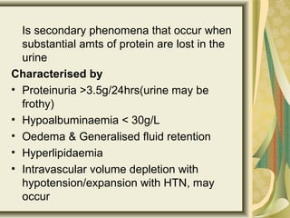
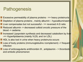
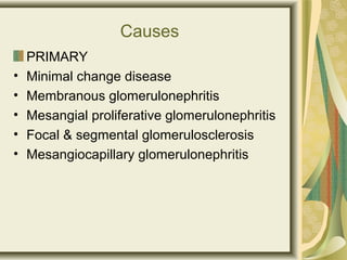
Nephrotic Syndrome
Is secondary phenomena that occur when
substantial amts of protein are lost in the
urine
Characterised by
• Proteinuria >3.5g/24hrs(urine may be
frothy)
• Hypoalbuminaemia < 30g/L
• Oedema & Generalised fluid retention
• Hyperlipidaemia
• Intravascular volume depletion with
hypotension/expansion with HTN, may
occur
Pathogenesis
Excessive permeability of plasma proteins - >> heavy proteinuria
Depletion of plasma proteins , mainly albumin – hypoalbuminaemia
Liver compensates but not successful - >> reversed A:G ratio
Reduced albumin -> decreased colloid oncotic pressure of the
blood -> oedema
Increased Lipoprotein synthesis and decreased catabolism by liver
->> Hyperlipidaemia (mainly VLDL and /or LDL)
HDL is also lost in urine when heavy proteinuria occurs
Loss of body proteins (immunoglobulins /complement) -> frequent
infection
Loss of anticoagulants antithrombin III , antiplasmin - > thrombotic
and embolic phenomenon
Causes
PRIMARY
• Minimal change disease
• Membranous glomerulonephritis
• Mesangial proliferative glomerulonephritis
• Focal & segmental glomerulosclerosis
• Mesangiocapillary glomerulonephritis
Causes
Secondary
• Infection: Bacteria
endocarditis,Malaria,Syphilis,Hepatitis
B,HIV
• Connective tissue diseases:
SLE,Rheumatoid arthritis
• Neoplasms: Hodgkin’s
lymphoma,Carcinomas,Leukaemias
• Metabolic: DM,Amyloidsis
• Drugs & toxins: Captopril,gold,Mercury
The diseases the cause Nephrotic
syndrome all affect the glomerulus
Mechanism:
• injury to podocytes
• Changed architecture
Scarring
Deposition of matrix or other
elements
Clinical Presentations
fluid retention ->abdominal
distention,ascites,edema,puffy eyelids
,scrotal swelling,weight gain,shortness of
breath
Anorexia
Hypertension
Oliguria
Orthostatic hypertension
Foamy urine
Periorbital edema
Pitting edema of lower limbs
Investigations
1.Urine analysis:
Protein : creatinine ratio in a spot
sample of urine (PCR>100mg/mL -
substantial)
proteinuria > 3.5g/L
Estimate 24 hour albumin excretion
(ACR>70 mg/ml)
RBC casts and WBC in
glomerulonephritis
Urea and creatinine may be elevated
<- renal failure
2.Estimate serum cholesterol
concentrations
3.Total cholesterol & LDL- cholesterol
elevated in most patients. HDL-
cholesterol is normal or decreased.
4. Blood sugar & glycosylated
hemoglobin tests for diabetes.
5. Hepatitis B and C serology, HIV
serology
6. Renal biopsy if cause is not clear
especially in an adult patient.
Management
• Treatment of nephrotic syndrome
involves three steps:
1.Measures to reduce proteinuria
2.Measures to control complications of
nephrotic syndrome
3.Treatment of underlying cause
Measures to reduce proteinuria
• If immunosuppressive drugs and other
measures against the underlying cause
do not benefit the patient
1.ACE inhibitors -> proteinuria,
intraglomerular pressure -> slows rate of
progression of renal failure. Prevents
development of haemodynamically
mediated FSG
2.NSAIDs -> proteinuria
Measures to control complications
of nephrotic syndrome
• Oedema -> salt restriction, rest,
judicious use of diuretics
• In severe cases -> I.V. salt-poor albumin
• Dietary proteins 0.8-1.0g/kg
• Vitamin D supplementation
• Hyperlipidaemia -> dietary restrictions,
lipid-lowering drugs
• Anticoagulants in patients with DVT or
arterial thrombosis
Measures to treat the underlying
disease
Minimal Change Disease
•Children – corticosteroids (prednisolone
60mg/m2 x 4wks, followed by alternate
day prednisolone 35mg/m2 for 4 additional
wks)
Children responding to steroid treatment in
1st
4 wks – “steroid responsive”, those who
relapse on withrawal –” steroid dependent”
•Patients with frequent relapses ->
cyclophosphamide 2mg/kg/day x 6 wks
Focal and segmental glomerulosclerosis
•Steroids (20-30% cases)
•Cylophosphamide, cyclosporin
Membranous glomerulonephritis
•Spontaneous remission (40%)
•30-40% cases remit and relapse repeatedly
•Rest 10-20% patients develop progressive
renal failure -> cyclophosphamide, cyclosporin
and chlorambucil in combination with steroids
may retard progression

Nephrotic syndrome

  • 1.
  • 2.
    Is secondary phenomenathat occur when substantial amts of protein are lost in the urine Characterised by • Proteinuria >3.5g/24hrs(urine may be frothy) • Hypoalbuminaemia < 30g/L • Oedema & Generalised fluid retention • Hyperlipidaemia • Intravascular volume depletion with hypotension/expansion with HTN, may occur
  • 3.
    Pathogenesis Excessive permeability ofplasma proteins - >> heavy proteinuria Depletion of plasma proteins , mainly albumin – hypoalbuminaemia Liver compensates but not successful - >> reversed A:G ratio Reduced albumin -> decreased colloid oncotic pressure of the blood -> oedema Increased Lipoprotein synthesis and decreased catabolism by liver ->> Hyperlipidaemia (mainly VLDL and /or LDL) HDL is also lost in urine when heavy proteinuria occurs Loss of body proteins (immunoglobulins /complement) -> frequent infection Loss of anticoagulants antithrombin III , antiplasmin - > thrombotic and embolic phenomenon
  • 4.
    Causes PRIMARY • Minimal changedisease • Membranous glomerulonephritis • Mesangial proliferative glomerulonephritis • Focal & segmental glomerulosclerosis • Mesangiocapillary glomerulonephritis
  • 5.
    Causes Secondary • Infection: Bacteria endocarditis,Malaria,Syphilis,Hepatitis B,HIV •Connective tissue diseases: SLE,Rheumatoid arthritis • Neoplasms: Hodgkin’s lymphoma,Carcinomas,Leukaemias • Metabolic: DM,Amyloidsis • Drugs & toxins: Captopril,gold,Mercury
  • 6.
    The diseases thecause Nephrotic syndrome all affect the glomerulus Mechanism: • injury to podocytes • Changed architecture Scarring Deposition of matrix or other elements
  • 7.
    Clinical Presentations fluid retention->abdominal distention,ascites,edema,puffy eyelids ,scrotal swelling,weight gain,shortness of breath Anorexia Hypertension Oliguria Orthostatic hypertension Foamy urine Periorbital edema Pitting edema of lower limbs
  • 8.
    Investigations 1.Urine analysis: Protein :creatinine ratio in a spot sample of urine (PCR>100mg/mL - substantial) proteinuria > 3.5g/L Estimate 24 hour albumin excretion (ACR>70 mg/ml) RBC casts and WBC in glomerulonephritis Urea and creatinine may be elevated <- renal failure
  • 9.
    2.Estimate serum cholesterol concentrations 3.Totalcholesterol & LDL- cholesterol elevated in most patients. HDL- cholesterol is normal or decreased. 4. Blood sugar & glycosylated hemoglobin tests for diabetes. 5. Hepatitis B and C serology, HIV serology 6. Renal biopsy if cause is not clear especially in an adult patient.
  • 10.
    Management • Treatment ofnephrotic syndrome involves three steps: 1.Measures to reduce proteinuria 2.Measures to control complications of nephrotic syndrome 3.Treatment of underlying cause
  • 11.
    Measures to reduceproteinuria • If immunosuppressive drugs and other measures against the underlying cause do not benefit the patient 1.ACE inhibitors -> proteinuria, intraglomerular pressure -> slows rate of progression of renal failure. Prevents development of haemodynamically mediated FSG 2.NSAIDs -> proteinuria
  • 12.
    Measures to controlcomplications of nephrotic syndrome • Oedema -> salt restriction, rest, judicious use of diuretics • In severe cases -> I.V. salt-poor albumin • Dietary proteins 0.8-1.0g/kg • Vitamin D supplementation • Hyperlipidaemia -> dietary restrictions, lipid-lowering drugs • Anticoagulants in patients with DVT or arterial thrombosis
  • 13.
    Measures to treatthe underlying disease Minimal Change Disease •Children – corticosteroids (prednisolone 60mg/m2 x 4wks, followed by alternate day prednisolone 35mg/m2 for 4 additional wks) Children responding to steroid treatment in 1st 4 wks – “steroid responsive”, those who relapse on withrawal –” steroid dependent” •Patients with frequent relapses -> cyclophosphamide 2mg/kg/day x 6 wks
  • 14.
    Focal and segmentalglomerulosclerosis •Steroids (20-30% cases) •Cylophosphamide, cyclosporin Membranous glomerulonephritis •Spontaneous remission (40%) •30-40% cases remit and relapse repeatedly •Rest 10-20% patients develop progressive renal failure -> cyclophosphamide, cyclosporin and chlorambucil in combination with steroids may retard progression