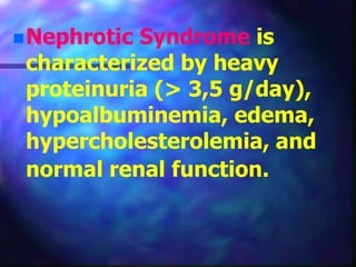
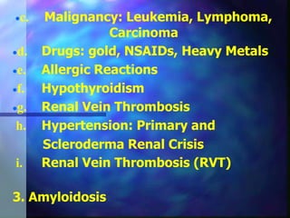
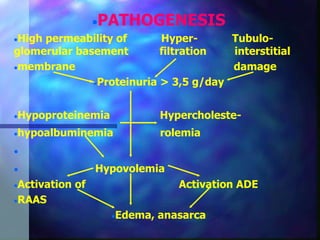
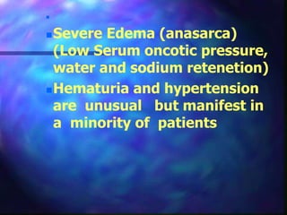
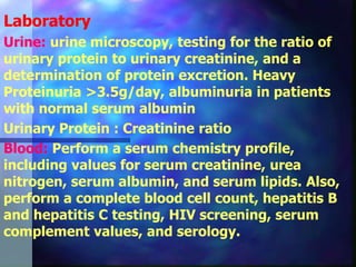
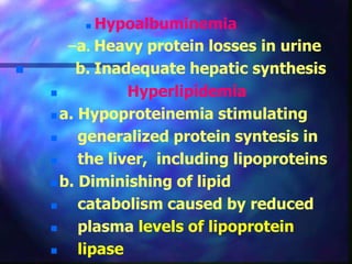
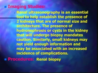
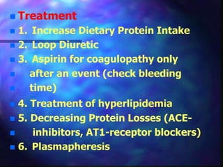
NEPHROTIC
SYNDROME
Nephrotic Syndrome is
characterized by heavy
proteinuria (> 3,5 g/day),
hypoalbuminemia, edema,
hypercholesterolemia, and
normal renal function.
 Etiology
 1. Primary
 a. Membranous Glomerulonephritis
Proliferative Glomerulonephritis
 b. Minimal Change Disease (mostly
 children)
c. Focal and Segmental Sclerosing
 Glomerulonephritis (FSGS)
 d. IgA Nephropathy
2. Secondary
a. Diabetes Mellitus
b. Immune Complex Diseases: Systemic
Lupus Erythematosus, Systemic
Vasculitis
Infection:
 a. Bacterial (Group A beta-hemolytic
streptococci, Syphilis,
Malaria, Tuberculosis)
 b. Viral infections (eg, varicella,
 hepatitis B, HIV type 1, infectious
 mononucleosis)
c. Malignancy: Leukemia, Lymphoma,
Carcinoma
d. Drugs: gold, NSAIDs, Heavy Metals
e. Allergic Reactions
f. Hypothyroidism
g. Renal Vein Thrombosis
h. Hypertension: Primary and
Scleroderma Renal Crisis
i. Renal Vein Thrombosis (RVT)
3. Amyloidosis
PATHOGENESIS
High permeability of Hyper- Tubulo-
glomerular basement filtration interstitial
membrane damage
Proteinuria > 3,5 g/day
Hypoproteinemia Hypercholeste-
hypoalbuminemia rolemia

 Hypovolemia
Activation of Activation ADE
RAAS
Edema, anasarca
History:
 NS can present with edema of
dependent parts. In most cases, this
includes the ankles or legs.
 Facial swelling or anasarca can be the
presenting symptom.
 patients notice frothy urine, which leads
to investigations that reveal evidence of
nephrotic syndrome.
 A hypercoagulable state leading to
thrombotic complications, such as deep
vein thrombosis of the calf veins or the
renal vein, may be the first clue
indicating nephrotic syndrome.
 Patients present with increasing edema
over a few days or weeks, lethargy, poor
appetite, weakness, and occasional
abdominal pain.
 The initial episode and the subsequent
relapses may follow an apparent viral
upper respiratory tract infection.
 Edema is the predominant feature and
initially develops around the eyes and
lower extremities. With time, the edema
becomes generalized and may be
associated with an increase in weight, the
development of an ascitic or pleural
effusion, and a decline in urine output.

Severe Edema (anasarca)
(Low Serum oncotic pressure,
water and sodium retenetion)
Hematuria and hypertension
are unusual but manifest in
a minority of patients
Laboratory
Urine: urine microscopy, testing for the ratio of
urinary protein to urinary creatinine, and a
determination of protein excretion. Heavy
Proteinuria >3.5g/day, albuminuria in patients
with normal serum albumin
Urinary Protein : Creatinine ratio
Blood: Perform a serum chemistry profile,
including values for serum creatinine, urea
nitrogen, serum albumin, and serum lipids. Also,
perform a complete blood cell count, hepatitis B
and hepatitis C testing, HIV screening, serum
complement values, and serology.
 Hypoalbuminemia
–a. Heavy protein losses in urine
 b. Inadequate hepatic synthesis
 Hyperlipidemia
 a. Hypoproteinemia stimulating
 generalized protein syntesis in
 the liver, including lipoproteins
 b. Diminishing of lipid
 catabolism caused by reduced
 plasma levels of lipoprotein
 lipase
Coagulopathy (~50% of
patients)
a. Hypercoagulable state
b. Loss of anti-thrombin III
 c. Reduced synthesis of
 proteins C and S, loss of
 other plasma proteins
 Other tests: testing
cryoglobulins and performing
serum protein
electrophoresis or urine
protein electrophoresis can
be useful for detecting the
etiology of nephrotic
syndrome. Antistreptolysin O
titers
Antinuclear antibodies
 Imaging Studies:
 Renal ultrasonography is an essential
tool to help establish the presence of
2 kidneys that are of normal size and
architecture. The presence of
hydronephrosis or cysts in the kidney
that will undergo biopsy mandates
caution. Similarly, small kidneys may
not yield enough information and
may be associated with an increased
incidence of complications.
 Procedures: Renal biopsy
 Treatment
 1. Underlying Disease therapy
 2. Immunosuppressive agents
 a. Prednisone 1-1.5mg/kg/d may be
 effective
 Use these high doses for 1-2
 months, then slowly taper
 b. Cytotoxic Therapy in selected
 cases - eg. cyclophosphamide,
 chlorambucil, mycofenolat mofetil
 cyclosporin
 Treatment
 1. Increase Dietary Protein Intake
 2. Loop Diuretic
 3. Aspirin for coagulopathy only
 after an event (check bleeding
 time)
 4. Treatment of hyperlipidemia
 5. Decreasing Protein Losses (ACE-
 inhibitors, AT1-receptor blockers)
 6. Plasmapheresis
 Complications:
 Infection
 Thromboembolism
 Hypovolemia
 Acute renal failure
 Hypertension
 Tetany
 Adverse effects of treatment
Prognosis:
 The prognosis for patients
with primary nephrotic
syndrome varies depending
on the histological type.
 The prognosis for children
with minimal-change
glomerulonephritis is
excellent.

Nervous. System nephrilogy. System clinics

  • 1.
  • 2.
    Nephrotic Syndrome is characterizedby heavy proteinuria (> 3,5 g/day), hypoalbuminemia, edema, hypercholesterolemia, and normal renal function.
  • 3.
     Etiology  1.Primary  a. Membranous Glomerulonephritis Proliferative Glomerulonephritis  b. Minimal Change Disease (mostly  children) c. Focal and Segmental Sclerosing  Glomerulonephritis (FSGS)  d. IgA Nephropathy
  • 4.
    2. Secondary a. DiabetesMellitus b. Immune Complex Diseases: Systemic Lupus Erythematosus, Systemic Vasculitis Infection:  a. Bacterial (Group A beta-hemolytic streptococci, Syphilis, Malaria, Tuberculosis)  b. Viral infections (eg, varicella,  hepatitis B, HIV type 1, infectious  mononucleosis)
  • 5.
    c. Malignancy: Leukemia,Lymphoma, Carcinoma d. Drugs: gold, NSAIDs, Heavy Metals e. Allergic Reactions f. Hypothyroidism g. Renal Vein Thrombosis h. Hypertension: Primary and Scleroderma Renal Crisis i. Renal Vein Thrombosis (RVT) 3. Amyloidosis
  • 6.
    PATHOGENESIS High permeability ofHyper- Tubulo- glomerular basement filtration interstitial membrane damage Proteinuria > 3,5 g/day Hypoproteinemia Hypercholeste- hypoalbuminemia rolemia   Hypovolemia Activation of Activation ADE RAAS Edema, anasarca
  • 7.
    History:  NS canpresent with edema of dependent parts. In most cases, this includes the ankles or legs.  Facial swelling or anasarca can be the presenting symptom.  patients notice frothy urine, which leads to investigations that reveal evidence of nephrotic syndrome.  A hypercoagulable state leading to thrombotic complications, such as deep vein thrombosis of the calf veins or the renal vein, may be the first clue indicating nephrotic syndrome.
  • 8.
     Patients presentwith increasing edema over a few days or weeks, lethargy, poor appetite, weakness, and occasional abdominal pain.  The initial episode and the subsequent relapses may follow an apparent viral upper respiratory tract infection.  Edema is the predominant feature and initially develops around the eyes and lower extremities. With time, the edema becomes generalized and may be associated with an increase in weight, the development of an ascitic or pleural effusion, and a decline in urine output.
  • 9.
     Severe Edema (anasarca) (LowSerum oncotic pressure, water and sodium retenetion) Hematuria and hypertension are unusual but manifest in a minority of patients
  • 10.
    Laboratory Urine: urine microscopy,testing for the ratio of urinary protein to urinary creatinine, and a determination of protein excretion. Heavy Proteinuria >3.5g/day, albuminuria in patients with normal serum albumin Urinary Protein : Creatinine ratio Blood: Perform a serum chemistry profile, including values for serum creatinine, urea nitrogen, serum albumin, and serum lipids. Also, perform a complete blood cell count, hepatitis B and hepatitis C testing, HIV screening, serum complement values, and serology.
  • 11.
     Hypoalbuminemia –a. Heavyprotein losses in urine  b. Inadequate hepatic synthesis  Hyperlipidemia  a. Hypoproteinemia stimulating  generalized protein syntesis in  the liver, including lipoproteins  b. Diminishing of lipid  catabolism caused by reduced  plasma levels of lipoprotein  lipase
  • 12.
    Coagulopathy (~50% of patients) a.Hypercoagulable state b. Loss of anti-thrombin III  c. Reduced synthesis of  proteins C and S, loss of  other plasma proteins
  • 13.
     Other tests:testing cryoglobulins and performing serum protein electrophoresis or urine protein electrophoresis can be useful for detecting the etiology of nephrotic syndrome. Antistreptolysin O titers Antinuclear antibodies
  • 14.
     Imaging Studies: Renal ultrasonography is an essential tool to help establish the presence of 2 kidneys that are of normal size and architecture. The presence of hydronephrosis or cysts in the kidney that will undergo biopsy mandates caution. Similarly, small kidneys may not yield enough information and may be associated with an increased incidence of complications.  Procedures: Renal biopsy
  • 15.
     Treatment  1.Underlying Disease therapy  2. Immunosuppressive agents  a. Prednisone 1-1.5mg/kg/d may be  effective  Use these high doses for 1-2  months, then slowly taper  b. Cytotoxic Therapy in selected  cases - eg. cyclophosphamide,  chlorambucil, mycofenolat mofetil  cyclosporin
  • 16.
     Treatment  1.Increase Dietary Protein Intake  2. Loop Diuretic  3. Aspirin for coagulopathy only  after an event (check bleeding  time)  4. Treatment of hyperlipidemia  5. Decreasing Protein Losses (ACE-  inhibitors, AT1-receptor blockers)  6. Plasmapheresis
  • 17.
     Complications:  Infection Thromboembolism  Hypovolemia  Acute renal failure  Hypertension  Tetany  Adverse effects of treatment
  • 18.
    Prognosis:  The prognosisfor patients with primary nephrotic syndrome varies depending on the histological type.  The prognosis for children with minimal-change glomerulonephritis is excellent.