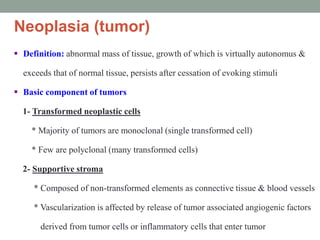
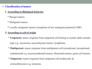
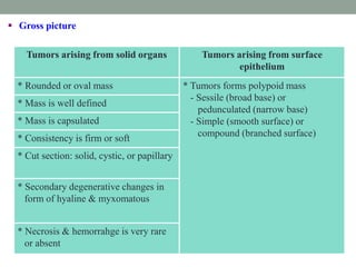
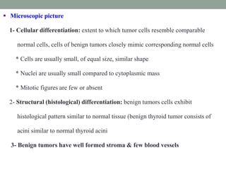
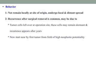
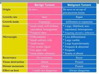
Neoplasia
Mohammed Fathy Bayomy, MSc, MD
Lecturer
Clinical Oncology & Nuclear Medicine
Faculty of Medicine
Zagazig University
Neoplasia (tumor)
 Definition: abnormal mass of tissue, growth of which is virtually autonomus &
exceeds that of normal tissue, persists after cessation of evoking stimuli
 Basic component of tumors
1- Transformed neoplastic cells
* Majority of tumors are monoclonal (single transformed cell)
* Few are polyclonal (many transformed cells)
2- Supportive stroma
* Composed of non-transformed elements as connective tissue & blood vessels
* Vascularization is affected by release of tumor associated angiogenic factors
derived from tumor cells or inflammatory cells that enter tumor
 Classification of tumors
1- According to Biological behavior
* Benign tumors
* Malignant tumors
* Locally malignant tumors (neoplasm of low malignant potential LMP)
2- According to cell of origin
* Unipotent: tumor originate from unipotent cell belong to certain adult somatic
type e.g. carcinoma, mesenchymal tumors, lymphoma
* Multipotent: tumor originate from multipotent cell (ectodermal, mesodermal,
endodermal) e.g. neuroectodermal tumors, blastemal tumors, germ cell tumors
* Totipotent: tumor originate from totipotent cell (embryonic &
extraembryonic) e.g. teratoma
3- According to tumor cell differentiation (cell type)
* Resemble adult somatic cell: carcinoma, mesenchymal tumors, lymphoma
* Resemble embryonic cells: neuroectodermal tumors, blastemal tumors, germ
cell tumors, tumors of vestigial remnants
* Undetermined origin: sarcoma
* Undifferentiated tumors: composed of extremely primitive cells
* Mixed tumors: composed of two cell types (biphasic) or multiple cell types
(multiphasic)
 General features
1- Arise de novo
2- Rate of growth is often slow & rarely reach large size
3- Mode of growth
* In solid structure: expansible, central cell mass proliferate pushing peripheral
cells, tumor expands compressing surrounding structures
* Tumors growing from surface project to outside & from papillary mass
4- Encapsulation: often capsulated, tumor is surrounded by fibrous capsule
consisting of compressed connective tissue, capsule is derived form host tissues &
partially due to pressure atrophy of surrounding tissues
Benign Tumors
 Gross picture
Tumors arising from solid organs Tumors arising from surface
epithelium
* Rounded or oval mass * Tumors forms polypoid mass
- Sessile (broad base) or
pedunculated (narrow base)
- Simple (smooth surface) or
compound (branched surface)
* Mass is well defined
* Mass is capsulated
* Consistency is firm or soft
* Cut section: solid, cystic, or papillary
* Secondary degenerative changes in
form of hyaline & myxomatous
* Necrosis & hemorrahge is very rare
or absent
 Microscopic picture
1- Cellular differentiation: extent to which tumor cells resemble comparable
normal cells, cells of benign tumors closely mimic corresponding normal cells
* Cells are usually small, of equal size, similar shape
* Nuclei are usually small compared to cytoplasmic mass
* Mitotic figures are few or absent
2- Structural (histological) differentiation: benign tumors cells exhibit
histological pattern similar to normal tissue (benign thyroid tumor consists of
acini similar to normal thyroid acini
3- Benign tumors have well formed stroma & few blood vessels
 Behavior
1- Localized at site of origin, not spread
2- Not recur after complete excision
3- May kill host when compressing vital organ e.g. brain
4- May cause obstruction when rise in hollow organ e.g. intestine
5- May produce hormones e.g. tumors of endocrine glands
6- Malignant transformation may occur & evident by
* Increase rate of growth
* Loss of cellular differentiation
* Inability to form intercellular ground substance
* Invasion of capsule & surrounding tissues
 General features
1- May recur locally after excision
2- Unable to produce distant metastasis
 Include
1- Epithelial tumors: verrucous carcinoma, inverted papilloma, villous adenoma,
choriocacinoma destruens, carcinoid, ameloblastoma,
craniopharyngioma
2- Mesenchymal tumors: fibromatosis, atypical fibrous histiocytoma, atypical
meningioma, hemangioblastoma, gaint cell tumor,
osteoblastoma, myxoma
Locally Malignant Tumors
 General features
1- Arise de novo or on top of precancerous lesion
2- Rate of growth is rapid, reach large size in short time
3- Mode of growth: mainly invasion (infiltration), some degree of expansion
Mechanism of invasion extracellular matrix
* Detachment of tumor cells from each other (downregulation E-cadherin)
* Attachment of matrix components (cell receptor bind to laminin & fibronectin)
* Degeneration of extracellular matrix (proteolytic enzymes: type IV collagenase,
cathepsin D, urokinase-type plasminogen activator)
* Migration of cells (autocrine motility factor, cleavage products, repulsion)
Malignant Tumors
 Gross picture
Tumors arising
from solid organs
Tumors arising from surface epithelium
Fungating Ulcerative Infiltrating
* Large ill-defined
mass
* Cauliflower like
mass
* Large size ulcer
* Uncapsulated,
false capsule
from compressed
surrounding
* Surface is
irregular
* Edges are raised
& everted
* Tubular organ:
- Annular
- Diffuse
* Infiltrate
surrounding
* Infiltrate
underlying tissue
* Consistency is
firm
* Friable
consistency
* Sharp borders
* Cut section:
necrosis &
hemorrahge
* Cut section:
necrosis &
hemorrahge
* Floor: necrosis &
hemorrahge
 Microscopic picture
1- Cellular Anaplasia
* Change in cellular morphology
- Large size cells
- Variable in size
- Nuclei: large, variable size & positions within cell
- Hyperchromatic nuclei due to duplication of DNA
- Nucleocytoplasmic ratio is increased ( 1 : 1 instead of 1 : 6)
- Nucleoli: large, prominent & variable position
-Abundant mitoses & abnormal mitotic figure (tripolar spindles)
- Tumor giant cell containing large polypoid nucleus or multiple nuclei
* Loss of polarity: malignant tumors show loss of normal relation between each
cell & neighboring ones & surrounding tissues e.g. malignant acini are
variable in size & shape, their wall may be formed of single or multiple layers,
lumen may be eccentric & irregular of absent
2- Structural (histological) differentiation: malignant tumors fail to reproduce
exact appearance & pattern of parent tissue, according to degree of resemblance
of structural pattern of tumor to normal tissue carcinoma is graded as well
differentiated (Grade I), moderate differentiated (grade II), poorly differentiated
(grade II), undifferentiated (grade IV)
3- Highly vascular stroma
4- Vessels may be thin walled as in sarcoma
5- Secondary changes as myxoid degeneration may occur
6- Areas of hemorrhage & necrosis are common
 Behavior
1- Not remain locally at site of origin, undergo local & distant spread
2- Recurrence after surgical removal is common, may be due to
* Tumor cells left over at operation site, these cells may remain dormant &
recurrence appears after years
* New start near by first tumor from field of high neoplastic potentiality
Benign Tumors Malignant Tumors
Origin De novo De novo or on top of
precancerous lesion
Growth rate Slow Rapid
Growth mode Expansion Infiltration or expansion
Gross * Small sized, well defined,
capsulated, homogenous
* Polypoid mass
* Large, illdefined, non-
capsulated, heterogenous
* Fungating, ulcerative, infiltrative
Microscopic * Differentiated
* Small uniform cells
* Small nuclei
* Few mitotic figure
* Few giant cells
* Intracellular substance
* Less differentiated
* Large varible
* Large hyperchromatic
* Frequent & abnormal
* Frequent
* Scanty or absent
Recurrence No recurrence Recur
Tissue destruction Absent Present
Distant metastasis Absent Present
Effect on host Usually not dangerous Always dangerous

Neoplasia

  • 1.
    Neoplasia Mohammed Fathy Bayomy,MSc, MD Lecturer Clinical Oncology & Nuclear Medicine Faculty of Medicine Zagazig University
  • 2.
    Neoplasia (tumor)  Definition:abnormal mass of tissue, growth of which is virtually autonomus & exceeds that of normal tissue, persists after cessation of evoking stimuli  Basic component of tumors 1- Transformed neoplastic cells * Majority of tumors are monoclonal (single transformed cell) * Few are polyclonal (many transformed cells) 2- Supportive stroma * Composed of non-transformed elements as connective tissue & blood vessels * Vascularization is affected by release of tumor associated angiogenic factors derived from tumor cells or inflammatory cells that enter tumor
  • 3.
     Classification oftumors 1- According to Biological behavior * Benign tumors * Malignant tumors * Locally malignant tumors (neoplasm of low malignant potential LMP) 2- According to cell of origin * Unipotent: tumor originate from unipotent cell belong to certain adult somatic type e.g. carcinoma, mesenchymal tumors, lymphoma * Multipotent: tumor originate from multipotent cell (ectodermal, mesodermal, endodermal) e.g. neuroectodermal tumors, blastemal tumors, germ cell tumors * Totipotent: tumor originate from totipotent cell (embryonic & extraembryonic) e.g. teratoma
  • 4.
    3- According totumor cell differentiation (cell type) * Resemble adult somatic cell: carcinoma, mesenchymal tumors, lymphoma * Resemble embryonic cells: neuroectodermal tumors, blastemal tumors, germ cell tumors, tumors of vestigial remnants * Undetermined origin: sarcoma * Undifferentiated tumors: composed of extremely primitive cells * Mixed tumors: composed of two cell types (biphasic) or multiple cell types (multiphasic)
  • 5.
     General features 1-Arise de novo 2- Rate of growth is often slow & rarely reach large size 3- Mode of growth * In solid structure: expansible, central cell mass proliferate pushing peripheral cells, tumor expands compressing surrounding structures * Tumors growing from surface project to outside & from papillary mass 4- Encapsulation: often capsulated, tumor is surrounded by fibrous capsule consisting of compressed connective tissue, capsule is derived form host tissues & partially due to pressure atrophy of surrounding tissues Benign Tumors
  • 6.
     Gross picture Tumorsarising from solid organs Tumors arising from surface epithelium * Rounded or oval mass * Tumors forms polypoid mass - Sessile (broad base) or pedunculated (narrow base) - Simple (smooth surface) or compound (branched surface) * Mass is well defined * Mass is capsulated * Consistency is firm or soft * Cut section: solid, cystic, or papillary * Secondary degenerative changes in form of hyaline & myxomatous * Necrosis & hemorrahge is very rare or absent
  • 7.
     Microscopic picture 1-Cellular differentiation: extent to which tumor cells resemble comparable normal cells, cells of benign tumors closely mimic corresponding normal cells * Cells are usually small, of equal size, similar shape * Nuclei are usually small compared to cytoplasmic mass * Mitotic figures are few or absent 2- Structural (histological) differentiation: benign tumors cells exhibit histological pattern similar to normal tissue (benign thyroid tumor consists of acini similar to normal thyroid acini 3- Benign tumors have well formed stroma & few blood vessels
  • 8.
     Behavior 1- Localizedat site of origin, not spread 2- Not recur after complete excision 3- May kill host when compressing vital organ e.g. brain 4- May cause obstruction when rise in hollow organ e.g. intestine 5- May produce hormones e.g. tumors of endocrine glands 6- Malignant transformation may occur & evident by * Increase rate of growth * Loss of cellular differentiation * Inability to form intercellular ground substance * Invasion of capsule & surrounding tissues
  • 9.
     General features 1-May recur locally after excision 2- Unable to produce distant metastasis  Include 1- Epithelial tumors: verrucous carcinoma, inverted papilloma, villous adenoma, choriocacinoma destruens, carcinoid, ameloblastoma, craniopharyngioma 2- Mesenchymal tumors: fibromatosis, atypical fibrous histiocytoma, atypical meningioma, hemangioblastoma, gaint cell tumor, osteoblastoma, myxoma Locally Malignant Tumors
  • 10.
     General features 1-Arise de novo or on top of precancerous lesion 2- Rate of growth is rapid, reach large size in short time 3- Mode of growth: mainly invasion (infiltration), some degree of expansion Mechanism of invasion extracellular matrix * Detachment of tumor cells from each other (downregulation E-cadherin) * Attachment of matrix components (cell receptor bind to laminin & fibronectin) * Degeneration of extracellular matrix (proteolytic enzymes: type IV collagenase, cathepsin D, urokinase-type plasminogen activator) * Migration of cells (autocrine motility factor, cleavage products, repulsion) Malignant Tumors
  • 11.
     Gross picture Tumorsarising from solid organs Tumors arising from surface epithelium Fungating Ulcerative Infiltrating * Large ill-defined mass * Cauliflower like mass * Large size ulcer * Uncapsulated, false capsule from compressed surrounding * Surface is irregular * Edges are raised & everted * Tubular organ: - Annular - Diffuse * Infiltrate surrounding * Infiltrate underlying tissue * Consistency is firm * Friable consistency * Sharp borders * Cut section: necrosis & hemorrahge * Cut section: necrosis & hemorrahge * Floor: necrosis & hemorrahge
  • 12.
     Microscopic picture 1-Cellular Anaplasia * Change in cellular morphology - Large size cells - Variable in size - Nuclei: large, variable size & positions within cell - Hyperchromatic nuclei due to duplication of DNA - Nucleocytoplasmic ratio is increased ( 1 : 1 instead of 1 : 6) - Nucleoli: large, prominent & variable position -Abundant mitoses & abnormal mitotic figure (tripolar spindles) - Tumor giant cell containing large polypoid nucleus or multiple nuclei * Loss of polarity: malignant tumors show loss of normal relation between each cell & neighboring ones & surrounding tissues e.g. malignant acini are variable in size & shape, their wall may be formed of single or multiple layers, lumen may be eccentric & irregular of absent
  • 13.
    2- Structural (histological)differentiation: malignant tumors fail to reproduce exact appearance & pattern of parent tissue, according to degree of resemblance of structural pattern of tumor to normal tissue carcinoma is graded as well differentiated (Grade I), moderate differentiated (grade II), poorly differentiated (grade II), undifferentiated (grade IV) 3- Highly vascular stroma 4- Vessels may be thin walled as in sarcoma 5- Secondary changes as myxoid degeneration may occur 6- Areas of hemorrhage & necrosis are common
  • 14.
     Behavior 1- Notremain locally at site of origin, undergo local & distant spread 2- Recurrence after surgical removal is common, may be due to * Tumor cells left over at operation site, these cells may remain dormant & recurrence appears after years * New start near by first tumor from field of high neoplastic potentiality
  • 15.
    Benign Tumors MalignantTumors Origin De novo De novo or on top of precancerous lesion Growth rate Slow Rapid Growth mode Expansion Infiltration or expansion Gross * Small sized, well defined, capsulated, homogenous * Polypoid mass * Large, illdefined, non- capsulated, heterogenous * Fungating, ulcerative, infiltrative Microscopic * Differentiated * Small uniform cells * Small nuclei * Few mitotic figure * Few giant cells * Intracellular substance * Less differentiated * Large varible * Large hyperchromatic * Frequent & abnormal * Frequent * Scanty or absent Recurrence No recurrence Recur Tissue destruction Absent Present Distant metastasis Absent Present Effect on host Usually not dangerous Always dangerous