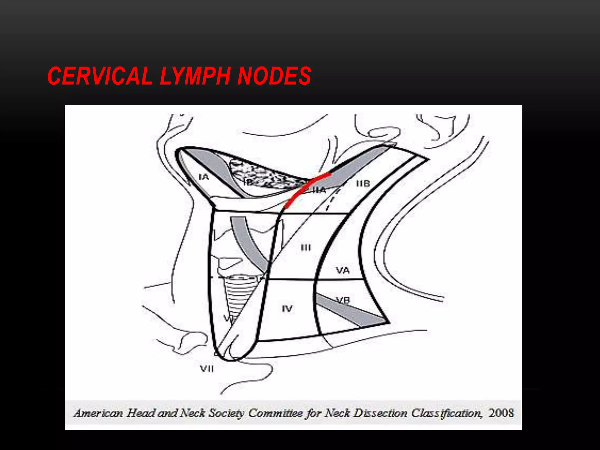
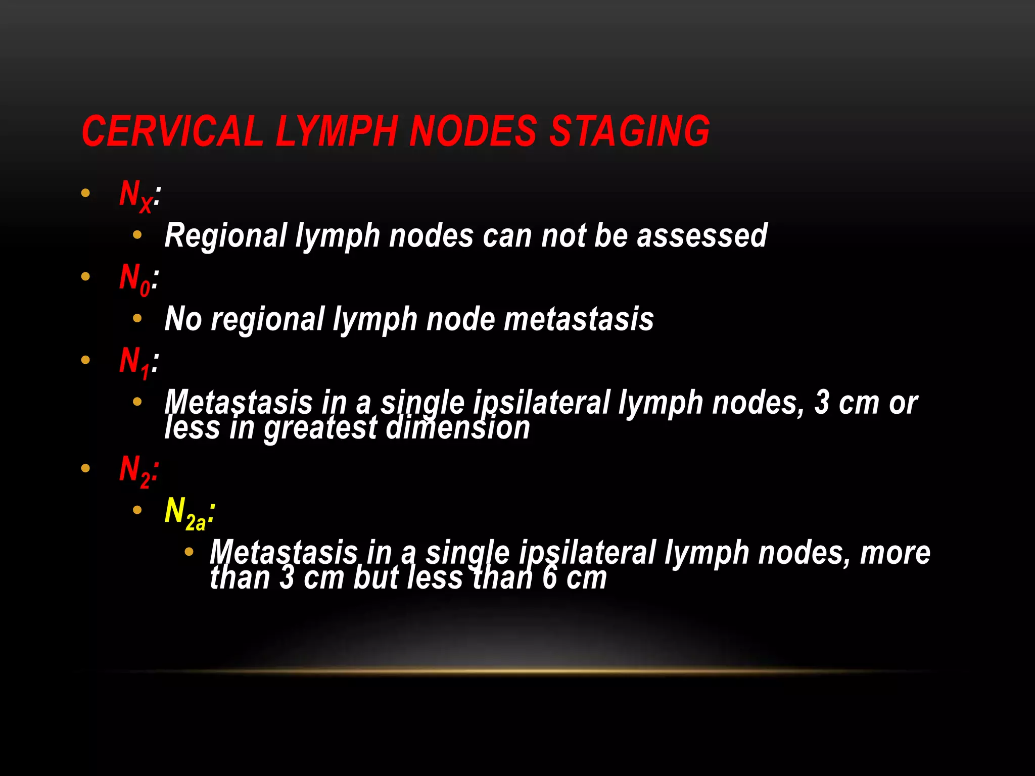
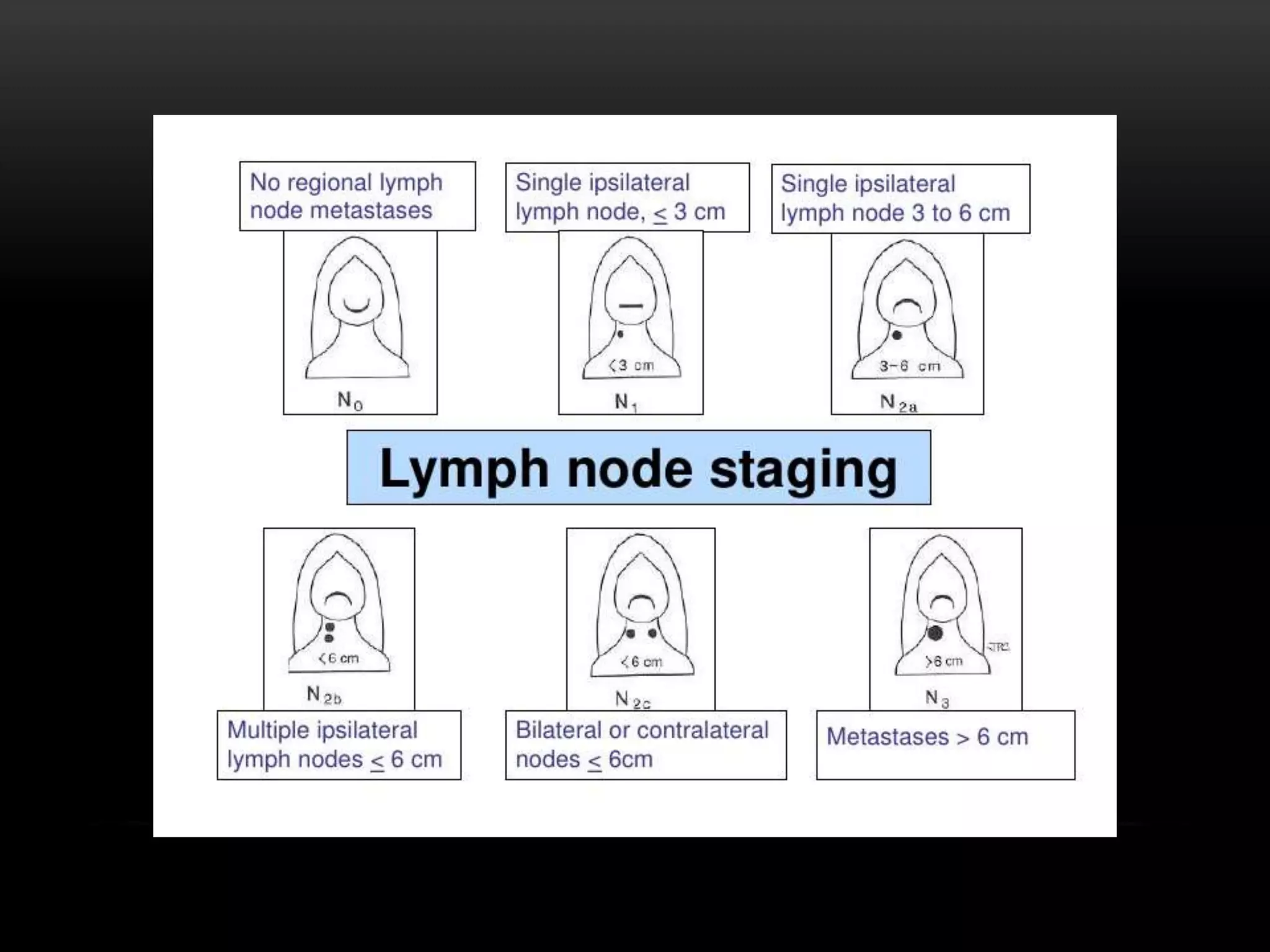
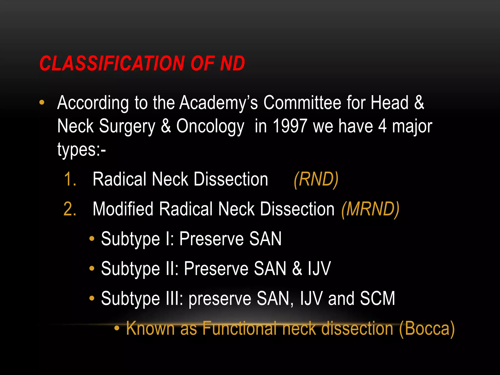
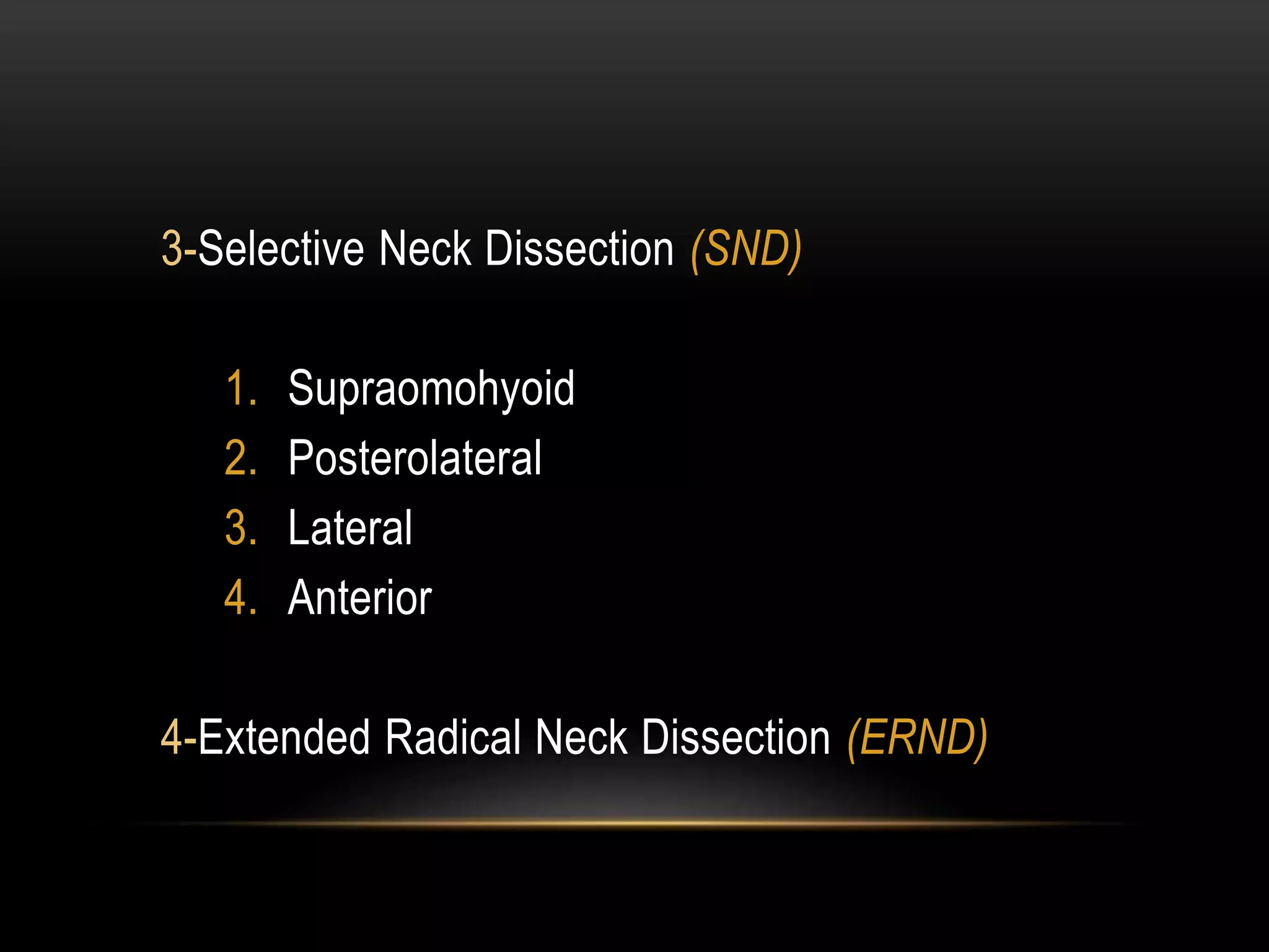
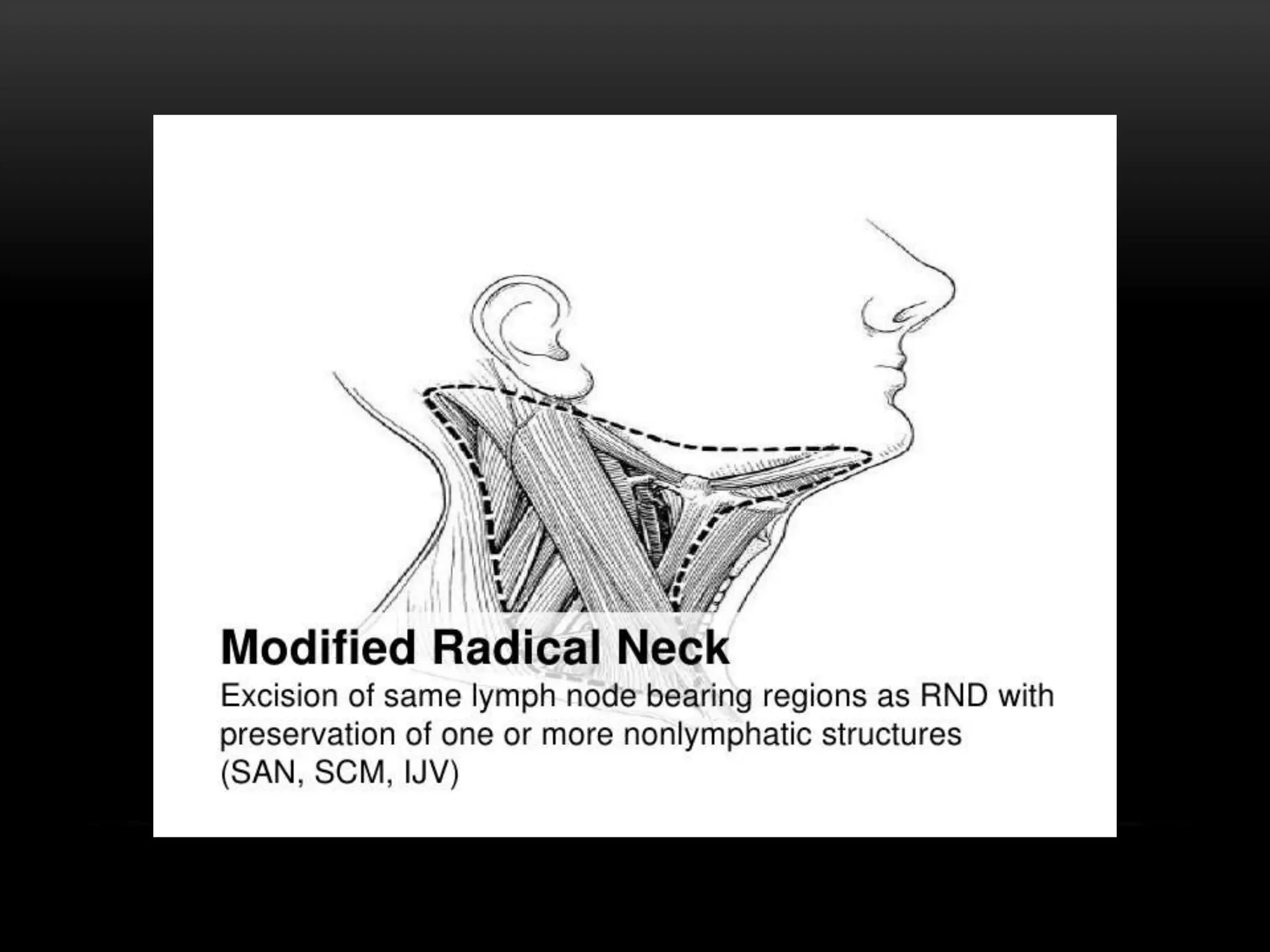
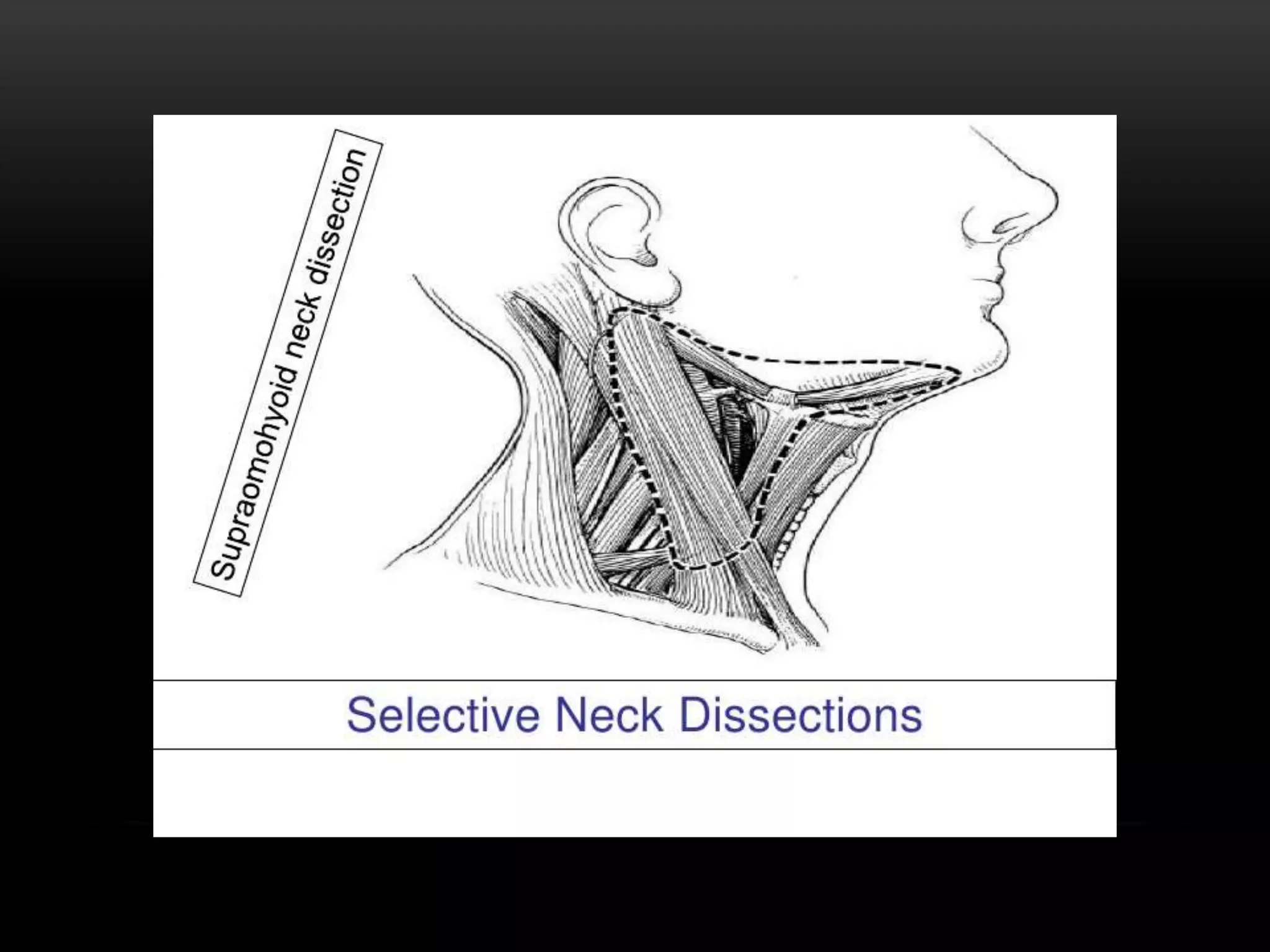
This document discusses types of neck dissection procedures performed to treat neck metastases from head and neck cancers. It describes radical neck dissection, modified radical neck dissection, and selective neck dissection. Radical neck dissection removes all lymph nodes from levels I-V, while modified versions preserve one or more non-lymphatic structures like the spinal accessory nerve or internal jugular vein. Selective neck dissection removes only the lymph nodes at levels that drain the primary tumor site. The indications, techniques, and complications of these neck dissection procedures are examined in detail.
Mr. Sunil, a 72-year-old male, presented with a 3-month history of a left neck swelling. Further examinations revealed metastatic squamous cell carcinoma in the left neck lymph nodes. He was diagnosed with carcinoma of unknown primary (CUP) and underwent radical neck dissection, followed by chemotherapy and radiotherapy. CUP describes metastatic cancers where the primary site cannot be identified despite various examinations and evaluations. Treatment options for CUP include surgery, radiation therapy, chemotherapy, or concurrent chemoradiation depending on the lymph node involvement and other factors. Prognosis depends on the stage and presence of extracapsular extension, with 5-year survival rates ranging from 30% for upper cervical nodes to 5%
This document describes the anatomy of the neck relevant to neck dissection surgery. It outlines the boundaries of the neck, key muscles and structures like the platysma, sternocleidomastoid, trapezius and vessels. It discusses the lymph node levels and types of neck dissection surgeries like radical and selective dissections. The document provides details of the surgical approach including skin incisions and dissection of structures to completely remove lymph nodes while preserving nearby nerves and vessels.
The document provides an overview of neck dissections, including their history, surgical anatomy of the neck, lymph node levels and staging, classifications of neck dissections, and indications for different types of neck dissections. It discusses the evolution of neck dissections from the radical neck dissection to more selective procedures. Key classifications covered are the radical, modified radical, and selective neck dissections, with the latter including supraomohyoid, lateral, and posterolateral variants. Indications and structures preserved for each type of dissection are outlined.
1. The document discusses the anatomy and classification of neck dissections for oral squamous cell carcinoma. It reviews the history and developments of neck dissection techniques from radical to functional approaches.
2. It describes the important structures and lymph node groups in the neck, including the spinal accessory nerve, internal jugular vein, and lymph node levels IA-III.
3. Performing neck dissection requires knowledge of the cervical lymph node drainage patterns and surgical anatomy to adequately remove metastatic lymph nodes while preserving important structures to minimize morbidity.
Metastasis of Neck Node with Unknown Primary Himanshu Soni
1) An unknown primary is defined as squamous cell carcinoma presenting in cervical lymph nodes with no identifiable primary tumor site after examination. This clinical entity is known as carcinoma of unknown primary (CUP).
2) Evaluation involves physical examination, imaging like PET-CT, and panendoscopy with biopsies of suspicious sites to identify the occult primary tumor. Bilateral tonsillectomy and tongue base biopsy can identify occult tumors in the tonsillar crypts in many cases.
3) Treatment depends on tumor stage but often involves combined modality treatment with surgery, radiation, and/or chemotherapy aimed at locoregional control while minimizing morbidity.
Neck node management of unknown primaryDr Rekha Arya
The document discusses the management of neck nodes with an occult primary tumor. It begins by defining an occult primary tumor as one that presents with lymph node or distant metastases when investigations fail to identify the primary site. It then discusses the diagnostic workup, which includes history, physical exam, imaging studies like PET CT, and biopsy of lymph nodes. Treatment depends on the lymph node level and stage. For early stage disease, neck dissection may be sufficient, while advanced disease requires chemoradiation. Post-treatment neck dissection may be needed depending on response. Radiation techniques like IMRT can help reduce toxicity compared to conventional radiation. Complications of treatment include risks of surgery and side effects of high-dose radiation.
This document provides an overview of neck dissection. It begins with definitions of neck dissection and discusses the rationale for performing neck dissections when treating head and neck cancers. It then covers topics like the classification of neck dissections, the surgical anatomy involved, different types of incisions used, potential complications, and the history and development of neck dissection techniques over time.
managment of neck nodes with occult primaryBharti Devnani
This document discusses the management of neck nodes with an occult primary tumor. It defines this condition as biopsy-proven cancer of the neck that cannot be linked to a primary lesion after a full clinical and radiological workup. It notes the estimated incidence is 3-7% of head and neck cancers initially presenting with cervical lymph node metastases. Risk of lymph node metastases depends on factors like density of lymphatics in the potential primary site and histologic characteristics of the lesion. Diagnostic workup involves imaging, biopsies of suspicious areas, and examination under anesthesia. Treatment involves neck dissection, with options like radical, modified radical or selective dissection depending on the extent of disease. Post-surgery management considers disease
Elevate the skin flaps superiorly to the level of the ramus of mandible. Identify and protect the external jugular vein.
Surgeon: Okay, elevating the skin flaps now. Carefully dissecting in the plane just deep to the platysma muscle. There's the external jugular vein, I'm protecting that as I elevate the flap. Almost to the level of the ramus now.
This document provides an overview of neck dissection procedures, including:
- A classification system for neck dissections and descriptions of radical, modified radical, extended, and selective neck dissections.
- Generic steps for all neck dissections including incision, exposure, and lymph node removal.
- Detailed descriptions of performing a radical neck dissection, focusing on three areas of special attention: the lower end of the internal jugular vein, the junction of the clavicle and trapezius muscle, and the upper end of the internal jugular vein.
This document discusses the history and anatomy of neck dissections for head and neck cancer. It traces developments from the late 19th century of increasingly conservative neck dissections to preserve important structures. It describes the levels and boundaries of lymph node groups involved in the neck dissection staging system, including levels I-V. Key structures like the hypoglossal nerve and spinal accessory nerve are discussed in relation to the lymph node groups.
The document discusses the management of salivary gland tumours, including an overview of the different salivary glands and tumours that can occur in each, the workup, staging, treatment options of surgery, radiation therapy and chemotherapy, with a focus on the evidence for use of adjuvant radiation therapy to improve local control based on several studies. Adjuvant radiation therapy significantly increases local control for high-risk features like advanced T and N stage, close or positive margins, nerve involvement and perineural invasion. Elective nodal radiation is also recommended for high-grade tumours but not for adenoid cystic or ac
The document discusses various complications that can occur following head and neck surgery like neck dissection. It classifies complications as major/minor, early/late, local/systemic. It describes in detail immediate local complications like bleeding, shock, airway obstruction, increased intracranial pressure, and nerve injury and their management. Intermediate complications discussed include chylous fistula, seroma, wound infection, and flap failure. Late complications mentioned are recurrence, parotid gland hypertrophy, lymphedema, and hypertrophic scars. Prevention and management strategies are provided for many of the complications.
Local flaps in head & neack reconstructionMd Roohia
A flap is a unit of tissue transferred from one site to another while maintaining its own blood supply. Flaps are classified based on their location as local or distant. Local flaps can be random, which rely on subdermal vessels, or axial, relying on named vessels. Flaps are also classified based on their composition, such as skin, muscle, or bone flaps. When planning local flaps, goals include color and thickness match, sensory preservation, and minimal secondary defects. Common local flap techniques include advancement, rotation, and transposition flaps to close facial defects. Complications can be prevented through proper planning to avoid tension and including a flap margin.
This document provides information about neck dissection procedures. It discusses the history and evolution of neck dissection, defines the different types of neck dissections including radical, modified radical and selective neck dissections, and describes the key steps in performing a modified radical neck dissection. Complications of neck dissection such as hemorrhage, wound infection, and carotid artery rupture are also summarized.
This document discusses maxillectomy, which is the surgical removal of part or all of the maxilla bone. It provides a history of maxillectomy and describes the anatomy of the maxilla bone. It also discusses different classifications of maxillectomy procedures based on the extent of bone removed. The common indications for maxillectomy are malignant tumors like squamous cell carcinoma. The approaches used include lateral rhinotomy, Weber-Ferguson, and transoral-transpalatal. Reconstruction options involve dental prosthetics, maxillofacial prosthetics, and titanium implants.
The document discusses different incision techniques for lip splitting surgeries. It describes various incisions used for lower and upper lip splitting, including the Roux-Trotter, McGregor, Robson, Bhatt, Hayter, Rassekh, Weber-Ferguson, and Altemir incisions. It emphasizes that incisions should follow relaxed skin tension lines to minimize scarring, protect important structures like nerves and vessels, and allow for adequate exposure while achieving good functional and cosmetic outcomes. The Hayter incision, which incorporates a chevron shape into the vermilion border, is highlighted as one of the best techniques.
This document provides an overview of neck dissection procedures. It discusses the history and evolution of neck dissection, from Kocher's initial proposal in 1880 to remove nodal metastases to more modern classifications. The surgical anatomy of neck structures is described in detail. Levels of cervical lymph nodes are defined based on boundaries of bones, muscles, blood vessels and nerves. Staging of head and neck cancers using the TNM system is explained. Factors affecting nodal metastasis and techniques for assessing cervical lymph nodes are also summarized.
Esthesioneuroblastoma (ENB) is a rare malignant tumor that arises from the olfactory epithelium in the nasal cavity. Imaging such as CT and MRI are used to determine the extent of the tumor. Histopathological examination shows small round blue cells forming rosettes. Treatment involves surgery such as craniofacial resection along with radiation therapy. For advanced disease, chemotherapy may be given as part of multimodality treatment. With aggressive treatment, 5-year survival rates for ENB exceed 60%.
Maxillary sinus carcinoma arises from the maxillary sinus and can spread locally and to lymph nodes. Diagnosis involves physical exam, CT/MRI imaging, and biopsy. Staging evaluates tumor size, lymph node involvement, and distant spread. Treatment depends on stage but may include surgery such as maxillectomy, radiation therapy such as IMRT, and chemotherapy such as cisplatin for locally advanced cases. The goal of treatment is a complete resection with negative margins or effective control with radiation with or without chemotherapy while minimizing side effects to nearby structures like the optic nerves and chiasm. Outcomes depend on stage, with earlier stages having higher survival rates treated with surgery or surgery plus radiation.
This document discusses cancer of the nasopharynx (NPC). Some key points:
- NPC is uncommon globally but more common in certain regions like Southern China.
- Risk factors include EBV infection, consumption of salted fish.
- Staging involves MRI and biopsy. Treatment depends on stage but often involves chemotherapy and radiation therapy.
- Advanced stages may receive neoadjuvant chemo followed by concurrent chemo-radiation. IMRT has improved treatment.
- Sequelae can include cranial neuropathy, xerostomia, endocrine issues. Lifelong follow up is needed due to risk of recurrence or second cancers. Outcomes have improved but salvage options after relapse present challenges
This document discusses neoadjuvant chemotherapy in head and neck cancer. It provides background on when neoadjuvant chemotherapy is given, what regimens are used, and evidence from studies comparing neoadjuvant chemotherapy plus radiation/surgery versus radiation/surgery alone. Several large studies found that adding docetaxel, cisplatin and fluorouracil as neoadjuvant chemotherapy improved overall and progression-free survival compared to cisplatin and fluorouracil alone. However, other studies found no difference in outcomes between neoadjuvant chemotherapy followed by chemoradiation versus chemoradiation alone. Concomitant chemoradiation appears superior to induction chemotherapy for larynx preservation.
This powerpoint describes the types of maxillectomy & operative steps for total maxillectomy. It also enumerates various flaps used for reconstruction of maxillectomy defect.
This document discusses management of the clinically node-negative neck in early-stage oral squamous cell carcinoma. The risk of occult nodal involvement is estimated to be 20-30% for T1/T2 tumors. Main management approaches are upfront elective neck dissection, watch and wait, or sentinel node biopsy. Tumor depth of invasion is a better predictor of nodal metastasis than thickness. Elective neck dissection provides accurate staging and optimal locoregional control, while sentinel node biopsy carries risk of missing occult disease and delaying adjuvant treatment. Floor of mouth tumors have early nodal spread necessitating bilateral neck dissection.
This document provides an overview of selective neck dissection for lymph node staging and treatment of head and neck cancers. It describes the anatomy of neck lymph node levels and drainage patterns. It also outlines the different types of selective neck dissections based on the lymph node levels removed, such as supraomohyoid (levels I-III) and posterolateral (levels II-V). The indications, contraindications, surgical technique, complications, and postoperative care of selective neck dissection are discussed in detail.
Local and regional flaps in head and neck reconstructionSaleh Bakry
Local and regional flaps are used for head and neck reconstruction. There are several types of flaps classified by blood supply, location, configuration, tissue content, and transfer method. Local flaps use adjacent tissue while regional flaps are near but not adjacent. Free flaps completely detach tissue and reconnect blood vessels microsurgically. Examples of local and regional flaps discussed are advancement, rotation, and transposition flaps as well as pedicled and free myocutaneous flaps.
This document discusses neck dissection, which refers to the systematic resection of lymph nodes and surrounding tissue from the neck. It describes the 7 levels of neck nodes and provides details on different types of neck dissections like radical, modified radical, and selective dissections. Complications of neck dissection like hemorrhage, nerve injury, chyle leak, and wound issues are outlined. The document also briefly discusses sentinel lymph node biopsy, minimally invasive approaches, and robotic neck surgery.
Neck dissection is the surgical removal of lymph nodes in the neck to treat cancer that has spread from primary sites in the head and neck region. The document discusses the history and evolution of neck dissection techniques from radical to more selective approaches. It describes the levels and classification of lymph nodes in the neck according to drainage patterns. Guidelines are provided for preoperative evaluation and planning, surgical approaches, classifications of neck dissections, and complications. The goal of neck dissection is to remove lymph nodes that may contain cancer metastases while preserving important structures like nerves and vessels.
This document provides information about neck dissection procedures. It describes the different types of neck dissections including radical neck dissection, modified radical neck dissection, and selective neck dissection. It discusses the levels and sublevels of lymph nodes in the neck and provides details on the technique, indications, and steps for performing a modified radical neck dissection.
This document discusses different types of neck dissection procedures used to treat cervical lymphadenopathy. It describes the classic radical neck dissection procedure and modifications like the modified radical neck dissection, which preserves certain non-lymphatic structures like the spinal accessory nerve or internal jugular vein. Selective neck dissections that remove only certain lymph node levels are also discussed. The document provides details of different incision types and complications that can occur with neck dissection procedures.
This document discusses the management of neck nodes in head and neck cancer. It covers lymph node levels, risk factors for metastasis, diagnostic workup, staging, surgical and radiation treatment options. For clinically negative nodes, elective neck irradiation or dissection are equally effective at controlling subclinical disease. For clinically positive nodes, factors like number of positive nodes and size influence treatment planning. Combined modality treatment with surgery and radiation provides better control than either alone for more advanced neck disease.
This document describes different types of neck dissections, including radical neck dissection where lymph nodes levels I-V and non-lymphatic structures are removed. Modified radical neck dissection involves removal of lymph node levels I-V while preserving one or more non-lymphatic structures. Selective neck dissection preserves lymph node groups and non-lymphatic structures. The document outlines the surgical procedure for radical neck dissection, including important anatomical structures and potential complications.
This document discusses different types of mastectomy procedures. It begins with defining mastectomy as the surgical removal of breast tissue, partially or completely. It then describes the different types of mastectomies, including simple/total mastectomy, modified radical mastectomy, radical mastectomy, skin sparing mastectomy, and breast conserving surgery. For each type of mastectomy, it outlines the details of the procedure, including the tissues removed and surgical approach. It emphasizes that the goal is to remove the breast tissue as well as lymph nodes, while preserving important structures like nerves and muscles. The best procedure depends on factors like tumor size and stage of cancer.
This document discusses various laryngeal surgeries including:
1. Endoscopic resections, vertical partial laryngectomies, and total laryngectomy are discussed as options for glottic cancer treatment. Complications can include bleeding, airway obstruction, and laryngeal stenosis.
2. Supraglottic laryngectomy and supracricoid partial laryngectomy are options for supraglottic cancers while preserving the larynx. Patient selection is important due to risk of aspiration.
3. Voice rehabilitation options after total laryngectomy include electrolarynx, esophageal voice, or tracheoesophageal voice via a puncture and fistula between the trachea and es
This document discusses the management of early laryngeal cancer. It covers diagnosis using laryngoscopy, radiological imaging like CT scans and MRI, and staging of laryngeal malignancies. Recommended treatments for early and late stage cancers are transoral laser microsurgery, radiotherapy, open partial laryngectomy, and total laryngectomy. Transoral laser microsurgery is described as the standard treatment for mid-cord glottic cancers and offers advantages like better voice quality and minimal swallowing difficulty compared to radiotherapy. Radiotherapy is an alternative organ-preserving option for early laryngeal cancers. Open partial laryngectomies include vertical and horizontal procedures tailored to the location and size of the tumor.
1. Management of metastatic squamous neck cancer with unknown primary depends on nodal stage, presence of extracapsular spread, and risk of primary sites. Excision biopsy and neck dissection followed by postoperative radiotherapy is often recommended.
2. Intensity modulated radiotherapy has improved outcomes over conventional radiotherapy by better targeting tumor tissue and reducing dose to surrounding organs.
3. The addition of chemotherapy to surgery or radiotherapy can provide even better survival results.
This document provides information on conservative laryngectomy procedures for early stage laryngeal cancers. It discusses the history and principles of partial laryngectomy surgeries which aim to preserve laryngeal functions like speech and swallowing while completely removing the tumor. Specific procedures covered include laryngofissure with cordectomy, vertical partial laryngectomy, and supracricoid partial laryngectomy. Assessment of tumor extent, classification of open partial laryngeal surgeries, reconstruction techniques, and oncological outcomes are also summarized.
This document discusses neck dissection procedures for treating head and neck cancers. It covers:
1. The types of neck dissections including radical neck dissection (RND), modified radical neck dissection (MRND), and selective neck dissection (SND). RND removes the highest number of lymph nodes while SND removes the fewest.
2. Factors that influence the risk of lymph node metastasis including primary tumor site, size, and histology. The first echelon lymph nodes that drain different primary sites are also described.
3. Staging of neck cancers using the TNM system and guidelines for indications of different neck dissection procedures based on tumor staging.
This document discusses the surgical approach to falcine meningiomas. Key points include:
- Falcine meningiomas account for 5-9% of intracranial meningiomas and can grow along the falx cerebri and invade the superior sagittal sinus.
- Presentation depends on tumor location along the falx, with symptoms ranging from frontal lobe syndrome to visual deficits.
- MRI is the gold standard for diagnosis, showing tumor location, size, and involvement of surrounding structures.
- Surgical resection aims to completely remove the tumor while preserving venous structures like the superior sagittal sinus. This may involve coagulating or reconstructing invaded sinus walls.
This document discusses different types of neck dissection procedures used to remove cervical lymph nodes. It describes the standard radical neck dissection, which removes lymph nodes from levels I through V, as well as the internal jugular vein and sternocleidomastoid muscle. Modified radical neck dissection spares one or more of these structures. Selective neck dissection removes lymph nodes from certain levels based on the primary cancer site, preserving other lymph node groups. Extended radical neck dissection removes additional lymph node groups or tissues beyond a standard radical neck dissection.
Carcinoma Buccal Mucosa- Anatomy to ManagementDrAyush Garg
This document provides information on carcinoma of the buccal mucosa, including its anatomy, epidemiology, risk factors, clinical features, diagnosis, staging, treatment, and prognosis. Carcinoma of the buccal mucosa is most common in India, Malaysia, and Taiwan, where it affects men more than women. Risk factors include tobacco and betel nut chewing. Treatment depends on the stage, with surgery or radiation typically used for early stages and chemoradiation for advanced stages. Prognosis depends on factors like tumor size and nodal involvement.
1) Mastectomy is the surgical removal of breast tissue, either partially or completely. It is commonly performed to treat breast cancer.
2) There are several types of mastectomy procedures including simple/total mastectomy, modified radical mastectomy, and breast conserving surgery.
3) Factors such as tumor size, lymph node involvement, and patient preferences help determine which mastectomy procedure is most suitable. Post-operative care and follow up is also important after mastectomy.
Radiotherapy techniques, indications and evidences in oral cavity and oropha...Dr.Amrita Rakesh
This document discusses indications, evidence, and radiation therapy techniques for oral cavity and oropharyngeal cancers. It covers:
- Anatomy of the oral cavity and oropharynx.
- Staging principles and indications for surgery vs systemic therapy.
- Principles of surgery including adequate resection margins and neck management.
- Use of adjuvant radiation therapy or chemoradiation to improve local control, especially for high-risk features like positive margins or extracapsular extension.
- Radiation techniques for oral cavity cancers including field design, dose recommendations, and advantages of IMRT for sparing parotid glands.
Recent guidelines in management of oral and oropharyngeal carcinoma barun kumar
This document provides guidelines for the management of oral and oropharyngeal cancer. It discusses the multi-disciplinary approach, TNM staging, diagnostic workup, treatment options based on stage, principles of surgery including resection, reconstruction and management of neck nodes, as well as follow up care. The guidelines emphasize a team-based approach and tailored treatment strategies based on specific tumor characteristics and stage.
1. Radiation techniques for treating esophageal cancer include EBRT using 3D-CRT, IMRT, or brachytherapy. IMRT allows for better sparing of organs at risk like the spinal cord, heart, and lungs compared to 3D-CRT.
2. For treatment planning, the gross tumor volume (GTV) and clinical target volume (CTV) must be accurately delineated using imaging like CT, PET, and endoscopy. The CTV includes margins around the GTV to account for microscopic disease.
3. Radiation fields typically cover 3-5cm above and below the tumor with a 2cm radial margin. Enlarged fields covering the whole esophagus
Rasamanikya is a excellent preparation in the field of Rasashastra, it is used in various Kushtha Roga, Shwasa, Vicharchika, Bhagandara, Vatarakta, and Phiranga Roga. In this article Preparation& Comparative analytical profile for both Formulationon i.e Rasamanikya prepared by Kushmanda swarasa & Churnodhaka Shodita Haratala. The study aims to provide insights into the comparative efficacy and analytical aspects of these formulations for enhanced therapeutic outcomes.
micro teaching on communication m.sc nursing.pdfAnurag Sharma
Microteaching is a unique model of practice teaching. It is a viable instrument for the. desired change in the teaching behavior or the behavior potential which, in specified types of real. classroom situations, tends to facilitate the achievement of specified types of objectives.
Title: Sense of Smell
Presenter: Dr. Faiza, Assistant Professor of Physiology
Qualifications:
MBBS (Best Graduate, AIMC Lahore)
FCPS Physiology
ICMT, CHPE, DHPE (STMU)
MPH (GC University, Faisalabad)
MBA (Virtual University of Pakistan)
Learning Objectives:
Describe the primary categories of smells and the concept of odor blindness.
Explain the structure and location of the olfactory membrane and mucosa, including the types and roles of cells involved in olfaction.
Describe the pathway and mechanisms of olfactory signal transmission from the olfactory receptors to the brain.
Illustrate the biochemical cascade triggered by odorant binding to olfactory receptors, including the role of G-proteins and second messengers in generating an action potential.
Identify different types of olfactory disorders such as anosmia, hyposmia, hyperosmia, and dysosmia, including their potential causes.
Key Topics:
Olfactory Genes:
3% of the human genome accounts for olfactory genes.
400 genes for odorant receptors.
Olfactory Membrane:
Located in the superior part of the nasal cavity.
Medially: Folds downward along the superior septum.
Laterally: Folds over the superior turbinate and upper surface of the middle turbinate.
Total surface area: 5-10 square centimeters.
Olfactory Mucosa:
Olfactory Cells: Bipolar nerve cells derived from the CNS (100 million), with 4-25 olfactory cilia per cell.
Sustentacular Cells: Produce mucus and maintain ionic and molecular environment.
Basal Cells: Replace worn-out olfactory cells with an average lifespan of 1-2 months.
Bowman’s Gland: Secretes mucus.
Stimulation of Olfactory Cells:
Odorant dissolves in mucus and attaches to receptors on olfactory cilia.
Involves a cascade effect through G-proteins and second messengers, leading to depolarization and action potential generation in the olfactory nerve.
Quality of a Good Odorant:
Small (3-20 Carbon atoms), volatile, water-soluble, and lipid-soluble.
Facilitated by odorant-binding proteins in mucus.
Membrane Potential and Action Potential:
Resting membrane potential: -55mV.
Action potential frequency in the olfactory nerve increases with odorant strength.
Adaptation Towards the Sense of Smell:
Rapid adaptation within the first second, with further slow adaptation.
Psychological adaptation greater than receptor adaptation, involving feedback inhibition from the central nervous system.
Primary Sensations of Smell:
Camphoraceous, Musky, Floral, Pepperminty, Ethereal, Pungent, Putrid.
Odor Detection Threshold:
Examples: Hydrogen sulfide (0.0005 ppm), Methyl-mercaptan (0.002 ppm).
Some toxic substances are odorless at lethal concentrations.
Characteristics of Smell:
Odor blindness for single substances due to lack of appropriate receptor protein.
Behavioral and emotional influences of smell.
Transmission of Olfactory Signals:
From olfactory cells to glomeruli in the olfactory bulb, involving lateral inhibition.
Primitive, less old, and new olfactory systems with different path
share - Lions, tigers, AI and health misinformation, oh my!.pptxTina Purnat
• Pitfalls and pivots needed to use AI effectively in public health
• Evidence-based strategies to address health misinformation effectively
• Building trust with communities online and offline
• Equipping health professionals to address questions, concerns and health misinformation
• Assessing risk and mitigating harm from adverse health narratives in communities, health workforce and health system
These simplified slides by Dr. Sidra Arshad present an overview of the non-respiratory functions of the respiratory tract.
Learning objectives:
1. Enlist the non-respiratory functions of the respiratory tract
2. Briefly explain how these functions are carried out
3. Discuss the significance of dead space
4. Differentiate between minute ventilation and alveolar ventilation
5. Describe the cough and sneeze reflexes
Study Resources:
1. Chapter 39, Guyton and Hall Textbook of Medical Physiology, 14th edition
2. Chapter 34, Ganong’s Review of Medical Physiology, 26th edition
3. Chapter 17, Human Physiology by Lauralee Sherwood, 9th edition
4. Non-respiratory functions of the lungs https://academic.oup.com/bjaed/article/13/3/98/278874
3. INTRODUCTION
Neck dissection(cervical lymphadenectomy)
mean systemic removal of lymph nodes along
with their surrounding fibrofatty tissue in
different compartments of the neck to eradicate
metastatic disease to the regional lymph nodes
of the neck. these metastases originate from
primary lesions that involve mucosal sites of
the upper aerodigestive tract.
5. ELECTIVELY NECK DISSECTION IN N0
• Depended on:-
• 1-Histology.
• 2-Tumor (T stage) classification
• 3- Location of the primary tumor.
• 4-Overall treatment plan.
6. HISTORICAL REVIEW
• Kocher 1880
• Advocate wide marginal lymphadenectomy
• In 1888, Jawdynski
• described en bloc resection of cervical lymph nodes with
• resection of IJV ,SCM and SAN with very high rate of mortality.
• In 1906, George W. Crile of the Cleveland Clinic
• described the radical neck dissection.
• In 1967 - Oscar Suarez and E. Bocca
• described a more conservative operation with MRND
17. CERVICAL LYMPH NODES STAGING
• NX:
• Regional lymph nodes can not be assessed
• N0:
• No regional lymph node metastasis
• N1:
• Metastasis in a single ipsilateral lymph nodes, 3 cm or
less in greatest dimension
• N2:
• N2a:
• Metastasis in a single ipsilateral lymph nodes, more
than 3 cm but less than 6 cm
18. • N2b:
• Metastasis in multiple ipsilateral lymph nodes, not
more than 6 cm
• N2c:
• Metastasis in bilateral or contralateral nodes not
more than 6 cm in diameter
• N3:
• Metastasis in lymph nodes more than 6 cm in in greatest
diameter
19.
20. CLASSIFICATION OF ND
• According to the Academy’s Committee for Head &
Neck Surgery & Oncology in 1997 we have 4 major
types:-
1. Radical Neck Dissection (RND)
2. Modified Radical Neck Dissection (MRND)
• Subtype I: Preserve SAN
• Subtype II: Preserve SAN & IJV
• Subtype III: preserve SAN, IJV and SCM
• Known as Functional neck dissection (Bocca)
23. RADICAL NECK DISSECTION
• This operation is defined as the en bloc removal
of the lymph node-bearing tissues of one side of
the neck from the inferior border of the
mandible to the clavicle and from the
contralateral anterior belly of digastric and
lateral border of the strap muscles to the anterior
border of the trapezius that include level l----V.
with spinal accessory nerve, SCM and IJV.
24.
25.
26. INDICATION OF RND
.
• 1- RND is indicated for patients with extensive lymph node metastases
with operable neck dis(N2a,N2b,N3)
• 2- extension beyond the capsule of the node or nodes that involves the
spinal accessory nerve , IJV and SCM.
• 3- access prior to pedicled flab reconstruction.
• 4- large tumor with risk or presence of occult metastasis.
• 5- Recurrence after RT.
27. CONTRAINDICATION OF RND
• 1- those patients whose primary tumors are untreatable.
• 2-any patient who is unfit for major surgery because of a serious
medical condition which would render anesthesia and major
surgery unsafe.
• 3- patients with distant metastases.
• In those patients who are deemed inoperable, radical
radiotherapy with or without adjuvant chemotherapy is seldom
curative but may provide excellent palliation.
28. TECHNIQUE OF RND
• 1-preoperative preparation of the patient.
• 2-position.
• 3-incision planning:-
• a- factors.
• b- criteria
• c- types(advantage and disadvantage).
• d- biopsy
29.
30. RADICAL NECK DISSECTION AS PART OF A COMBINED
PROCEDURE
• When a primary tumour is removed in continuity with a neck dissection, a
band of continuity may be kept between the neck dissection and the
primary growth.
• Laryngeal cancer
• In a total laryngectomy, the neck dissection should b e left attached along
the whole length of the larynx to include the superior and inferior lymphatic
pedicles. A neck dissection is never carried out with a hemilaryngectomy
but for a supraglottic laryngectomy, it is pedicled on the thyrohyoid
membrane.
• Pharyngeal cancer
• When a pharyngectomy i s performed, the pedicle should be as broad as
possible and is best left along the whole length o f the pharynx.
31. • Oral cancer
• Oral cancers drain to the submandibular, submental and upper deep
cervical lymph nodes in levels I, II and III. The specimen should be left
attached along the lower border of the mandible and include the inner
layer of periosteum to preserve continuity if possible when neck
dissection is combined with radical resections.
• Oropharyngeal cancer
• Tumors of the oropharynx drain by a pedicle to the upper deep cervical
nodes in levels II, III and IV. Therefore, leave a specimen attached near
the tail of the parotid gland if possible.
32. COMPLICATION
• 1-major and minor.
2-early, intermediate and late.
• 3-local and systemic.
• 4- general and specific.
• Up to 20 %of patients will have a major
complication.
• the mortality rate 1 %
33. • The complication rate may be increase :-
• 1- patient receive radiotherapy
• 2-chemotherapy.
• 3-associted with upper aerodigestive tract
procedure
34. A- general complication:-
• Atelectasis
• Basal collapse
• Pneumonia
• Urinary retention
• Deep venous thrombosis.
35. • B- specific complication
• Air lake
• Hemorrhage
• Wound infection
• Chylous leak and fistula.
• Pneumothorax
• Nerve injury
• Facial and cerebral edema
• Blindness
• Carotid artery rupture
36. MODIFIED NECK DISSECTION
• This operation consists of removal of all lymph nodes
groups (levels I-V) with preservation of one or more non
lymphatic structures
• major purpose of these modifications relates to the
morbidity encountered when the spinal accessory nerve is
removed. Although the degree of morbidity is less for
removal of the SCM and the IJV, this issue becomes far
more important if bilateral neck dissections are required.
Simultaneous sacrifice of both IJVs may result in severe
swelling of the face with increased intracranial pressure.
37.
38. • We have three types MRND:-
• Modified radical neck dissection (type 1 ) . This operation is
indicated for patients with cervical metastases where the spinal
accessory nerve is not involved.
• It may be used for elective treatment of the NO neck but, as
previously described, there are more conservative procedures
currently available to treat this condition
39.
40. • Modified radical neck dissection (type 2) :-
• Preservation of the internal jugular vein in squamous cell
carcinoma is often not carried out when operating for palpable
disease .
• In addition to the indications described above for a type 1
dissection.
• A type 2 dissection may be carried out where preservation of the
internal jugular vein is important either when performing a
second side operation, for microvascular anastomosis or when
histology dictates that the vein need not be resected, i.e.
differentiated thyroid cancer.
41.
42. • Modified radical neck dissection (type 3):-
• This operation, otherwise known as a comprehensive or
functional neck dissection.
• Has been used for elective treatment for the NO neck in patients
with squamous cell carcinoma of the upper aerodigestive tract.
• This is appropriate treatment if the patient has had previous
radiotherapy.
• It is also indicated for patients with skin tumors such as
melanoma, squamous cell carcinoma and Merkel cell carcinoma
that originate in the narrow band of the scalp within the confines
of the anterior and posterior aspects of the auricle.
43.
44. Type dissection Indications
1-type 1
2-type 2
3-type3
-Operable palpable neck disea se ( N1, N 2a,
N2b) not i nvolving the SAN
-Can occasionally be done for N0 neck.
-the same indications as for a type 1 –
procedure particularly for a second side
opera tion when there is need for
microvascular anastomosis or when histology
dictate IJV not need be resected , i.e.
differentiated thyroid cancer.
-Treatment of the NO neck.
-Treatment of differentiated thyroid cancer.
-Skin tumors such as melanoma , squamou s
cell carcinoma and M erkel cell carcinom a.
45. SELECTIVE NECK DISSECTION
• SND is performed for patients who are at risk for early lymph
node metastases.
• The procedure consists of the en bloc removal of one or more
lymph node groups at risk for harboring metastatic cancer, an
assessment that is based on the location of the primary tumor.
Thus the levels removed depend on the location of the primary
lesion and its known pattern of spread.
• More functionally and cosmetically type.
• Used for staging the neck in disease tumor amenable to remove
by surgery alone.
47. • 1-supraomohyoid and extended supraomohyoid type:-
• used for oral cavity cancer, the procedure of choice is SND
(levels I through III)
• The procedure involves removal of the lymph nodes in the
levels I, II, III .
• The cutaneous branches of the cervical plexus and the
posterior border of the SCM mark the posterior limit of the
dissection. The inferior limit is the junction between the
superior belly of the omohyoid muscle and the IJV.
48.
49. • Indication:-
• 1-oral cavity cancer with high risk of occult metastasis.
• 2-low volume LN disease N1 with indication of post/op radiotherapy.
• When the there is tongue C.A so the dissection involve level I, II, III,IV.
• The first echlon of the oral cavity is I.II.III.
• If the oral C.A with N0 so the treatment either:-
• 1-surgery of the primary tumor with SND
• 2-RT so the SND not recommented.
• If oral C.A with N+ so the treatment either:-
• 1-MRND
• 2-SND I,II.III.IV If the disease limited to the level I AND II.
50. • Indication of the contralateral SND:-
• 1-primary tumors of floor of mouth.
• 2-primary tumors on the lateral or vental surface of the
tongue.
• 3-patient planned for ipsilateral SND without post op
RT.
• 4-With all patient with N2c.
51. • 2-lateral neck dissection.
• for Oropharyngeal, Hypopharyngeal, and Laryngeal Cancer .
• The procedure refers to the removal of level II, III ,IV.
• The superior limit of dissection is the skull base, and the inferior limit
is the clavicle; the anterior (medial) limit is the lateral border of the
sternohyoid muscle and the stylohyoid muscle, and the posterior
(lateral) limit of the dissection is marked by the cutaneous branches of
the cervical plexus and the posterior border of SCM.
52.
53. • 3-posteriolateral Neck dissection
• it is primarily used to eradicate nodal metastasis associated with cutaneous
malignancies and soft tissue sarcomas.
• Posterolateral neck dissection involves the removal of the suboccipital,
retroauricular, LEVELS II ,III, IV,V.
• The superior limit of dissection is the skull base anteriorly and the nuchal
ridge posteriorly; the inferior limit is the clavicle; the medial (anterior) limit is
the lateral border of the sternohyoid muscle and the stylohyoid muscle; and
the lateral (posterior) limit is the anterior border of the trapezius muscle
inferiorly and the midline of the neck superior .
• it is important to remove the intervening subdermal fat and underlying fascia
between the lymph node groups and the primary disease, which ensures the
removal of smaller nests of metastasizing tumor cells characteristic of
malignancies that originate in cutaneous soft tissue.
54. INDICATION
• A- when the primary lesion in the posterior scalp and upper neck:
• the procedure of choice is an SND of levels II through V (postauricular and
suboccipital LN).
• B-cutaneous malignancies that arise on the preauricular, anterior
scalp, and temporal regions.
• the elective neck dissection of choice is SND that includes the parotid
and facial nodes; levels IIA, IIB, III, and VA; and the external jugular
nodes.
• C-cutaneous malignancies that arise on the anterior and lateral
face.
• the elective neck dissection of choice is SND of the parotid and facial
nodes in levels IA, IB, II, and III.
55. 4-ANTERIOR NECK DISSECTION
• Selective Neck Dissection for Cancer of the Midline Structures of the
Anterior Lower Neck
• The procedure is most often indicated, with or without dissection of
other neck levels, for cancer of the thyroid, advanced glottic and
subglottic larynx cancer, advanced piriform sinus cancer, and cervical
esophageal/tracheal cancer.
• Including the paratracheal, precricoid (Delphian), and perithyroid
nodes and the nodes located along the recurrent laryngeal nerves.
• The superior limit of dissection is the body of the hyoid bone, and the
inferior limit is the suprasternal notch; the lateral limits are defined by
the medial border of the carotid sheath (the common carotid artery).
56. INDICATION OF SND
type Level involve in
dissection
indication
supraomohyiod I-III T, -T4 N0 SCC oral cavity
Extended supraomohyiod I-IV Skin cancer (SCC and melanoma)
anterior to the line of the tragus.
Performed in conjunction with a
superficial parotidectomy.
lateral II,III,IV T2-T4: N0 SCC larynx, oropharynx and
hypopharynx.
posteriolateral II-V Skin cancer (SCC and melanoma)
posterior to the line of the tragus
central VI Differantiated thyroid carcinoma
Subglottic and hypopharyngea l SCC
57. EXTENDED NECK DISSECTION
• Any of the neck dissections described previously may be extended to
remove other lymph node groups or vascular, neural, or muscular
structures that are not routinely removed in other neck dissection
• It is indicated when the primary tumour arises in the parotid gland or
pharynx when a retropharyngeal node dissection is required. Finally, it
is indicated for transglottic and subglottic carcinomas along with
carcinomas of the cervical oesophagus and thyroid when it is
necessary to remove the paratracheal, pretracheal and anterior
compartment nodes with an associated neck dissection
58. SUMMARY
• Neck dissection is an operative procedure designed to remove
metastases that involve the regional cervical lymph nodes.
• The gold standard procedure is RND, which for most patients is too
extensive and results in excessive morbidity. Modifications of the RND
procedure have evolved, and these were designed to reduce morbidity
by sparing non lymphatic structures (modified RND) and to treat early
nodal disease by removing only the lymph node groups at greatest risk
for harboring metastases (selective neck dissection).
• To help the reader determine which type of neck dissection is most
appropriate for the management of nodal disease associated with the
three major sites of the upper aerodigestive tract .