Download to read offline

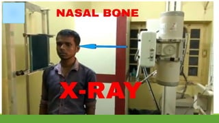
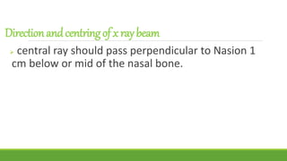
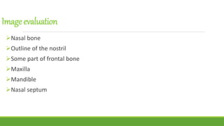
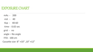
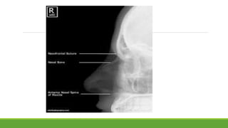

The document outlines the procedure for performing a nasal bone view X-ray, which is used to detect nasal bone fractures and foreign bodies. It provides details on patient positioning, X-ray beam direction, and image evaluation criteria, including specific exposure settings. Additionally, it references a positioning guide by Katharine 'Kitty' Clark.


![Radiography of skull [Autosaved].pptxriuyowioehgg](https://cdn.slidesharecdn.com/ss_thumbnails/radiographyofskullautosaved-251211014507-1d75cfe3-thumbnail.jpg?width=640&height=640&fit=bounds)


