Download to read offline
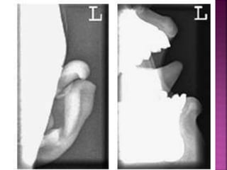
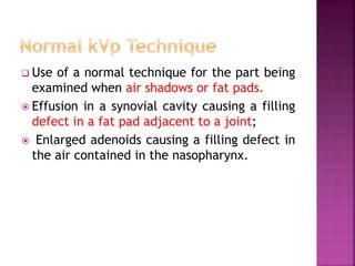
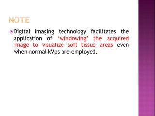


This document discusses soft tissue radiography, which assesses soft tissues like muscle, skin, and glands using low-energy x-rays without contrast media. It provides techniques for soft tissue radiography including using appropriate exposure and reducing scatter to improve contrast and image sharpness. Soft tissue radiography can use normal techniques when air or fat is present, two films, or wedge filters. It may also use subnormal, low, or high kVp settings depending on the body part and desired visualization. Digital imaging allows windowing acquired images to view soft tissues at normal kVp as well.


