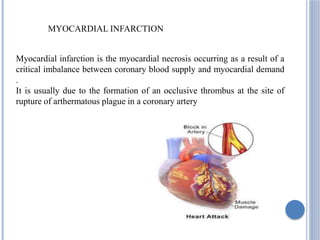
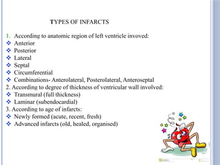
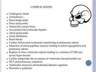
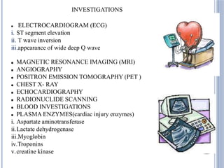
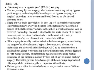
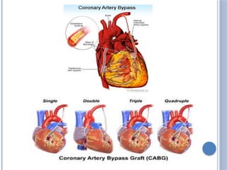
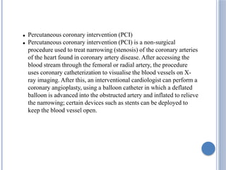
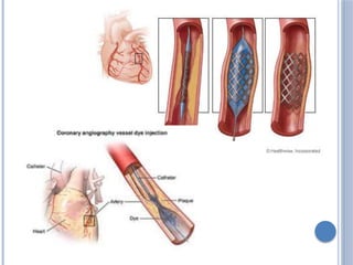
3. MYOCARDIAL INFARCTION
Myocardial infarction is the myocardial necrosis occurring as a result of a
critical imbalance between coronary blood supply and myocardial demand
.
It is usually due to the formation of an occlusive thrombus at the site of
rupture of arthermatous plague in a coronary artery
4. TYPES OF INFARCTS
1. According to anatomic region of left ventricle invoved:
Anterior
Posterior
Lateral
Septal
Circumferential
Combinations- Anterolateral, Posterolateral, Anteroseptal
2. According to degree of thickness of ventricular wall involved:
Transmural (full thickness)
Laminar (subendocardial)
3. According to age of infarcts:
Newly formed (acute, recent, fresh)
Advanced infarcts (old, healed, organised)
5. Tobacco smoking
Hypertension
Drug abuse
Obesity
Stress
Alcohol
Age
Gender
Diabetes
Hyperlipoproteinaemia
Family history of Ischaemic Heart
Disease
Hyperhomocysteinemia
Chronic kidney disease
ETIOLOGY
6. Atherosclerosis Arterial spasm Atherosclerosis+Plaque split+Thrombus
gradual Obstruction sudden reversible occlusion sudden not usually reversible
Ischemia
Hypoxia
Reduced oxygen demand
Thrombolysis
Permanent thrombus Necrosis MI
Unstable Angina
Angina
PATHOPHYSIOLOGY
7. CLINICAL FEATURES
Symptoms-
Pain in the area between shoulder blades, arm, chest, chest, jaw, left arm, or
upper abdomen
dizziness, fatigue, light-headedness, clammy skin, cold sweat, or sweating
Gastrointestinal: heartburn, indigestion, nausea, or vomiting
Chest: Chest pain, discomfort, fullness, or tightness
Neck: discomfort or tightness
Arm: discomfort or tightness anxiety, feeling of impending doom,
sensation of an abnormal heartbeat, shortness of breath, or shoulder discomfort
9. COMPLICATIONS
Cardiogenic shock
Arrhythmia's ;
Sinus bradycardia
Sinus tachycardia
Ventricular ectopic beats
Accelerated idioventricular rhythm
Atrial tachycardia
Atrial fibrillation
Heart blocks
Cardiac failure,most commonly manifesting as pulmonary edema
Infarction of mitral papillary muscles leading to mitral regurgitation and
pulmonary edema
Rupture of inter ventricular septum leading to a murmur of VSD and
severe hypo-tension
Cardiac tamponade due to rupture of ventricular into pericardial sac
DVT and pulmonary embolism
Ventricular aneurysm and dyskinetic/akinetic segments
Dresselar’s syndrome
10. INVESTIGATIONS
ELECTROCARDIOGRAM (ECG)
i. ST segment elevation
ii. T wave inversion
iii.appearance of wide deep Q wave
MAGNETIC RESONANCE IMAGING (MRI)
ANGIOGRAPHY
POSITRON EMISSION TOMOGRAPHY (PET )
CHEST X- RAY
ECHOCARDIOGRAPHY
RADIONUCLIDE SCANNING
BLOOD INVESTIGATIONS
PLASMA ENZYMES(cardiac injury enzymes)
i. Aspartate aminotransferase
ii.Lactate dehydrogenase
iii.Myoglobin
iv.Troponins
v.creatine kinase
12. SURGICAL
Coronary artery bypass graft (CABG) surgery
Coronary artery bypass surgery, also known as coronary artery bypass
graft surgery, and colloquially heart bypass or bypass surgery, is a
surgical procedure to restore normal blood flow to an obstructed
coronary artery.
There are two main approaches. In one, the left internal thoracic artery
(internal mammary artery) is diverted to the left anterior descending
branch of the left coronary artery. In the other, a great saphenous vein is
removed from a leg; one end is attached to the aorta or one of its major
branches, and the other end is attached to the obstructed artery
immediately after the obstruction to restore blood flow.
This surgery is usually performed with the heart stopped, necessitating
the usage of cardiopulmonary bypass; however, two alternative
techniques are also available allowing CABG to be performed on a
beating heart either without using the cardiopulmonary bypass deemed
as "off-pump" surgery or performing beating surgery using partial
assistance of the cardiopulmonary bypass called as "on-pump beating"
surgery. The latter gathers the advantages of the on-pump stopped and
off-pump while minimizing their respective side-effects.
This surgery is often indicated when coronary arteries have a 50% to
13.
14. Percutaneous coronary intervention (PCI)
Percutaneous coronary intervention (PCI) is a non-surgical
procedure used to treat narrowing (stenosis) of the coronary arteries
of the heart found in coronary artery disease. After accessing the
blood stream through the femoral or radial artery, the procedure
uses coronary catheterization to visualise the blood vessels on X-
ray imaging. After this, an interventional cardiologist can perform a
coronary angioplasty, using a balloon catheter in which a deflated
balloon is advanced into the obstructed artery and inflated to relieve
the narrowing; certain devices such as stents can be deployed to
keep the blood vessel open.
15.
16. Treatment after a myocardial infarction
Aspirin: to reduce the ’stickiness’ of platelets in the blood
which helps to prevent blood clots forming.
Clopidogrel in addition to aspirin, especially after
angioplasty with stenting.
A beta-blocker : to slow the heart rate, lower blood pressure
and to reduce the chance of abnormal heart rhythms
developing
An ACE inhibitor or angiotensin converting enzyme-
inhibitor : ACE inhibitors have a number of actions
including lowering the blood pressure, reducing the risk of
structural changes in the damaged heart muscle and having a
protective effect concerning future events
A statin drug : to lower the level of cholesterol and other
blood lipids in your blood. This helps to prevent the build-up
of atheroma and therefore reduces the risk in the future.