Downloaded 199 times


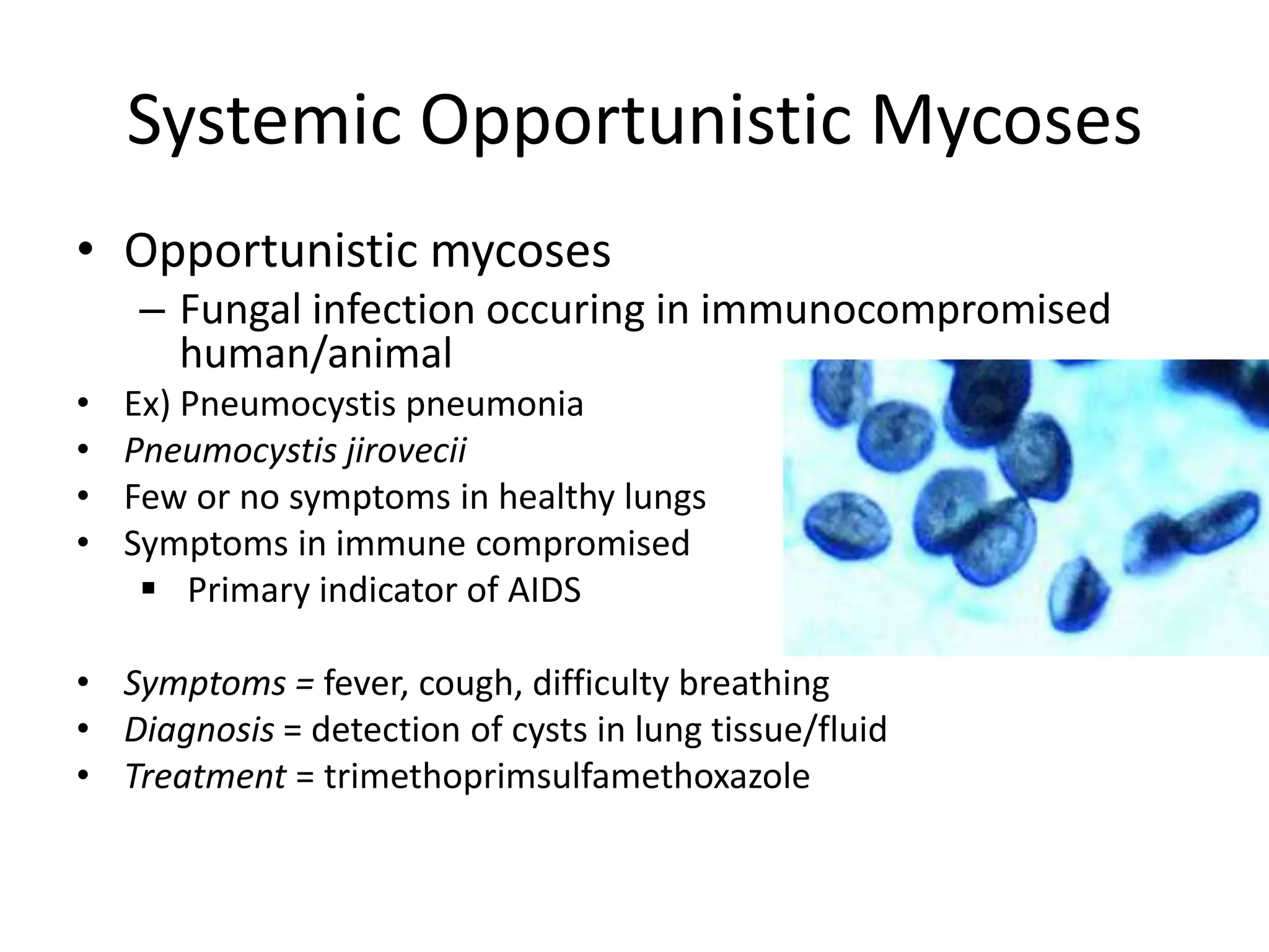

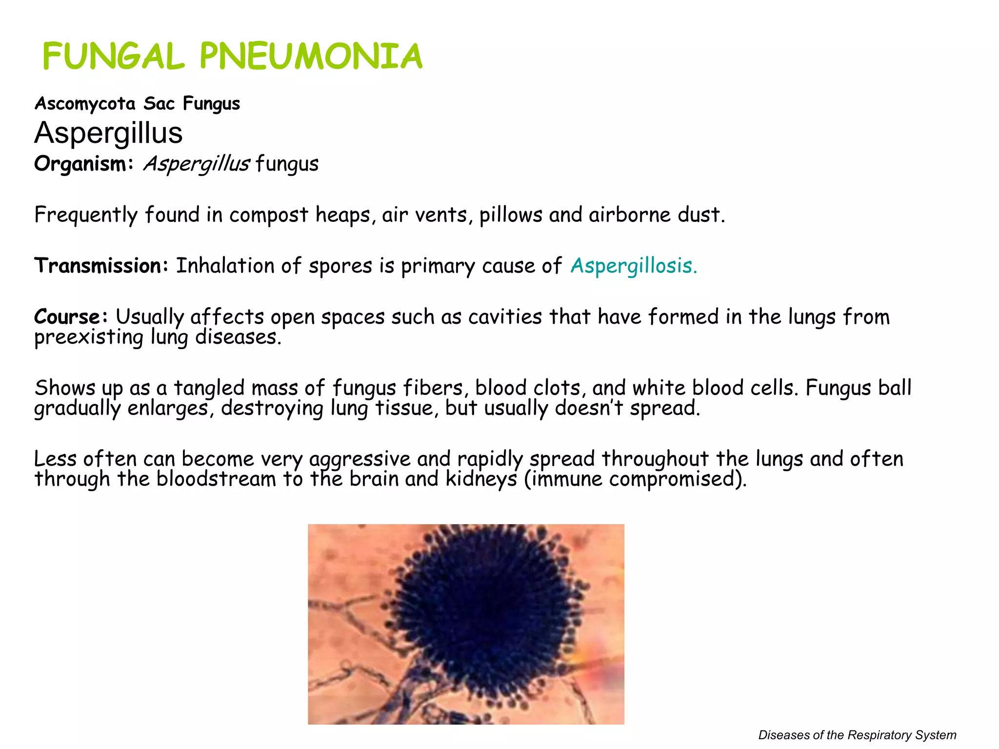


Mycology is the study of fungi, of which there are over 100,000 known species but only about 200 that can cause human disease. Fungi have cell walls containing chitin and reproduce both sexually and asexually. Common fungal infections include ringworm, athlete's foot, and nail fungus caused by dermatophytic fungi. Systemic fungal infections occur when fungi enter the bloodstream, often through the lungs, and can spread throughout the body. Opportunistic fungal infections mainly affect immunocompromised individuals. Aspergillosis is a common fungal pneumonia caused by inhaling Aspergillus spores. Pneumocystis pneumonia is seen primarily in AIDS patients. Candid


