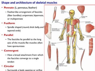
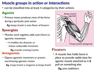
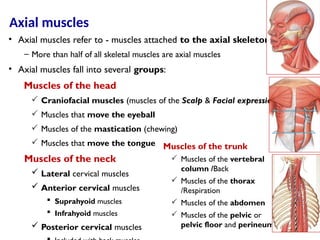
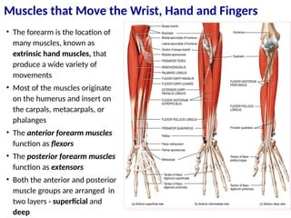
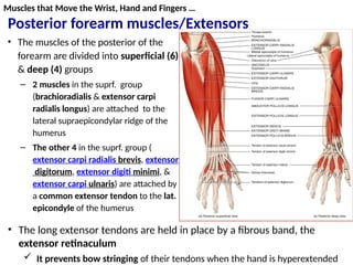
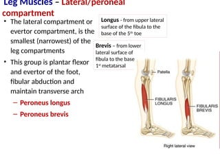
Muscular system
• Muscle is composed of cells which can contract to cause movement
 Produce movement
 Allow us to move, breath, transport materials and circulate our blood
etc
 Maintain posture and body position
 By resisting the pull of gravity and preventing unwanted movements
 They hold some articulating bones in place by maintaining tension on the
tendons
 Guard entrance and exits of the body
Myology - the study of muscles
 There are three different types of muscle;
1. Cardiac Muscle (striated, involuntary)
 Makes up the wall of the heart
2. Smooth Muscle (unstriated, involuntary)
 Found in structures which we do not have
control over such as blood vessels, stomach
and intestine, urethra, uterus, internal muscles
of the eye
3. Skeletal Muscle (striated, voluntary)
 This is the muscle attached to our skeletons
and allows us to move our bodies
Types of muscle tissue
Types of muscle tissue and their structure
Cardiac muscle
• found in the walls of the heart
• It contracts to force the blood around the
body
• Contracts without stimulation
 However, the strength and rate of the
contraction is modified by the
autonomic nervous system
• does not tire, fibers are short with a single
central nucleus; they are striated
• The cells join directly together and are
connected by connective tissue
• Cardiac muscle tissue found only in the
walls of the heart
• Fibres
 Contain single central nucleus
 Branch freely - connected via gap
junctions
 Have very high aerobic capacity
• Contract according to sliding filament
theory
Smooth muscle
• Smooth muscle does not tire
• Smooth muscle cells are spindle shaped and contain a single oval,
centrally located nucleus; they are unstriated
• They have no sheath but are connected by connective tissue
• found in the walls of the internal organs,
• Smooth muscle contracts without stimulation
 However, the strength and rate of the contraction
is modified by the autonomic nervous system
Skeletal muscle
– It is under voluntary control but tires
quickly
• Most of it is attached to bones, but
there are exceptions (attached to a
connective tissue via tendons) in
– The tongue, upper esophagus, some
facial muscles, and some sphincter
• is the muscle that moves our body and its limbs as well as other
parts
Connective tissues of a skeletal muscle
• Muscle fibres are surrounded by a loose
connective tissue called endomysium, this
contains capillaries which supply the muscle
cells with blood
– The cells with their surrounding endomysium
are bundled together into fasciculae
• The fasciculae are surrounded by a strong
connective tissue called perimysium
• A muscle is made up of many fasciculae
which are bound together by a dense
connective tissue called epimysium
• The epimysium grades imperceptibly into
connective tissue sheets called fasciae (deep
and superficial)
• Are attached to bones (mostly),
cartilages, ligaments, or fascia or to
some combination of …
 Direct = right onto bone
– Epimysium of muscle fibers
continuous with the periosteum a
bone
Example - intercostal muscles
 Indirect = via tendon/aponeurosis
– More common
– Leave bony markings = tubercle,
crest, ridge, etc.
– Sometimes attach to skin
Internal intercostals
External intercostals
Pectoralis
minor
Pectoralis
major
• origin : end of a muscle
attached to the less movable
bone
• insertion : end of a muscle
attached to the most movable
bone
• belly thicker middle region
b/n insertion and origin
 May have multiple origins, insertions
and bellies
General anatomy of skeletal muscles
Origin insertion and
belly
Shape and architecture of skeletal muscles
• Pennate (L. pennatus, feather)
– Feather-like arrangement of fascicles
(fiber bundles): unipennate, bipennate,
or multipennate
• Fusiform
– Spindle shaped (round, thick belly, and
tapered ends)
• Parallel
– The fascicles lie parallel to the long
axis of the muscle; flat muscles often
have aponeuroses
• Convergent
– Have a broad attachment from which
the fascicles converge to a single
tendon
• Circular
– Surrounds a body opening or orifice
Muscle groups in action or Interactions
Agonist
• Primary mover, produces most of the force
during a particular joint action
Eg. biceps brachii is main flexor of forearm
Synergists
• Muscles work together, adds extra force to
agonistic movement
– It modifies the direction or
reduce undesirable movement
Eg. muscles crossing 2 joints
Antagonists
• Oppose agonistic movement or prevent
overshooting agonistic motion
Eg. triceps brachii is antagonist to biceps brachii
• can be classified into at least 4 categories by their actions
Fixators
• A muscle that holds bone in
place to provide stable base for
agonist muscle attached to it &
pull on something else
Eg. joint stablilizers
 Location: (eg) brachialis =
arm
 Shape: (eg) deltoid =
triangle
 Relative Size: (eg)
minimus, maximus, longus
 Direction of Fascicles:
(eg) oblique, rectus
 Location of Attachment:
(eg) brachioradialis
 Number of Origins: (eg)
biceps, quadriceps
 Action: (eg) flexor,
adductor, extensor
Naming of Skeletal Muscle
• Muscle names describe
one or more attributes
of the muscle
Principal of Muscle Naming
• They are classified based on the skeletal
position on which they are attached
– Axial muscles
– Appendicular muscles
Axial muscles
– Muscles of the head
– Muscles of the neck
– Muscles of the trunk
 Muscles of the vertebral column, thorax, abdomen,
and pelvic and perineum
Appendicular muscles
– Muscles of the pectoral girdle & upper limb
– Muscles of the pelvic girdle & lower limb
Classification of Skeletal muscles
60% of skeletal muscles
40% of skeletal muscles
Axial muscles
• Axial muscles refer to - muscles attached to the axial skeleton
– More than half of all skeletal muscles are axial muscles
• Axial muscles fall into several groups:
Muscles of the head
 Craniofacial muscles (muscles of the Scalp & Facial expression)
 Muscles that move the eyeball
 Muscles of the mastication (chewing)
 Muscles that move the tongue
Muscles of the neck
 Lateral cervical muscles
 Anterior cervical muscles
 Suprahyoid muscles
 Infrahyoid muscles
 Posterior cervical muscles
Muscles of the trunk
 Muscles of the vertebral
column /Back
 Muscles of the thorax
/Respiration
 Muscles of the abdomen
 Muscles of the pelvic or
pelvic floor and perineum
• responsible for the
variety and subtlety of
human facial expression
• around the orbit, nose,
mouth and auricles
Origin
• skull bones or fascia
Insersion
• They attach to the skin
of the face
Muscles of Facial
Expression
Muscles of the Scalp
Occipitofrontalis
• The occipitofrontalis is a helmet
like strap of muscle covering the
scalp
• It is divided into
– The frontalis of the forehead
and
– The occipitalis at the rear of the
head
– They are connected to each
other by a broad aponeurosis,
the galea aponeurotica
• The occipitofrontalis moves the
scalp, forehead skin, and
eyebrows
Axial Muscles … muscles of the head
Facial muscles around the mouth
Muscles moving the eyeball
• Known as extrinsic eye muscles (six pairs)
– These muscles are fast and precisely controlled
• Originate on skull bones and attach onto
the sclera
• Movements include pulling toward the
muscle origin, and rotation of the eyeball
Muscles of the Head: Muscles of Mastication
• The muscles of mastication
are strong muscles which
open & close the mouth
for chewing
• They move the jaw
(mandible) forwards,
backwards and from side
to side at the TMJ
(temporomandibular joint)
Muscles of Mastication Location Action
Temporalis
A large triangular muscle on the
side of the head.
Closes the jaw.
Masseter
Rectangular muscle from the
cheek to the outside of the jaw.
Closes the jaw.
Medial and Lateral
Pterygoids
Two muscles deep inside the jaw;
from the pterygoid plate to the
mandible.
Open and closes the jaw
Moves the jaw from side to side and
forwards and backwards.
Buccinator (???) In the soft tissue of the cheeks.
Flattens cheeks to press food under the
molars, and in sucking and blowing
 Movement of the tongue is vital during speech, mastication
(chewing), deglutition (swallowing)
 Extrinsic tongue muscles move the tongue, while intrinsic tongue
muscles change the shape of the tongue
Muscles of the Head: Moving the Tongue
Genioglossus
Muscles of the Pharynx
Pharyngeal constrictors
• External circular layer of
pharyngeal muscles … 3
• Constrict walls of pharynx during
swallowing
– Move food into esophagus
Laryngeal elevators
• Internal, longitudinal
layer of muscles … 2
• Elevate the larynx and
shorten the pharynx
during swallowing
and speaking
Palatal muscles
• Elevate the soft palate
Esophagus
Posterior view
Muscles of the neck
Lateral cervical muscles
Muscles Location Action
Sternocleido
mastoid
Long thick
muscle at the
side of the
neck
Both muscles together
flex the neck
An individual muscle will
draw the head to that
shoulder.
Scalenus
anterior
posterior
border of the
SCM
lateral flexion and
rotation of neck
Scalenus
medius
Lateral cervical
region
lateral flexion of neck
Scalenus
posterior
Lateral cervical
region
lateral flexion of neck
• Two groups of muscles
stabilize the hyoid bone,
and allow movements of
the tongue and larynx
A. Suprahyoid muscles
• Muscles superior to the hyoid
– Stylohyoid
– Digastric
– Mylohyoid
– Geniohyoid
B. Infrahyoid muscles
• Muscles inferior to the hyoid
– Omohyoid
– Sternohyoid
– Thyrohyoid
– Sternothyroid
Anterior cervical muscles
• Most body weight lies anterior to the
vertebral column, especially in obese
people
– Consequently, many strong muscles are
attached to the spinous and transverse
processes necessary to support & move
the vertebral column
• There are two major groups of muscles
in the back
– Extrinsic Muscles of the Back
– Intrinsic Muscles of the Back
• The extrinsic back muscles
control limb movements
• The intrinsic (deep) back muscles
include muscles that specifically
act on the vertebral column, producing
its movements and maintaining posture
Muscles of the Back
• The intrinsic or deep muscles of
the back are three layers
1. Superficial (Splenius) group
• Cranial portion - splenius capitis
• Cervical portion- splenius cervicis
• Lumbar portion- quadratus lumborum
2. Intermediate (Erector Spinae) group -
3 parts
• Iliocostalis (lateral column)
• Longissimus (intermediate)
• Spinalis (medial column)
3. Deep (Transversospinal) group
• Semispinalis (capitis, cervicis, & thoracis)
• Multifidus (best developed in the lumbar
region)
• Rotatores (developed in the thoracic
region)
Deep Muscles of the Back
Muscles of the thorax/Respiration
1.External intercostals
2. Internal intercostals
3. Innermost intercostal
4. Diaphragm
Muscles of the Anterior Abdominal Wall
 4 major abdominal muscles
• The 2 minors are;
 Pyramidalis
 Cremaster
• The Four majors are; " TIRE around
the abdomen"
 Transversus abdominis
 Internal abdominal obliq.
 Rectus abdominis
 External abdominal obliq.
 3 layers of muscles
 Pelvic diaphragm – deepest
 levator ani and coccygeus
 Urogenital diaphragm –
middle
 External urethral sphincter
 External anal sphincter
 Deep transverse perineus
 Superficial perineal space -
just deep to the skin contains 3
muscles
 Ischiocavernosus
 Bulbospongiosus
 Superfi.transverse perineus
Pelvis floor muscles
• Muscles of the upper limb
 Muscles Acting on the
Scapula
 Muscles Acting on the
Humerus
 Muscles Acting on the
Forearm
 Muscles Acting on the
Wrist and Hand
• Muscles of the lower limb
 Muscles Acting on the Hip
and Femur
 Muscles Acting on the Knee
 Muscles Acting on the Foot
Appendicular muscles
Muscles that Move the Pectoral Girdle
• Several muscles
stabilize the scapula
during movements of
the humerus
• Movements of the
scapula increase the
range of motion at the
shoulder
• Anterior muscles
– Pectoralis minor
– Subclavius
– Serratus anterior
• Posterior muscles
(1) The superficial extrinsic muscles - Trapezius
(2) The deep extrinsic muscles - Levator scapulae, rhomboids major and
minor
Muscles that Move the Humerus
• Muscles with origins on
the axial skeleton
– Pectoralis major
– Latissimus dorsi
• Muscles with origins on
the scapula
– Deltoid Muscle
– Teres Major Muscle
– Rotator cuff muscles (SITS)
• Supraspinatus
• Infraspinatus
• Teres Minor Muscle
• Subscapularis
– Supraspinatus
– infraspinatus
– teres minor
– subscapularis
• They are named rotator
cuff because they form a
cuff around the
glenohumeral joint
Muscles that move the humerus … Scapular origins
Rotator cuff muscles (SITS)
Anterior view
• They are 4 scapulohumeral muscles (referred to as SITS)
• All except the supraspinatus
are rotators of the humerus
 The supraspinatus, besides
being part of the rotator
cuff, initiates and assists the
deltoid in the first stage of
abduction of the arm
Muscles that Move the Radius and Ulna
• Forearm flexors
– Biceps brachii
– Brachialis
– Coracobrachialis
• Forearm extensors
– Triceps brachi
– Anconeus
Muscles for Pronation and Supination
• Wrist movements
contribute to our
great flexibility in
our use of our
hands
• Pronators
– Pronator teres
– Pronator
quadratus
• Supinators
– Supinator
Muscles that Move the Wrist, Hand and Fingers
• The forearm is the location of
many muscles, known as
extrinsic hand muscles, that
produce a wide variety of
movements
• Most of the muscles originate
on the humerus and insert on
the carpals, metacarpals, or
phalanges
• The anterior forearm muscles
function as flexors
• The posterior forearm muscles
function as extensors
• Both the anterior and posterior
muscle groups are arranged in
two layers - superficial and
deep
Anterior forearm muscles/Flexors
• The anterior forearm muscles
are divided into 3 layers
– Deep, intermediate and
superficial layers
• A septum of deep fascia
separates the deep layer
from the superficial &
intermediate layers
• The tendons of most flexor
muscles pass across the anterior
surface of the wrist
• These tendons are held in place
by the flexor retinaculum
– This retinaculum prevents bow
stringing of the tendons when
Muscles that Move the Wrist, Hand and Fingers
• The muscles of the posterior of the
forearm are divided into superficial (6)
& deep (4) groups
– 2 muscles in the suprf. group
(brachioradialis & extensor carpi
radialis longus) are attached to the
lateral supraepicondylar ridge of the
humerus
– The other 4 in the suprf. group (
extensor carpi radialis brevis, extensor
digitorum, extensor digiti minimi, &
extensor carpi ulnaris) are attached by
a common extensor tendon to the lat.
epicondyle of the humerus
• The long extensor tendons are held in place by a fibrous band, the
extensor retinaculum
 It prevents bow stringing of their tendons when the hand is hyperextended
Muscles that Move the Wrist, Hand and Fingers …
Posterior forearm muscles/Extensors
Intrinsic Muscles of the hand
• Intrinsic hand muscles both
originate and insert within the
hand
• are innervated by branches of the
ulnar and median nerves
• They can be divided into 4 groups:
A. Thenar muscles in the thenar
compartment
B. Hypothenar muscles in the
hypothenar compartment
C. Lumbrical muscles in the central
compartment
D. Interosseous muscles b/n the
metacarpal bones
• The flexibility of the hand is due
to the large number of bones
and joints, as well as to the
many muscles controlling them
Intrinsic Muscles of the Hand
Thenar muscles
Name Description Action
Thenar (thumb)
group
Muscles of the thumb.
Thumb
movements
Adductor Pollicis
A triangular muscle with 2 heads; it arises from the
3rd metacarpal and inserts onto the thumb. •Adducts thumb.
Abductor Pollicis
Brevis
A long thin muscle; it arises from the ulna and
radius and inserts onto the thumb. •Abducts thumb.
Opponens Pollicis
The fleshy part of the thumb; it arises from the
flexor retinaculum and trapezium and inserts onto
the thumb.
•Opposes thumb.
Flexor Pollicis
Brevis
A thin muscle, it arises from the flexor retinaculum
and carpal bones (trapezium, trapezoid and
capitate) and inserts onto the thumb.
•Flexes thumb.
Muscles of the hand …
Hypothenar muscles
• These muscles deal with
the movement of the
5th digit
• They lie in the
hypothenar
compartment of the
palm with the
5th metacarpal bone
• The hypothenar muscles
produce the hypothenar
eminence, or ball of the
5th digit
• They are all supplied by
the deep branch of the
ulnar nerve
Hypothenar (little
finger) Group
Muscles of the little finger. Little finger movements.
Abductor Digiti
Minimi
A small muscle arising from the flexor
retinaculum and pisiform and inserting
onto the little finger.
•Abducts little finger.
Flexor Digiti
Minimi Brevis
A small thin muscle arising from the flexor
retinaculum and hamate and inserting
onto the little finger.
•Flexes little finger
Opponens Digiti
Minimi
A small muscle arising from the flexor
retinaculum and hamate and inserting
onto the little finger.
•Opposes little finger to
thumb.
Hypothenar muscles
Muscles of the hand …
Lumbrical muscles
• There are 4 slender lumbrical (L.
lumbricus, earthworm) muscles-
one for each digit
Action
– Flex & extend digits at the
metacarpophalangeal &
interphalangeal joints respectively
Interosseous muscles
• There are 7 interossei muscles located b/n the metacarpal bones
• They are arranged in 2 layers: 3 palmar and 4 dorsal muscles
Action
– The dorsal interossei
abduct the digits
(DAB: Dorsal ABduct)
Palmar interossei 1 to 3
Interosseous muscles…
Action
– The palmar interossei adduct the digits
(PAD: Palmar ADduct)
– They also assist the actions of the
lumbrical muscles.
– These are important movements in
typing, writing, and playing the piano
Muscles of the lower limb
• They include the muscles that move the
thigh and leg and those that control
movement of the foot
‒ Muscles Acting on the Hip and Femur
‒ Muscles Acting on the Knee
‒ Muscles Acting on the Foot
• Many of these muscles cross two joints
and act on both
• Lower limb muscles are larger and
stronger - with posture and stability
being more important than range of
motion, are specialized for locomotion
and balance.
Appendicular Muscles …
Muscles Acting on the Hip and Femur
• Muscles crossing the
hip and knee can be
grouped into
– Hip flexors (4)
– Anterior compartment
(4 knee extensors)
– Gluteal muscles (3)
– Lateral rotators (5)
– Medial compartment
(6 adductors)
– Posterior compartment
(3 knee flexors)
Thigh muscles - Anterior compartment
Hip flexors
Psoas Muscles
Iliacus
Sartorius
Rectus femori
Action
• Iliopsoas is the strongest flexor
of the thigh at the hip joint
Anterior compartment - Quadriceps Muscle
• It is the great extensor muscle
of the leg, the biggest muscle in
the body
• It covers almost all of the
anterior surface and sides of
the femur
• It is divided into 4 parts:
– Rectus femoris
– Vastus lateralis
– Vastus medialis
– Vastus intermedius
• The names of the parts of the
quadriceps m. indicate their
form or location (L. vastus,
large) and (L. rectus, straight)
Thigh muscles …
Thigh muscles …
Gluteal muscles
• This is the group of muscles that form the buttocks
• They include gluteal muscles consist of three glutei mm.
– Gluteus maximus
– G. Medius
– G. Minimus
Lateral rotators
Piriformis
Quadratus femoris
Obturator internus
Superior gemillus
Inferior gemillus
Obturator externus
Thigh muscles …
• These muscles form the inside (medial side) of
the thigh
• They include
– Adductor magnus
– Adductor longus
– Adductor brevis
– Pectineus
– Gracilis
– Obturator externus
Three layers
A. Superficial- pectineus, Gracilis and adductor L
B. Middle- Adductor b
C. Deep- Adductor M & obturator externus
Thigh Muscles …
Medial compartment - Adductors
Posterior thigh muscles - Hamstring muscles
• Large bulk of 3 muscles on the back of
the thigh
• They arise from the ischial tuberosity
• Insert into the tibia or fibula
– Biceps Femoris (2 heads; long & short)
– Semitendinosus (has a long thin
tendon).
– Semimembranosus (it is membranous
at its origin)
• Extend hip and flex knee joint
Thigh Muscles …
• Muscles of the anterior
compartment are
– Tibialis anterior
– Extensor digitorum longus
– Extensor hallucis longus
– Peroneus tertius (eversion)
• These muscles pass anterior to
the transversely oriented axis
of the ankle (talocrural) joint to
insert on the metatarsals and
toes
– Therefore, are dorsiflexors of
the ankle joint, elevating the
forefoot and depressing the
heel
Muscles of the leg – Anterior Compartment
• The lateral compartment or
evertor compartment, is the
smallest (narrowest) of the
leg compartments
• This group is plantar flexor
and evertor of the foot,
fibular abduction and
maintain transverse arch
– Peroneus longus
– Peroneus brevis
Leg Muscles – Lateral/peroneal
compartment
Longus - from upper lateral
surface of the fibula to the
base of the 5th
toe
Brevis – from lower
lateral surface of
fibula to the base of
1st
metatarsal
Copyright 2010, John Wiley & Sons, Inc.
Muscles that Move the Foot and Toes
Leg Muscles – Posterior compartment
• Muscles of the
posterior compartment
are primarily plantar
flexors and flex the toes
• Superficial
– Gastrocnemius
– Soleus
– Plantaris
• Deep
– Tibialis posterior
– Flexor digitorum longus
– Flexor hallucis longus
– Popliteus
Leg Muscles – Posterior compartment
The dorsum of the foot
• There are two closely connected
muscles on the dorsum of the foot
• Extensor digitorum brevis
• Extensor hallucis brevis
( medial slip of the extensor
digitorum brevis )
• These broad thin muscles form a
fleshy mass on the lateral part of
the dorsum of the foot, anterior
to the lateral malleolus
• Actions: the extensor digitorum brevis extends the second to fourth digits
at the metatarsophalangeal joints, and the extensor hallucis brevis
extends the first digit or great toe at the metatarsophalangeal joint.
• These muscles help the long extensor muscles extend the toes
The sole of the foot
• Plantar aponeurosis - thickened deep fascia, tout when toes
are dorsiflexed (arch of foot increases)
– Attachments- posterior- tubercle of calcaneus; anteriorly divide in
to five slips and again each divide in to two and become
continuous with flexor sheaths of the digits
– Function – protection, keep the longitudinal arch
• There are four muscular layers in the sole of the foot
1. First layer – three muscles
2. Second layer – two muscles and two tendons
3. Third layer - Three muscles
4. Fourth layer - Two muscles and two tendons
• These muscles serve major roles in support of the body and locomotion
• Flexion of the toes and a role in the longitudinal arch of the foot are
specific roles of these muscles
First layer
1. Abductor hallucis inserts in to
the proximal phalanx of hallux.
2. Abductor digiti minimi inserts
in the proximal phalanx of toe
5
3. Flexor digitorum brevis inserts
in to the middle phalanges of
2- 5 by splitting in to 2 bands
• These muscles act as a
functional group that acts as an
elastic spring for supporting the
arches of the foot and
maintaining the concavity of
the foot.
• Two abductors and one flexor in b/n the abductors
• Origin – calcaneus
Second layer
• Muscles of the second layer flex,
adduct, and abduct the toes
• Muscles include
– Flexor digitorum accessorius
(quadratus plantae); has to
heads (medial and lateral);
– Four lumbricales- origin form
medial side of the long
tendons of flexor muscles
arranged from 1st
to 4th
from
medial to lateral. The lateral 3
are innervated by deep
division of lateral plantar and
the 1st
by medial p.
 Tendons – flexor hallucis longus
and flexor digotrum longus
Third layer
• This layer has 3 short muscles
of the great and small digits,
which lie in the anterior half of
the sole of the foot.
• Two act on the great digit and
one on the small digit.
• Includes:
– flexor hallucis brevis, adductor
hallucis and flexor digiti
minimi.
• A sesamoid bone adheres to
each of the tendons of the two
heads of flexor hallucis brevis.
Fourth layer
Tendons
• Tendons of tibialis posterior
and peroneus L
Muscles
1. Plantar interossei – 3
Action - adduct 3rd
, 4th
, and 5th
toes toward 2nd
toe
2. Dorsal interossei- 4
 Each arise from adjacent metatarsal (bipennate
arrangement)
Action- adduction of 2nd
, 3rd
&4th
digits away from line of
2nd
toe, flex metacarpophalangeal J. and extend
interphalangeal J

MUSCULAR SYSTEM and its classification.pptx

  • 1.
    Muscular system • Muscleis composed of cells which can contract to cause movement  Produce movement  Allow us to move, breath, transport materials and circulate our blood etc  Maintain posture and body position  By resisting the pull of gravity and preventing unwanted movements  They hold some articulating bones in place by maintaining tension on the tendons  Guard entrance and exits of the body Myology - the study of muscles
  • 2.
     There arethree different types of muscle; 1. Cardiac Muscle (striated, involuntary)  Makes up the wall of the heart 2. Smooth Muscle (unstriated, involuntary)  Found in structures which we do not have control over such as blood vessels, stomach and intestine, urethra, uterus, internal muscles of the eye 3. Skeletal Muscle (striated, voluntary)  This is the muscle attached to our skeletons and allows us to move our bodies Types of muscle tissue
  • 3.
    Types of muscletissue and their structure
  • 4.
    Cardiac muscle • foundin the walls of the heart • It contracts to force the blood around the body • Contracts without stimulation  However, the strength and rate of the contraction is modified by the autonomic nervous system • does not tire, fibers are short with a single central nucleus; they are striated • The cells join directly together and are connected by connective tissue • Cardiac muscle tissue found only in the walls of the heart • Fibres  Contain single central nucleus  Branch freely - connected via gap junctions  Have very high aerobic capacity • Contract according to sliding filament theory
  • 5.
    Smooth muscle • Smoothmuscle does not tire • Smooth muscle cells are spindle shaped and contain a single oval, centrally located nucleus; they are unstriated • They have no sheath but are connected by connective tissue • found in the walls of the internal organs, • Smooth muscle contracts without stimulation  However, the strength and rate of the contraction is modified by the autonomic nervous system
  • 6.
    Skeletal muscle – Itis under voluntary control but tires quickly • Most of it is attached to bones, but there are exceptions (attached to a connective tissue via tendons) in – The tongue, upper esophagus, some facial muscles, and some sphincter • is the muscle that moves our body and its limbs as well as other parts
  • 7.
    Connective tissues ofa skeletal muscle • Muscle fibres are surrounded by a loose connective tissue called endomysium, this contains capillaries which supply the muscle cells with blood – The cells with their surrounding endomysium are bundled together into fasciculae • The fasciculae are surrounded by a strong connective tissue called perimysium • A muscle is made up of many fasciculae which are bound together by a dense connective tissue called epimysium • The epimysium grades imperceptibly into connective tissue sheets called fasciae (deep and superficial)
  • 8.
    • Are attachedto bones (mostly), cartilages, ligaments, or fascia or to some combination of …  Direct = right onto bone – Epimysium of muscle fibers continuous with the periosteum a bone Example - intercostal muscles  Indirect = via tendon/aponeurosis – More common – Leave bony markings = tubercle, crest, ridge, etc. – Sometimes attach to skin Internal intercostals External intercostals Pectoralis minor Pectoralis major
  • 9.
    • origin :end of a muscle attached to the less movable bone • insertion : end of a muscle attached to the most movable bone • belly thicker middle region b/n insertion and origin  May have multiple origins, insertions and bellies General anatomy of skeletal muscles Origin insertion and belly
  • 10.
    Shape and architectureof skeletal muscles • Pennate (L. pennatus, feather) – Feather-like arrangement of fascicles (fiber bundles): unipennate, bipennate, or multipennate • Fusiform – Spindle shaped (round, thick belly, and tapered ends) • Parallel – The fascicles lie parallel to the long axis of the muscle; flat muscles often have aponeuroses • Convergent – Have a broad attachment from which the fascicles converge to a single tendon • Circular – Surrounds a body opening or orifice
  • 11.
    Muscle groups inaction or Interactions Agonist • Primary mover, produces most of the force during a particular joint action Eg. biceps brachii is main flexor of forearm Synergists • Muscles work together, adds extra force to agonistic movement – It modifies the direction or reduce undesirable movement Eg. muscles crossing 2 joints Antagonists • Oppose agonistic movement or prevent overshooting agonistic motion Eg. triceps brachii is antagonist to biceps brachii • can be classified into at least 4 categories by their actions Fixators • A muscle that holds bone in place to provide stable base for agonist muscle attached to it & pull on something else Eg. joint stablilizers
  • 12.
     Location: (eg)brachialis = arm  Shape: (eg) deltoid = triangle  Relative Size: (eg) minimus, maximus, longus  Direction of Fascicles: (eg) oblique, rectus  Location of Attachment: (eg) brachioradialis  Number of Origins: (eg) biceps, quadriceps  Action: (eg) flexor, adductor, extensor Naming of Skeletal Muscle • Muscle names describe one or more attributes of the muscle
  • 13.
  • 14.
  • 15.
    • They areclassified based on the skeletal position on which they are attached – Axial muscles – Appendicular muscles Axial muscles – Muscles of the head – Muscles of the neck – Muscles of the trunk  Muscles of the vertebral column, thorax, abdomen, and pelvic and perineum Appendicular muscles – Muscles of the pectoral girdle & upper limb – Muscles of the pelvic girdle & lower limb Classification of Skeletal muscles 60% of skeletal muscles 40% of skeletal muscles
  • 16.
    Axial muscles • Axialmuscles refer to - muscles attached to the axial skeleton – More than half of all skeletal muscles are axial muscles • Axial muscles fall into several groups: Muscles of the head  Craniofacial muscles (muscles of the Scalp & Facial expression)  Muscles that move the eyeball  Muscles of the mastication (chewing)  Muscles that move the tongue Muscles of the neck  Lateral cervical muscles  Anterior cervical muscles  Suprahyoid muscles  Infrahyoid muscles  Posterior cervical muscles Muscles of the trunk  Muscles of the vertebral column /Back  Muscles of the thorax /Respiration  Muscles of the abdomen  Muscles of the pelvic or pelvic floor and perineum
  • 17.
    • responsible forthe variety and subtlety of human facial expression • around the orbit, nose, mouth and auricles Origin • skull bones or fascia Insersion • They attach to the skin of the face Muscles of Facial Expression
  • 18.
    Muscles of theScalp Occipitofrontalis • The occipitofrontalis is a helmet like strap of muscle covering the scalp • It is divided into – The frontalis of the forehead and – The occipitalis at the rear of the head – They are connected to each other by a broad aponeurosis, the galea aponeurotica • The occipitofrontalis moves the scalp, forehead skin, and eyebrows Axial Muscles … muscles of the head
  • 19.
  • 20.
    Muscles moving theeyeball • Known as extrinsic eye muscles (six pairs) – These muscles are fast and precisely controlled • Originate on skull bones and attach onto the sclera • Movements include pulling toward the muscle origin, and rotation of the eyeball
  • 21.
    Muscles of theHead: Muscles of Mastication • The muscles of mastication are strong muscles which open & close the mouth for chewing • They move the jaw (mandible) forwards, backwards and from side to side at the TMJ (temporomandibular joint) Muscles of Mastication Location Action Temporalis A large triangular muscle on the side of the head. Closes the jaw. Masseter Rectangular muscle from the cheek to the outside of the jaw. Closes the jaw. Medial and Lateral Pterygoids Two muscles deep inside the jaw; from the pterygoid plate to the mandible. Open and closes the jaw Moves the jaw from side to side and forwards and backwards. Buccinator (???) In the soft tissue of the cheeks. Flattens cheeks to press food under the molars, and in sucking and blowing
  • 22.
     Movement ofthe tongue is vital during speech, mastication (chewing), deglutition (swallowing)  Extrinsic tongue muscles move the tongue, while intrinsic tongue muscles change the shape of the tongue Muscles of the Head: Moving the Tongue
  • 23.
    Muscles of theHead: Moving the Tongue Genioglossus
  • 24.
    Muscles of thePharynx Pharyngeal constrictors • External circular layer of pharyngeal muscles … 3 • Constrict walls of pharynx during swallowing – Move food into esophagus Laryngeal elevators • Internal, longitudinal layer of muscles … 2 • Elevate the larynx and shorten the pharynx during swallowing and speaking Palatal muscles • Elevate the soft palate Esophagus Posterior view
  • 25.
    Muscles of theneck Lateral cervical muscles Muscles Location Action Sternocleido mastoid Long thick muscle at the side of the neck Both muscles together flex the neck An individual muscle will draw the head to that shoulder. Scalenus anterior posterior border of the SCM lateral flexion and rotation of neck Scalenus medius Lateral cervical region lateral flexion of neck Scalenus posterior Lateral cervical region lateral flexion of neck
  • 26.
    • Two groupsof muscles stabilize the hyoid bone, and allow movements of the tongue and larynx A. Suprahyoid muscles • Muscles superior to the hyoid – Stylohyoid – Digastric – Mylohyoid – Geniohyoid B. Infrahyoid muscles • Muscles inferior to the hyoid – Omohyoid – Sternohyoid – Thyrohyoid – Sternothyroid Anterior cervical muscles
  • 27.
    • Most bodyweight lies anterior to the vertebral column, especially in obese people – Consequently, many strong muscles are attached to the spinous and transverse processes necessary to support & move the vertebral column • There are two major groups of muscles in the back – Extrinsic Muscles of the Back – Intrinsic Muscles of the Back • The extrinsic back muscles control limb movements • The intrinsic (deep) back muscles include muscles that specifically act on the vertebral column, producing its movements and maintaining posture Muscles of the Back
  • 28.
    • The intrinsicor deep muscles of the back are three layers 1. Superficial (Splenius) group • Cranial portion - splenius capitis • Cervical portion- splenius cervicis • Lumbar portion- quadratus lumborum 2. Intermediate (Erector Spinae) group - 3 parts • Iliocostalis (lateral column) • Longissimus (intermediate) • Spinalis (medial column) 3. Deep (Transversospinal) group • Semispinalis (capitis, cervicis, & thoracis) • Multifidus (best developed in the lumbar region) • Rotatores (developed in the thoracic region) Deep Muscles of the Back
  • 29.
    Muscles of thethorax/Respiration 1.External intercostals 2. Internal intercostals 3. Innermost intercostal 4. Diaphragm
  • 30.
    Muscles of theAnterior Abdominal Wall  4 major abdominal muscles • The 2 minors are;  Pyramidalis  Cremaster • The Four majors are; " TIRE around the abdomen"  Transversus abdominis  Internal abdominal obliq.  Rectus abdominis  External abdominal obliq.
  • 31.
     3 layersof muscles  Pelvic diaphragm – deepest  levator ani and coccygeus  Urogenital diaphragm – middle  External urethral sphincter  External anal sphincter  Deep transverse perineus  Superficial perineal space - just deep to the skin contains 3 muscles  Ischiocavernosus  Bulbospongiosus  Superfi.transverse perineus Pelvis floor muscles
  • 32.
    • Muscles ofthe upper limb  Muscles Acting on the Scapula  Muscles Acting on the Humerus  Muscles Acting on the Forearm  Muscles Acting on the Wrist and Hand • Muscles of the lower limb  Muscles Acting on the Hip and Femur  Muscles Acting on the Knee  Muscles Acting on the Foot Appendicular muscles
  • 33.
    Muscles that Movethe Pectoral Girdle • Several muscles stabilize the scapula during movements of the humerus • Movements of the scapula increase the range of motion at the shoulder • Anterior muscles – Pectoralis minor – Subclavius – Serratus anterior • Posterior muscles (1) The superficial extrinsic muscles - Trapezius (2) The deep extrinsic muscles - Levator scapulae, rhomboids major and minor
  • 34.
    Muscles that Movethe Humerus • Muscles with origins on the axial skeleton – Pectoralis major – Latissimus dorsi • Muscles with origins on the scapula – Deltoid Muscle – Teres Major Muscle – Rotator cuff muscles (SITS) • Supraspinatus • Infraspinatus • Teres Minor Muscle • Subscapularis
  • 35.
    – Supraspinatus – infraspinatus –teres minor – subscapularis • They are named rotator cuff because they form a cuff around the glenohumeral joint Muscles that move the humerus … Scapular origins Rotator cuff muscles (SITS) Anterior view • They are 4 scapulohumeral muscles (referred to as SITS) • All except the supraspinatus are rotators of the humerus  The supraspinatus, besides being part of the rotator cuff, initiates and assists the deltoid in the first stage of abduction of the arm
  • 36.
    Muscles that Movethe Radius and Ulna • Forearm flexors – Biceps brachii – Brachialis – Coracobrachialis • Forearm extensors – Triceps brachi – Anconeus
  • 37.
    Muscles for Pronationand Supination • Wrist movements contribute to our great flexibility in our use of our hands • Pronators – Pronator teres – Pronator quadratus • Supinators – Supinator
  • 38.
    Muscles that Movethe Wrist, Hand and Fingers • The forearm is the location of many muscles, known as extrinsic hand muscles, that produce a wide variety of movements • Most of the muscles originate on the humerus and insert on the carpals, metacarpals, or phalanges • The anterior forearm muscles function as flexors • The posterior forearm muscles function as extensors • Both the anterior and posterior muscle groups are arranged in two layers - superficial and deep
  • 39.
    Anterior forearm muscles/Flexors •The anterior forearm muscles are divided into 3 layers – Deep, intermediate and superficial layers • A septum of deep fascia separates the deep layer from the superficial & intermediate layers • The tendons of most flexor muscles pass across the anterior surface of the wrist • These tendons are held in place by the flexor retinaculum – This retinaculum prevents bow stringing of the tendons when Muscles that Move the Wrist, Hand and Fingers
  • 40.
    • The musclesof the posterior of the forearm are divided into superficial (6) & deep (4) groups – 2 muscles in the suprf. group (brachioradialis & extensor carpi radialis longus) are attached to the lateral supraepicondylar ridge of the humerus – The other 4 in the suprf. group ( extensor carpi radialis brevis, extensor digitorum, extensor digiti minimi, & extensor carpi ulnaris) are attached by a common extensor tendon to the lat. epicondyle of the humerus • The long extensor tendons are held in place by a fibrous band, the extensor retinaculum  It prevents bow stringing of their tendons when the hand is hyperextended Muscles that Move the Wrist, Hand and Fingers … Posterior forearm muscles/Extensors
  • 41.
    Intrinsic Muscles ofthe hand • Intrinsic hand muscles both originate and insert within the hand • are innervated by branches of the ulnar and median nerves • They can be divided into 4 groups: A. Thenar muscles in the thenar compartment B. Hypothenar muscles in the hypothenar compartment C. Lumbrical muscles in the central compartment D. Interosseous muscles b/n the metacarpal bones • The flexibility of the hand is due to the large number of bones and joints, as well as to the many muscles controlling them
  • 42.
  • 43.
    Thenar muscles Name DescriptionAction Thenar (thumb) group Muscles of the thumb. Thumb movements Adductor Pollicis A triangular muscle with 2 heads; it arises from the 3rd metacarpal and inserts onto the thumb. •Adducts thumb. Abductor Pollicis Brevis A long thin muscle; it arises from the ulna and radius and inserts onto the thumb. •Abducts thumb. Opponens Pollicis The fleshy part of the thumb; it arises from the flexor retinaculum and trapezium and inserts onto the thumb. •Opposes thumb. Flexor Pollicis Brevis A thin muscle, it arises from the flexor retinaculum and carpal bones (trapezium, trapezoid and capitate) and inserts onto the thumb. •Flexes thumb. Muscles of the hand …
  • 44.
    Hypothenar muscles • Thesemuscles deal with the movement of the 5th digit • They lie in the hypothenar compartment of the palm with the 5th metacarpal bone • The hypothenar muscles produce the hypothenar eminence, or ball of the 5th digit • They are all supplied by the deep branch of the ulnar nerve
  • 45.
    Hypothenar (little finger) Group Musclesof the little finger. Little finger movements. Abductor Digiti Minimi A small muscle arising from the flexor retinaculum and pisiform and inserting onto the little finger. •Abducts little finger. Flexor Digiti Minimi Brevis A small thin muscle arising from the flexor retinaculum and hamate and inserting onto the little finger. •Flexes little finger Opponens Digiti Minimi A small muscle arising from the flexor retinaculum and hamate and inserting onto the little finger. •Opposes little finger to thumb. Hypothenar muscles Muscles of the hand …
  • 46.
    Lumbrical muscles • Thereare 4 slender lumbrical (L. lumbricus, earthworm) muscles- one for each digit Action – Flex & extend digits at the metacarpophalangeal & interphalangeal joints respectively
  • 47.
    Interosseous muscles • Thereare 7 interossei muscles located b/n the metacarpal bones • They are arranged in 2 layers: 3 palmar and 4 dorsal muscles Action – The dorsal interossei abduct the digits (DAB: Dorsal ABduct)
  • 48.
    Palmar interossei 1to 3 Interosseous muscles… Action – The palmar interossei adduct the digits (PAD: Palmar ADduct) – They also assist the actions of the lumbrical muscles. – These are important movements in typing, writing, and playing the piano
  • 49.
    Muscles of thelower limb • They include the muscles that move the thigh and leg and those that control movement of the foot ‒ Muscles Acting on the Hip and Femur ‒ Muscles Acting on the Knee ‒ Muscles Acting on the Foot • Many of these muscles cross two joints and act on both • Lower limb muscles are larger and stronger - with posture and stability being more important than range of motion, are specialized for locomotion and balance. Appendicular Muscles …
  • 50.
    Muscles Acting onthe Hip and Femur • Muscles crossing the hip and knee can be grouped into – Hip flexors (4) – Anterior compartment (4 knee extensors) – Gluteal muscles (3) – Lateral rotators (5) – Medial compartment (6 adductors) – Posterior compartment (3 knee flexors)
  • 51.
    Thigh muscles -Anterior compartment Hip flexors Psoas Muscles Iliacus Sartorius Rectus femori Action • Iliopsoas is the strongest flexor of the thigh at the hip joint
  • 52.
    Anterior compartment -Quadriceps Muscle • It is the great extensor muscle of the leg, the biggest muscle in the body • It covers almost all of the anterior surface and sides of the femur • It is divided into 4 parts: – Rectus femoris – Vastus lateralis – Vastus medialis – Vastus intermedius • The names of the parts of the quadriceps m. indicate their form or location (L. vastus, large) and (L. rectus, straight) Thigh muscles …
  • 53.
    Thigh muscles … Glutealmuscles • This is the group of muscles that form the buttocks • They include gluteal muscles consist of three glutei mm. – Gluteus maximus – G. Medius – G. Minimus
  • 54.
    Lateral rotators Piriformis Quadratus femoris Obturatorinternus Superior gemillus Inferior gemillus Obturator externus Thigh muscles …
  • 55.
    • These musclesform the inside (medial side) of the thigh • They include – Adductor magnus – Adductor longus – Adductor brevis – Pectineus – Gracilis – Obturator externus Three layers A. Superficial- pectineus, Gracilis and adductor L B. Middle- Adductor b C. Deep- Adductor M & obturator externus Thigh Muscles … Medial compartment - Adductors
  • 56.
    Posterior thigh muscles- Hamstring muscles • Large bulk of 3 muscles on the back of the thigh • They arise from the ischial tuberosity • Insert into the tibia or fibula – Biceps Femoris (2 heads; long & short) – Semitendinosus (has a long thin tendon). – Semimembranosus (it is membranous at its origin) • Extend hip and flex knee joint Thigh Muscles …
  • 57.
    • Muscles ofthe anterior compartment are – Tibialis anterior – Extensor digitorum longus – Extensor hallucis longus – Peroneus tertius (eversion) • These muscles pass anterior to the transversely oriented axis of the ankle (talocrural) joint to insert on the metatarsals and toes – Therefore, are dorsiflexors of the ankle joint, elevating the forefoot and depressing the heel Muscles of the leg – Anterior Compartment
  • 58.
    • The lateralcompartment or evertor compartment, is the smallest (narrowest) of the leg compartments • This group is plantar flexor and evertor of the foot, fibular abduction and maintain transverse arch – Peroneus longus – Peroneus brevis Leg Muscles – Lateral/peroneal compartment Longus - from upper lateral surface of the fibula to the base of the 5th toe Brevis – from lower lateral surface of fibula to the base of 1st metatarsal
  • 59.
    Copyright 2010, JohnWiley & Sons, Inc. Muscles that Move the Foot and Toes Leg Muscles – Posterior compartment • Muscles of the posterior compartment are primarily plantar flexors and flex the toes • Superficial – Gastrocnemius – Soleus – Plantaris • Deep – Tibialis posterior – Flexor digitorum longus – Flexor hallucis longus – Popliteus
  • 60.
    Leg Muscles –Posterior compartment
  • 61.
    The dorsum ofthe foot • There are two closely connected muscles on the dorsum of the foot • Extensor digitorum brevis • Extensor hallucis brevis ( medial slip of the extensor digitorum brevis ) • These broad thin muscles form a fleshy mass on the lateral part of the dorsum of the foot, anterior to the lateral malleolus • Actions: the extensor digitorum brevis extends the second to fourth digits at the metatarsophalangeal joints, and the extensor hallucis brevis extends the first digit or great toe at the metatarsophalangeal joint. • These muscles help the long extensor muscles extend the toes
  • 62.
    The sole ofthe foot • Plantar aponeurosis - thickened deep fascia, tout when toes are dorsiflexed (arch of foot increases) – Attachments- posterior- tubercle of calcaneus; anteriorly divide in to five slips and again each divide in to two and become continuous with flexor sheaths of the digits – Function – protection, keep the longitudinal arch • There are four muscular layers in the sole of the foot 1. First layer – three muscles 2. Second layer – two muscles and two tendons 3. Third layer - Three muscles 4. Fourth layer - Two muscles and two tendons • These muscles serve major roles in support of the body and locomotion • Flexion of the toes and a role in the longitudinal arch of the foot are specific roles of these muscles
  • 63.
    First layer 1. Abductorhallucis inserts in to the proximal phalanx of hallux. 2. Abductor digiti minimi inserts in the proximal phalanx of toe 5 3. Flexor digitorum brevis inserts in to the middle phalanges of 2- 5 by splitting in to 2 bands • These muscles act as a functional group that acts as an elastic spring for supporting the arches of the foot and maintaining the concavity of the foot. • Two abductors and one flexor in b/n the abductors • Origin – calcaneus
  • 64.
    Second layer • Musclesof the second layer flex, adduct, and abduct the toes • Muscles include – Flexor digitorum accessorius (quadratus plantae); has to heads (medial and lateral); – Four lumbricales- origin form medial side of the long tendons of flexor muscles arranged from 1st to 4th from medial to lateral. The lateral 3 are innervated by deep division of lateral plantar and the 1st by medial p.  Tendons – flexor hallucis longus and flexor digotrum longus
  • 66.
    Third layer • Thislayer has 3 short muscles of the great and small digits, which lie in the anterior half of the sole of the foot. • Two act on the great digit and one on the small digit. • Includes: – flexor hallucis brevis, adductor hallucis and flexor digiti minimi. • A sesamoid bone adheres to each of the tendons of the two heads of flexor hallucis brevis.
  • 67.
    Fourth layer Tendons • Tendonsof tibialis posterior and peroneus L Muscles 1. Plantar interossei – 3 Action - adduct 3rd , 4th , and 5th toes toward 2nd toe 2. Dorsal interossei- 4  Each arise from adjacent metatarsal (bipennate arrangement) Action- adduction of 2nd , 3rd &4th digits away from line of 2nd toe, flex metacarpophalangeal J. and extend interphalangeal J

Editor's Notes

  • #52 The fascia lata divides the thigh muscles into three compartments, each with its own nerve and blood supply: the anterior (extensor) compartment, medial (adductor) compartment, and posterior (flexor) compartment. Muscles of the anterior compartment function mainly as extensors of the knee, those of the medial compartment as adductors of the femur, and those of the posterior compartment as extensors of the hip and flexors of the knee.
  • #54 The deep lateral rotators of the pelvic region (table 10.17; fig. 10.31) rotate the femur laterally, as when you cross your legs to rest an ankle on your knee. Thus, they oppose medial rotation by the gluteus medius and minimus. Most of them also abduct or adduct the femur. The abductors are important in walking because, when we lift one foot from the ground, they shift the body weight to the other leg and prevent us from falling over.
  • #56 The pit at the rear of the knee, called the popliteal fossa, is bordered by the biceps tendon on the lateral side and the tendons of the semimembranosus and semitendinosus on the medial side. When wolves attack large prey, they often attempt to sever the hamstring tendons, because this renders the prey helpless. Hamstring injuries are common among sprinters, soccer players, and other athletes who rely on quick acceleration