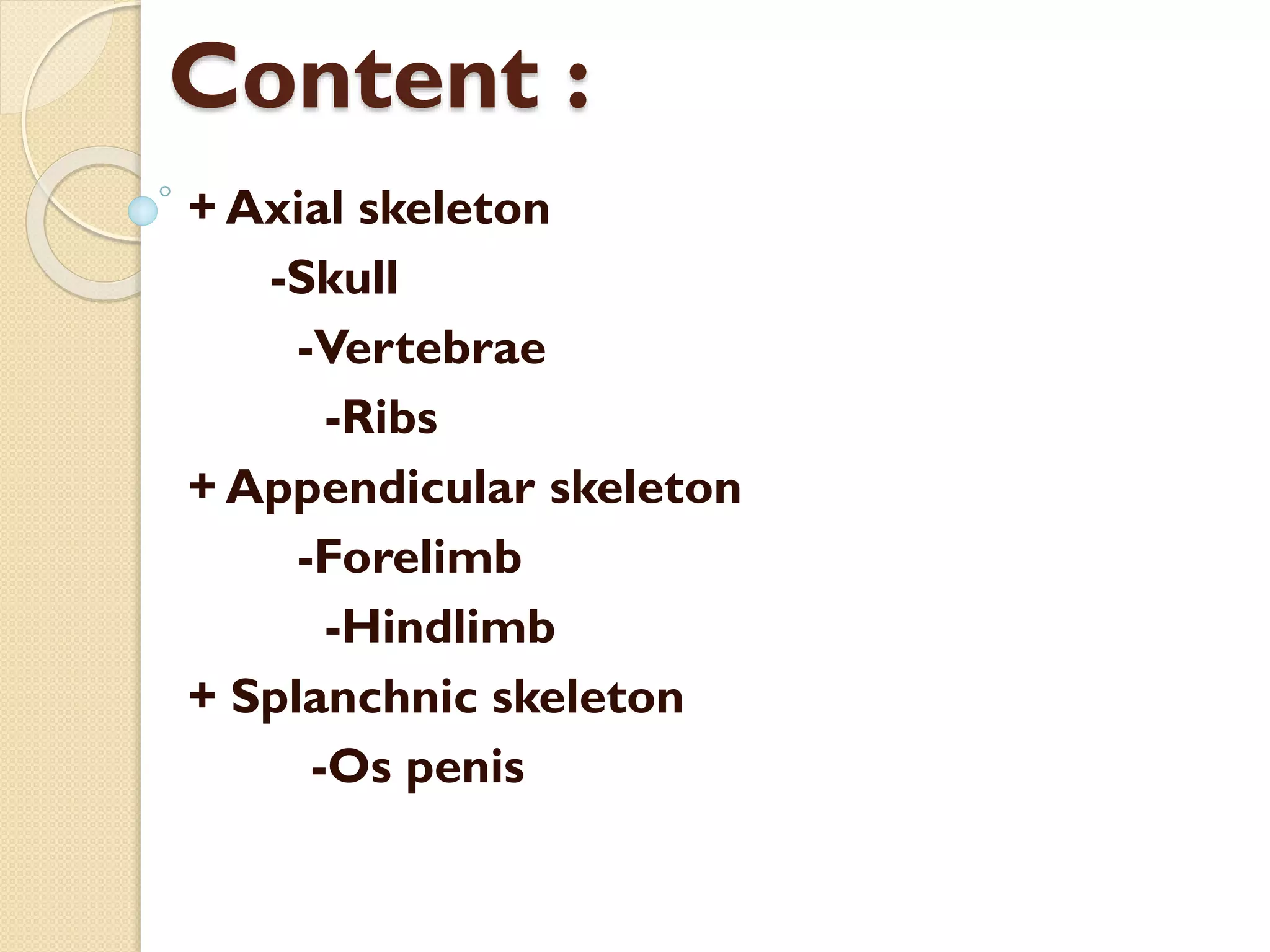
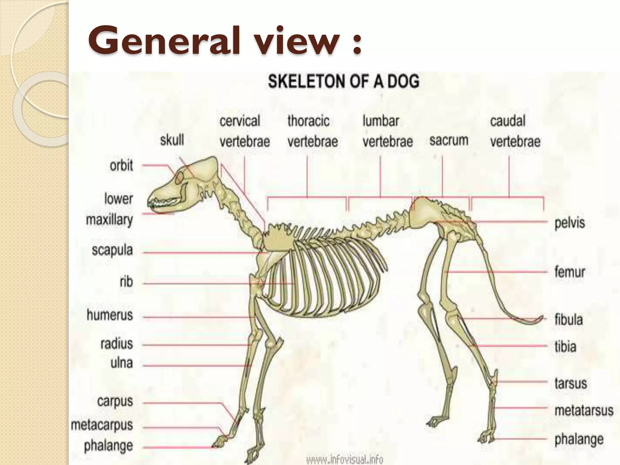
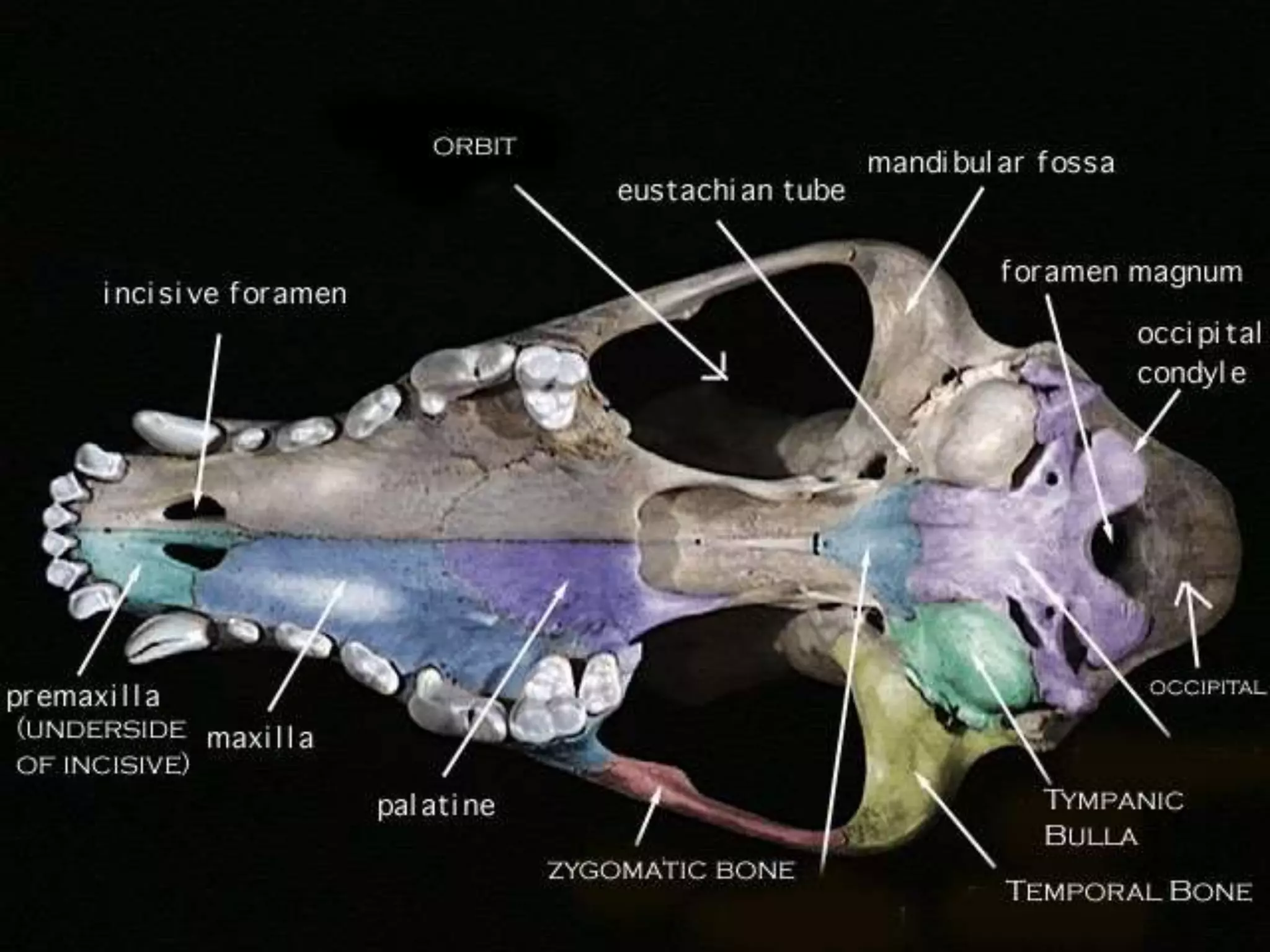
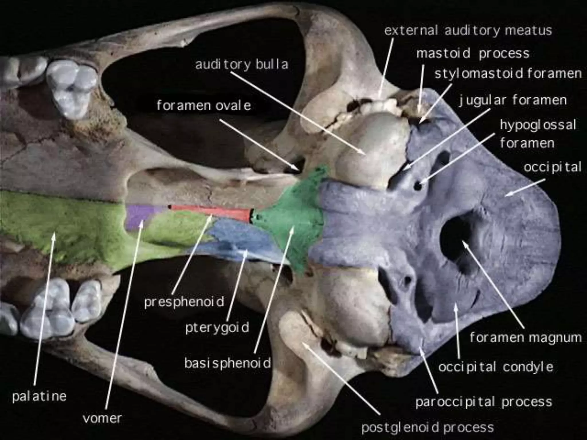
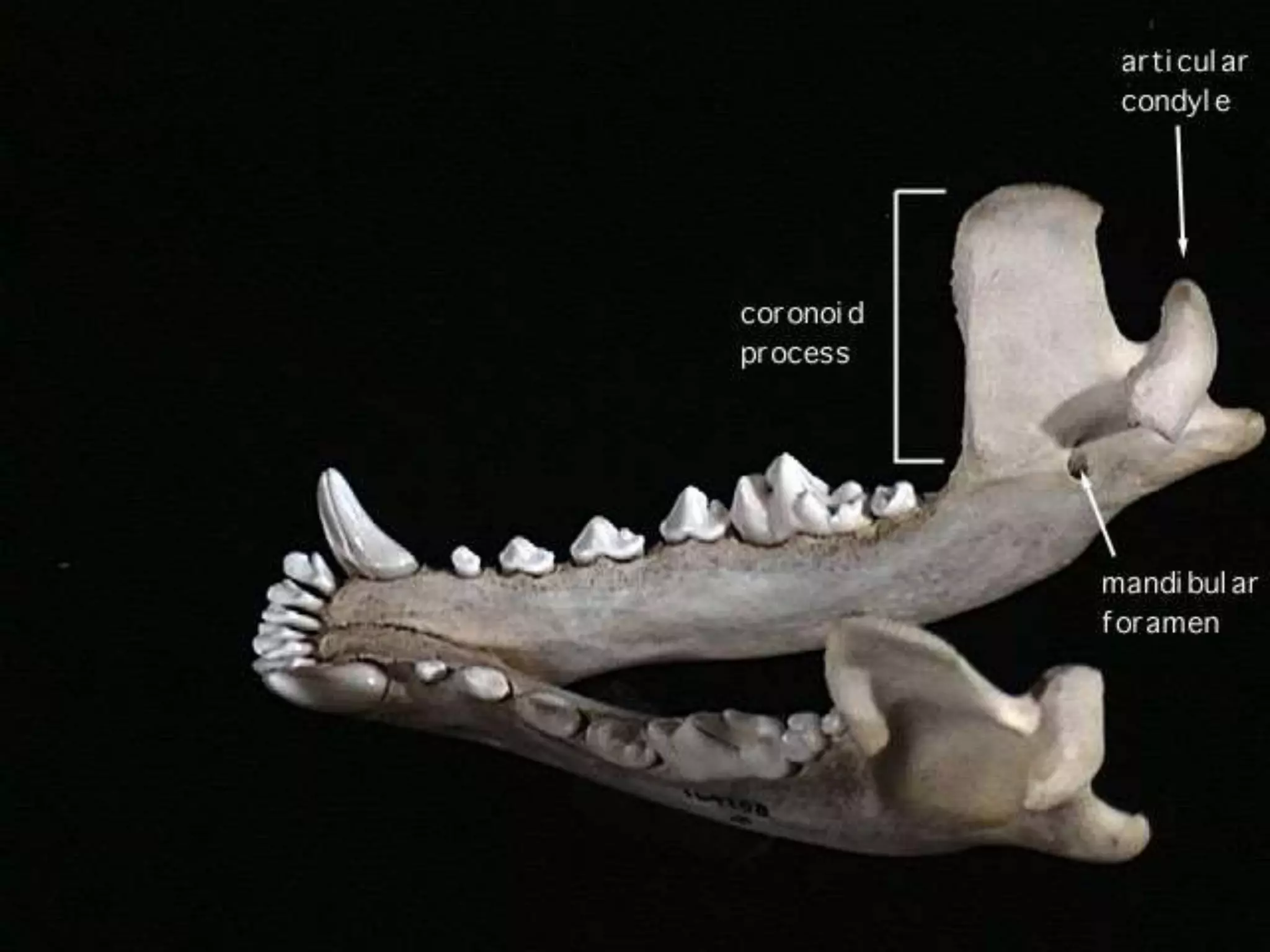
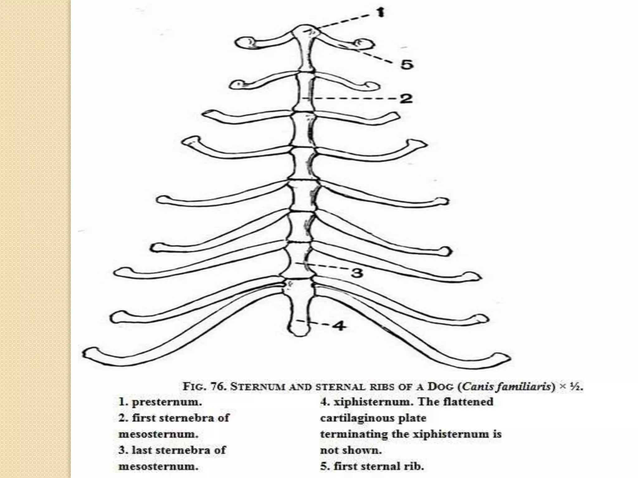
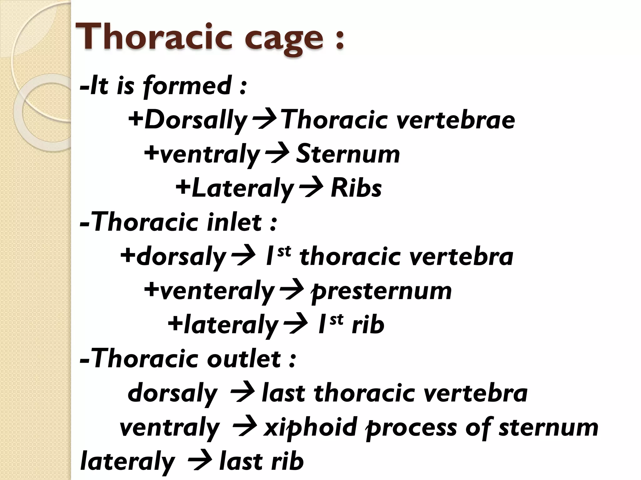
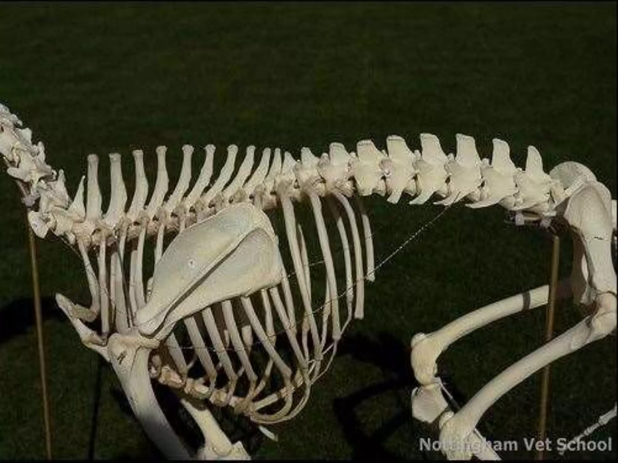
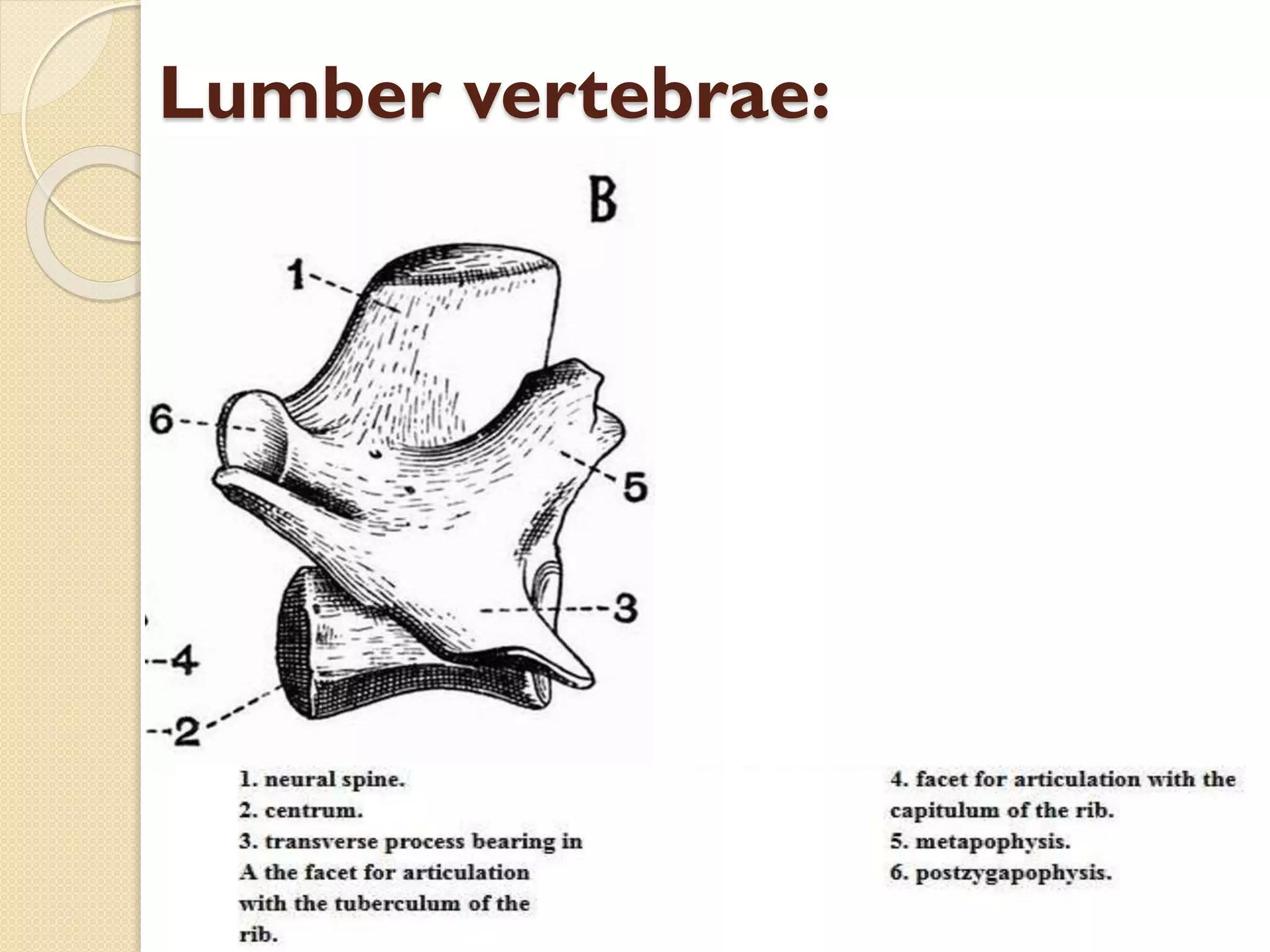
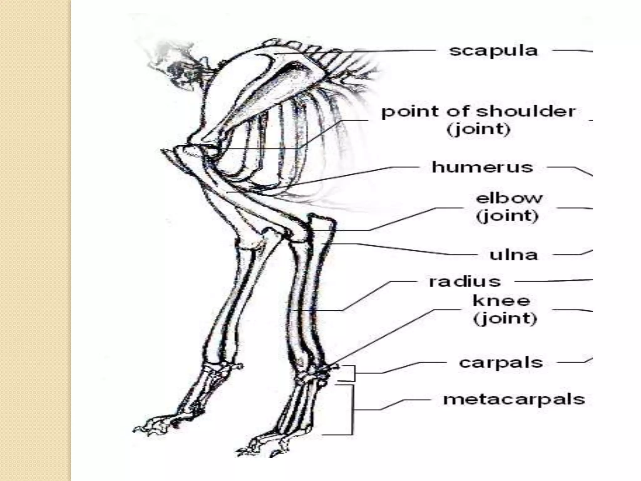
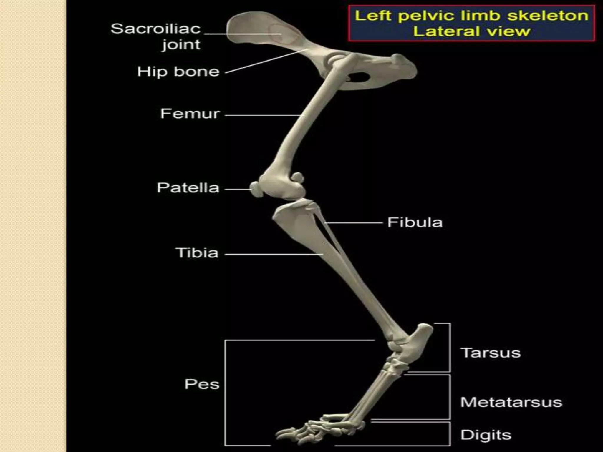
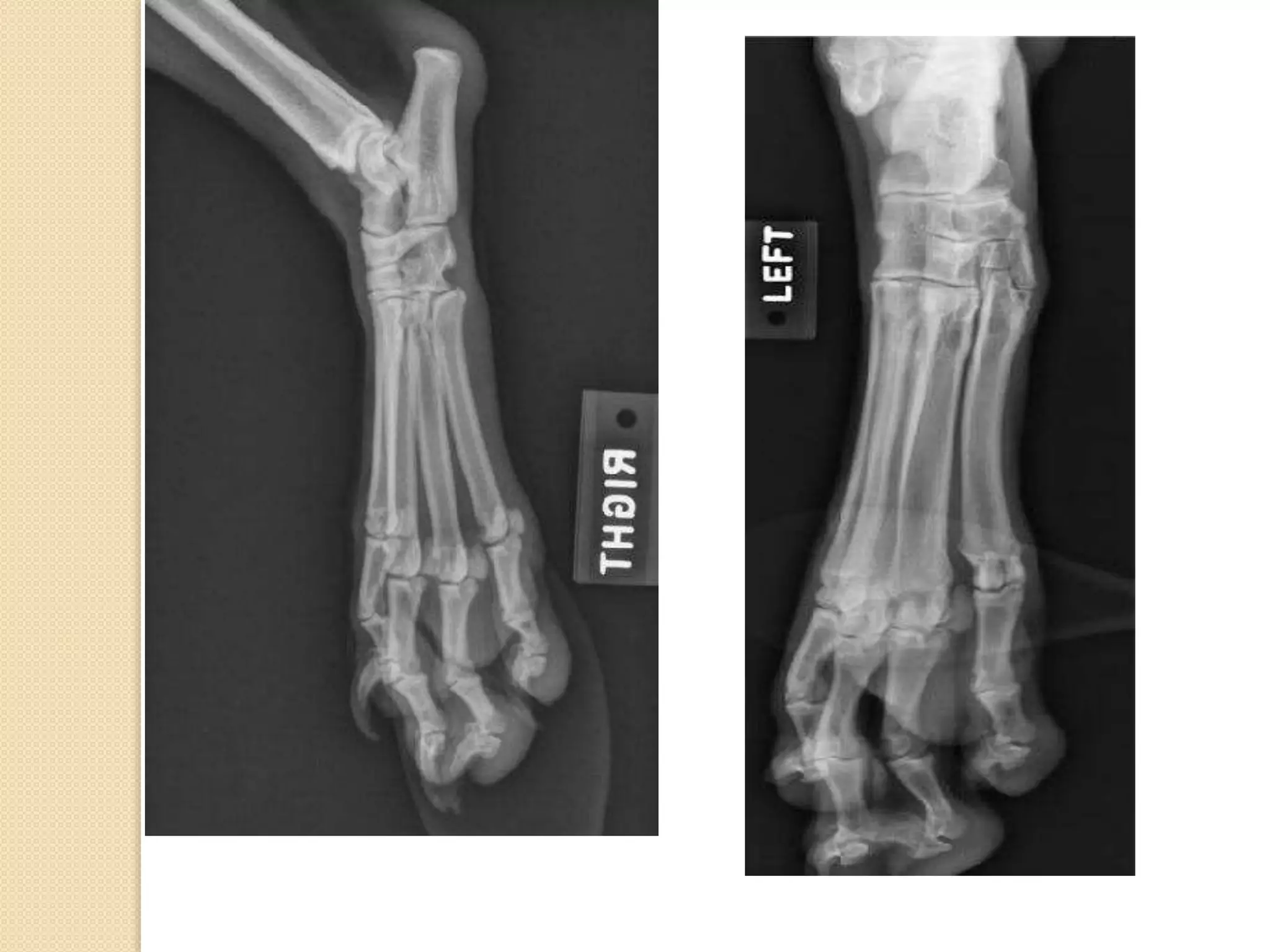
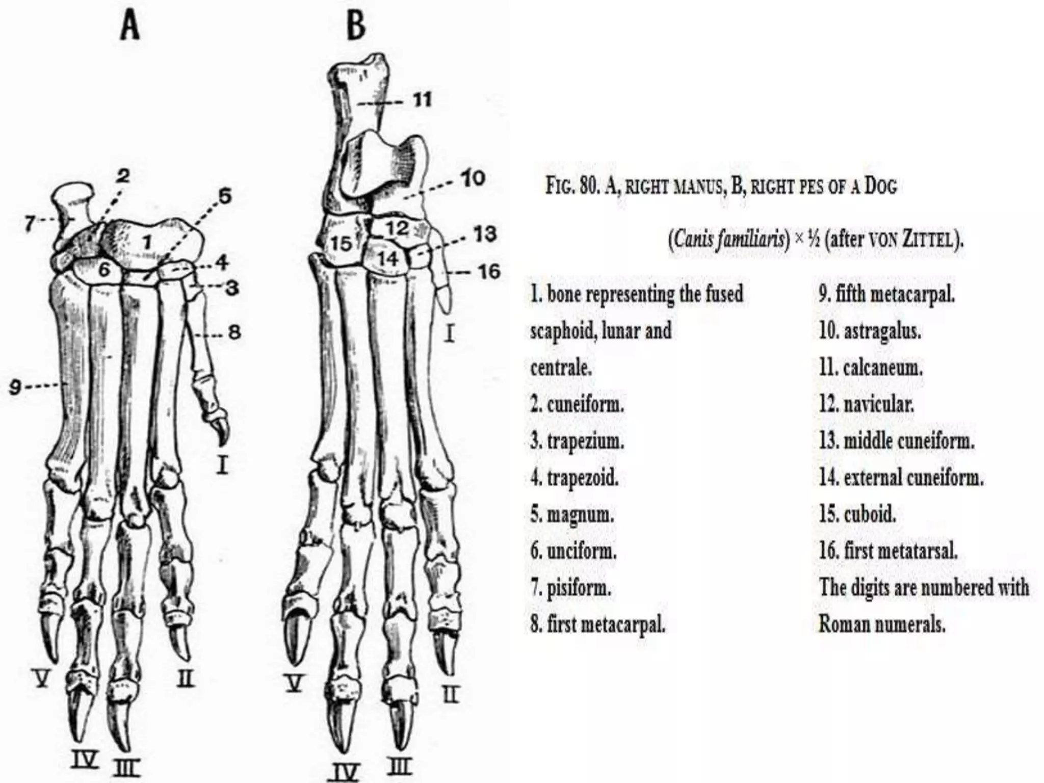
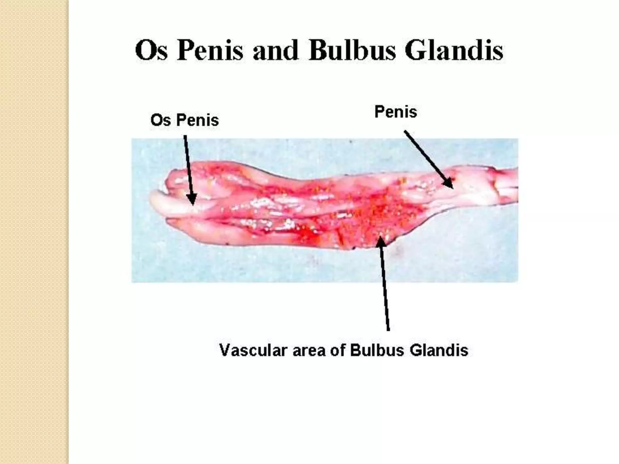
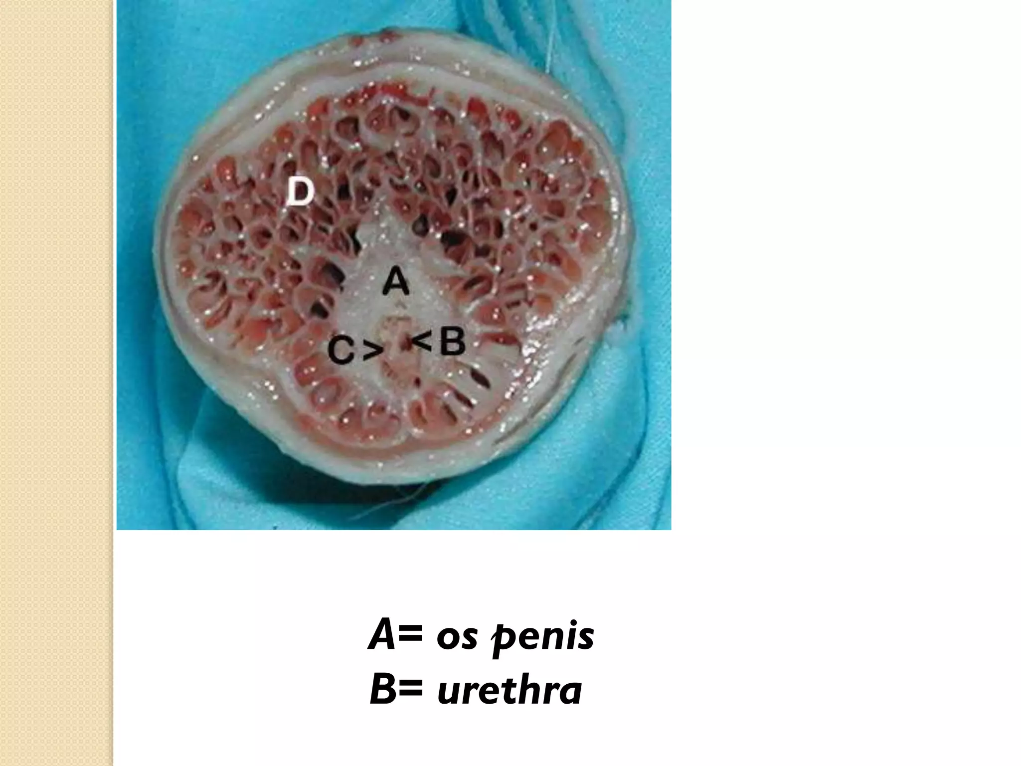
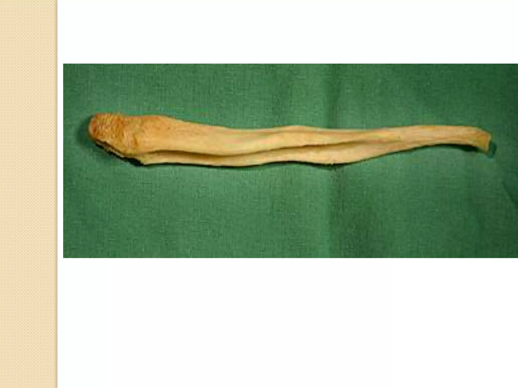
Content :
+ Axial skeleton
-Skull
-Vertebrae
-Ribs
+ Appendicular skeleton
-Forelimb
-Hindlimb
+ Splanchnic skeleton
-Os penis
General view :
Skull :
-The skull consist of 3 bones combined with
each other to form the skull.
-The skull consist of cranium , mandible and
hyoid bone.
-The cranium consist of many bones which
combine with each other to form the cranium.
-Those bones are :
Frontal bone-nasal bone-maxillary bone-occipital bone
sphenoid bone-temporal bone-parietal bone
Lacrimal bone-zygomatic bone-incisive bone
Palatine bone-vomer bone-pterygoid bone
Ethmoid bone
-The facial crest and facial tuberosity are absent
-Only the frontal paranasal sinus is observed
and divided into cranial –middle and caudal
part.

-The maxillary sinus is small and open directly
to the nasal cavity and called maxillary recess.
Mandible:
-The mandible consist of body and 2
ramus.
-The 2 halves of the mandible is not
completely fused with each other by the
mandibular symphysis.
-The masseteric fossa is very deep for
the insertion of the masseter muscle.
Skull radiology:
Tongue

Hyoid bone
Vertebrae:
-The vertebrae is a group of bones which
form the vertebral column in which the spinal
cord is found.
-It is divided into :
Cervical(C)
thoracic(T),lumber(L),sacrum(S),and
caudal(Cd)
-The vertebrae formula is :
C=7
T=13
L=7
S=3
Cd=20-23
Cervical vertebrae:

-1st vertebra is called atlas.
-2nd vertebra is called axis
-The last vertebra has articulation surface for 1st rib.
Atlas

Axis
Radiology:
Thoracic vertebrae:

-It is characterised by long spinous process
-The last 3 vertebrae have accessory
processes
-It has articular surface for the rib
articulation
A.Vertebral canal; B.Transverse process;
C. Dorsal spinous process;
D.Vertebral
arch; E. Body;
F. Cranial articular process; H. Cranial costal
fovea.
Ribs:
Each rib is a curved rod, which at its dorsal end is movably
articulated to the vertebra, and at its ventral end is either
connected with the sternum, or ends freely. In the dog there are
thirteen pairs of ribs, nine pairs of which are directly connected
with the sternum, while the remaining four end freely and are
known as floating ribs. Each rib is obviously divided into two
parts, a dorsal or vertebral part, and a ventral or sternal
part. The vertebral portion, which forms about two-thirds of
the whole rib, is a flattened, regularly curved rod, completely
ossified. Its dorsal end is rounded, forming the head or
capitulum, which articulates with a concave surface furnished
partly by the corresponding vertebra and partly by the vertebra
next in front. The last three or four however articulate with one
vertebra only
J. Head;

K.Tuberculum.
Articulation between ribs and thoracic
vertebrae
Sternum:
This is an elongated cylindrical structure lying in the mid-ventral
wall of the thorax, and is divided into eight segments or
sternebrae. The anterior segment, the presternum (fig. 76, 1)
or manubrium sterni is expanded in front; the next six
segments, which, together form the mesosternum are
elongated, somewhat contracted in the middle and expanded at
the ends. The last segment or xiphisternum (fig. 76, 4) is long
and narrow, and terminates in a flattened expanded plate of
cartilage. The first pair of sternal ribs articulate with the sides of
the presternum, and the remaining pairs between the successive
sternebrae. Between the last sternebra and the xiphisternum
two pairs articulate. Development shows that the sternum is
formed by the union in the middle line of two lateral portions;
this can be well seen in the presternum and xiphisternum of the
puppy, but no traces of this median division remain in the adult
dog.
Thoracic cage :
-It is formed :
+Dorsally Thoracic vertebrae
+ventraly Sternum
+Lateraly Ribs
-Thoracic inlet :
+dorsaly 1st thoracic vertebra
+venteraly presternum
+lateraly 1st rib
-Thoracic outlet :
dorsaly  last thoracic vertebra
ventraly  xiphoid process of sternum
lateraly  last rib
Lumber vertebrae:
B.Transverse process; C. Dorsal
spinous process;
E. Body; F. Cranial articular
process;
G. Caudal articular process.

A.Vertebral canal; B.Transverse process;
C. Dorsal spinous process;
D.Vertebral arch;
E. Body; F. Cranial articular process.
Sacral and caudal vertebrae:

sacrum (A)

coccygeal or caudal vertebrae (B).
From this dorsal view, notice the attachment of the wings of the sacrum
(C) to the ilium (D).
E is the pelvic symphysis
Forelimb:
-Forelimb bones are completely
separated from the axial skeleton due
to the absence of clavicle bone in dog;
while communication occur by
muscules.
-Forelimb bones are:
scapula-humerus-radius and ulna-carpusmetacarpus and phalanges.
Scapula:
-Scapula is a flat bone which form
the shoulder girdle.
-The ratio between supraspinous
fossa and infraspinous fossa is 1:1.

-The scapular spine end by wide
trunkated acromion and the
tubular spinae is absent.
The articular surface, the glenoid cavity (E) is at the distal end of the bone, and the
.supraspinous fossa (C) is cranial to the spinous process (A), or spine of the scapula
The distal end of the spinous process (A) of the dog has an acromium process (B) .
When we dissect the thoracic limb, we will find that the supraspinatous muscle sits in the
supraspinous fossa (C) and that the infraspinatous muscle sits in the infraspinous fossa (D).
Note that the supraspinatous is above (supra) the spine and the infraspinatous fossa is
below (infra) the spine.
This second image shows the medial aspect eht fo left scapula dionelgarpus eht taht ecitoN .
.(E) ytivac dionelg eht fo tcepsa lainarc eht (arpus) evoba si (F) elcrebut
The small process on the supraglenoid tubercle is the corocoid process (G).
The smooth surface corresponds to where the subscapularis muscle lies, and it is known as
the subscapular fossa (H) (don't worry about these muscle names for now). The roughened
surface is the serrated face (I), and it is where the serratus ventralis muscle attaches
Humerus:
-It is a long bone which is called also the brachium-The head of the humerus with the glenoid cavity
of the scapula form the shoulder joint.

-The presence of supratrochlear foramen
D=supratrochlear foramen
Shoulder joint:
Radius and ulna:
In case of dog the radius and ulna is separated
from each other like human , while in farm
domestic animals ( horse, cattle, sheep, goat,
camel ) the radius and ulna is fused.
B=Radius C=Ulna
Bone A is the radius and bone
B is the ulna. Image 1 is the
cranial view, and because the
olecranon (E) curves medially
and the styloid process (F) of
the ulna is found caudal and
lateral to the distal end of the
radius, we can identify these
bones as the left radius and
ulna. Image 2 is then a view of
the lateral aspect of these
bones.
The proximal extremity of the
radius is the head (C), and the
distal extremity is the trochlea
(D).
The ulna consists of the
olecranon (E) proximally and
the styloid process (F) distally.
Notice that the styloid process
resembles a stylus pen.
The trochlea of the humerus sits
within the trochlear notch
(G).
Elbow joint:
Carpus , metacarpus and
phalanges:
Carpus is a group of small bone which form the
wrist joint , it is arranged in rows which differ
from species to another .

No. of carpal bone : 7
No. of bones in the proximal row : 4
No. of bones in the distal row : 4
Metacarpal bones is a group of long bones
which form the palm in human hand, the number
of these bones differ from species to another.

No. of metacarpal bones : 5
The first one is the shortest while the III and IV is
the longest .
Phalanges is a group of 3 bones which form the
fingers ( except in the first finger 2 bones ) , they
are called 1st , 2nd ,3rd .

The 3rd phalange and the distal part of the 2nd
phalange is coverd by claws.
Hind limb:
The hind limb which is the lower limb in human is directly connected to the axial
skeleton through the joint between os coxae
and the sacrum
It consist of :
Os coxae – femur – patella – tibia – fibula – tarsus
– metatarsus –and digits
Os coxae :
It is the first bone in the hind limb which form the pelvis
The right and left bone fused with each other by the pelvic symphysis
It consist of 3 bones ilium- ischium and
pubis
The gluteal surface is concave ( spoon like)
Femur :

It is a long bone which form the thigh
region.
The head of the femur form the hip joint
with the acetabulum of the os coxae .
The caudal surface characterized by 2
rough lines diverge proximally and distally
towards the extremities called linea

Aspera
The present of the supracondyloid facets.
Hip joint:
Hip joint is formed of the head of the femur
with the acetabulum of os coxae.
Fibula , tibia and patella :
B= tibia
C= fibula
D = Patella
The fibula (B) is lateral to
the tibia (A), and the tibial
tuberocity (C) and tibial
crest (D) are on the cranial
aspect of the tibia. This
means that image one is a
cranial view and image two is
a lateral view of the left
tibia and fibula.
The tibial tuberosity is on
the proximal aspect of the
tibia
Stifle joint :
Stifle joint is a compound joint which is
formed of 3 bones (distal extremity of femur
– proximal extremity of tibia – patella)
Tarsus , metatarsus and
phalanges :

Tarsus
The cluster of bones are the tarsal bones (A), which
make up the tarsus, or hock. The tibial-tarsal joint is
the same as your ankle.
The calcaneus (B) is the large tarsal bone that makes
up the heel of man. Unfortunately, I could not get a
good view to show that the calcaneus is in the lateral
portion of the hock.
The metatarsal bones are labelled C.
Remember that at the tarsus, cranial becomes dorsal
(E) and caudal becomes plantar (F), just like when
you plant your foot on the ground.
The phalanges (D) are similar to those in the
thoracic limb.
Os penis (baculum) :
Os penis is a bone which is found in the
penis of the dog which help to prevent the
closure of the urethra during intercourse
due to the contraction of the vaginal
muscles of the bitch .
A= os penis
B= urethra
short notes in dog skeleton

short notes in dog skeleton

  • 2.
    Content : + Axialskeleton -Skull -Vertebrae -Ribs + Appendicular skeleton -Forelimb -Hindlimb + Splanchnic skeleton -Os penis
  • 3.
  • 5.
    Skull : -The skullconsist of 3 bones combined with each other to form the skull. -The skull consist of cranium , mandible and hyoid bone. -The cranium consist of many bones which combine with each other to form the cranium. -Those bones are : Frontal bone-nasal bone-maxillary bone-occipital bone sphenoid bone-temporal bone-parietal bone Lacrimal bone-zygomatic bone-incisive bone Palatine bone-vomer bone-pterygoid bone Ethmoid bone
  • 7.
    -The facial crestand facial tuberosity are absent
  • 11.
    -Only the frontalparanasal sinus is observed and divided into cranial –middle and caudal part. -The maxillary sinus is small and open directly to the nasal cavity and called maxillary recess.
  • 12.
    Mandible: -The mandible consistof body and 2 ramus. -The 2 halves of the mandible is not completely fused with each other by the mandibular symphysis. -The masseteric fossa is very deep for the insertion of the masseter muscle.
  • 15.
  • 17.
    Vertebrae: -The vertebrae isa group of bones which form the vertebral column in which the spinal cord is found. -It is divided into : Cervical(C) thoracic(T),lumber(L),sacrum(S),and caudal(Cd) -The vertebrae formula is : C=7 T=13 L=7 S=3 Cd=20-23
  • 18.
    Cervical vertebrae: -1st vertebrais called atlas. -2nd vertebra is called axis -The last vertebra has articulation surface for 1st rib.
  • 19.
  • 21.
  • 22.
    Thoracic vertebrae: -It ischaracterised by long spinous process -The last 3 vertebrae have accessory processes -It has articular surface for the rib articulation
  • 24.
    A.Vertebral canal; B.Transverseprocess; C. Dorsal spinous process; D.Vertebral arch; E. Body; F. Cranial articular process; H. Cranial costal fovea.
  • 27.
    Ribs: Each rib isa curved rod, which at its dorsal end is movably articulated to the vertebra, and at its ventral end is either connected with the sternum, or ends freely. In the dog there are thirteen pairs of ribs, nine pairs of which are directly connected with the sternum, while the remaining four end freely and are known as floating ribs. Each rib is obviously divided into two parts, a dorsal or vertebral part, and a ventral or sternal part. The vertebral portion, which forms about two-thirds of the whole rib, is a flattened, regularly curved rod, completely ossified. Its dorsal end is rounded, forming the head or capitulum, which articulates with a concave surface furnished partly by the corresponding vertebra and partly by the vertebra next in front. The last three or four however articulate with one vertebra only
  • 28.
  • 29.
    Articulation between ribsand thoracic vertebrae
  • 30.
    Sternum: This is anelongated cylindrical structure lying in the mid-ventral wall of the thorax, and is divided into eight segments or sternebrae. The anterior segment, the presternum (fig. 76, 1) or manubrium sterni is expanded in front; the next six segments, which, together form the mesosternum are elongated, somewhat contracted in the middle and expanded at the ends. The last segment or xiphisternum (fig. 76, 4) is long and narrow, and terminates in a flattened expanded plate of cartilage. The first pair of sternal ribs articulate with the sides of the presternum, and the remaining pairs between the successive sternebrae. Between the last sternebra and the xiphisternum two pairs articulate. Development shows that the sternum is formed by the union in the middle line of two lateral portions; this can be well seen in the presternum and xiphisternum of the puppy, but no traces of this median division remain in the adult dog.
  • 32.
    Thoracic cage : -Itis formed : +Dorsally Thoracic vertebrae +ventraly Sternum +Lateraly Ribs -Thoracic inlet : +dorsaly 1st thoracic vertebra +venteraly presternum +lateraly 1st rib -Thoracic outlet : dorsaly  last thoracic vertebra ventraly  xiphoid process of sternum lateraly  last rib
  • 34.
  • 35.
    B.Transverse process; C.Dorsal spinous process; E. Body; F. Cranial articular process; G. Caudal articular process. A.Vertebral canal; B.Transverse process; C. Dorsal spinous process; D.Vertebral arch; E. Body; F. Cranial articular process.
  • 36.
    Sacral and caudalvertebrae: sacrum (A) coccygeal or caudal vertebrae (B).
  • 37.
    From this dorsalview, notice the attachment of the wings of the sacrum (C) to the ilium (D). E is the pelvic symphysis
  • 40.
    Forelimb: -Forelimb bones arecompletely separated from the axial skeleton due to the absence of clavicle bone in dog; while communication occur by muscules. -Forelimb bones are: scapula-humerus-radius and ulna-carpusmetacarpus and phalanges.
  • 42.
    Scapula: -Scapula is aflat bone which form the shoulder girdle. -The ratio between supraspinous fossa and infraspinous fossa is 1:1. -The scapular spine end by wide trunkated acromion and the tubular spinae is absent.
  • 44.
    The articular surface,the glenoid cavity (E) is at the distal end of the bone, and the .supraspinous fossa (C) is cranial to the spinous process (A), or spine of the scapula The distal end of the spinous process (A) of the dog has an acromium process (B) . When we dissect the thoracic limb, we will find that the supraspinatous muscle sits in the supraspinous fossa (C) and that the infraspinatous muscle sits in the infraspinous fossa (D). Note that the supraspinatous is above (supra) the spine and the infraspinatous fossa is below (infra) the spine. This second image shows the medial aspect eht fo left scapula dionelgarpus eht taht ecitoN . .(E) ytivac dionelg eht fo tcepsa lainarc eht (arpus) evoba si (F) elcrebut The small process on the supraglenoid tubercle is the corocoid process (G). The smooth surface corresponds to where the subscapularis muscle lies, and it is known as the subscapular fossa (H) (don't worry about these muscle names for now). The roughened surface is the serrated face (I), and it is where the serratus ventralis muscle attaches
  • 45.
    Humerus: -It is along bone which is called also the brachium-The head of the humerus with the glenoid cavity of the scapula form the shoulder joint. -The presence of supratrochlear foramen
  • 47.
  • 49.
  • 52.
    Radius and ulna: Incase of dog the radius and ulna is separated from each other like human , while in farm domestic animals ( horse, cattle, sheep, goat, camel ) the radius and ulna is fused.
  • 53.
  • 54.
    Bone A isthe radius and bone B is the ulna. Image 1 is the cranial view, and because the olecranon (E) curves medially and the styloid process (F) of the ulna is found caudal and lateral to the distal end of the radius, we can identify these bones as the left radius and ulna. Image 2 is then a view of the lateral aspect of these bones. The proximal extremity of the radius is the head (C), and the distal extremity is the trochlea (D). The ulna consists of the olecranon (E) proximally and the styloid process (F) distally. Notice that the styloid process resembles a stylus pen. The trochlea of the humerus sits within the trochlear notch (G).
  • 56.
  • 58.
    Carpus , metacarpusand phalanges: Carpus is a group of small bone which form the wrist joint , it is arranged in rows which differ from species to another . No. of carpal bone : 7 No. of bones in the proximal row : 4 No. of bones in the distal row : 4
  • 61.
    Metacarpal bones isa group of long bones which form the palm in human hand, the number of these bones differ from species to another. No. of metacarpal bones : 5 The first one is the shortest while the III and IV is the longest .
  • 62.
    Phalanges is agroup of 3 bones which form the fingers ( except in the first finger 2 bones ) , they are called 1st , 2nd ,3rd . The 3rd phalange and the distal part of the 2nd phalange is coverd by claws.
  • 65.
    Hind limb: The hindlimb which is the lower limb in human is directly connected to the axial skeleton through the joint between os coxae and the sacrum It consist of : Os coxae – femur – patella – tibia – fibula – tarsus – metatarsus –and digits
  • 67.
    Os coxae : Itis the first bone in the hind limb which form the pelvis The right and left bone fused with each other by the pelvic symphysis It consist of 3 bones ilium- ischium and pubis The gluteal surface is concave ( spoon like)
  • 71.
    Femur : It isa long bone which form the thigh region. The head of the femur form the hip joint with the acetabulum of the os coxae . The caudal surface characterized by 2 rough lines diverge proximally and distally towards the extremities called linea Aspera The present of the supracondyloid facets.
  • 75.
    Hip joint: Hip jointis formed of the head of the femur with the acetabulum of os coxae.
  • 76.
    Fibula , tibiaand patella : B= tibia C= fibula D = Patella
  • 77.
    The fibula (B)is lateral to the tibia (A), and the tibial tuberocity (C) and tibial crest (D) are on the cranial aspect of the tibia. This means that image one is a cranial view and image two is a lateral view of the left tibia and fibula. The tibial tuberosity is on the proximal aspect of the tibia
  • 78.
    Stifle joint : Stiflejoint is a compound joint which is formed of 3 bones (distal extremity of femur – proximal extremity of tibia – patella)
  • 80.
    Tarsus , metatarsusand phalanges : Tarsus
  • 83.
    The cluster ofbones are the tarsal bones (A), which make up the tarsus, or hock. The tibial-tarsal joint is the same as your ankle. The calcaneus (B) is the large tarsal bone that makes up the heel of man. Unfortunately, I could not get a good view to show that the calcaneus is in the lateral portion of the hock. The metatarsal bones are labelled C. Remember that at the tarsus, cranial becomes dorsal (E) and caudal becomes plantar (F), just like when you plant your foot on the ground. The phalanges (D) are similar to those in the thoracic limb.
  • 87.
    Os penis (baculum): Os penis is a bone which is found in the penis of the dog which help to prevent the closure of the urethra during intercourse due to the contraction of the vaginal muscles of the bitch .
  • 89.