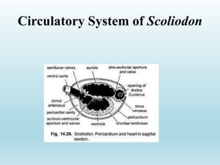
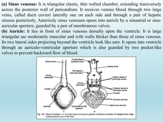
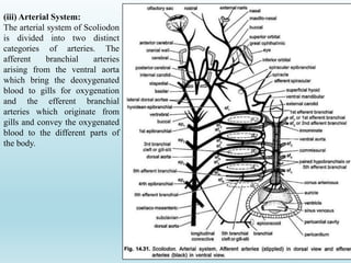
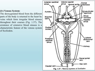
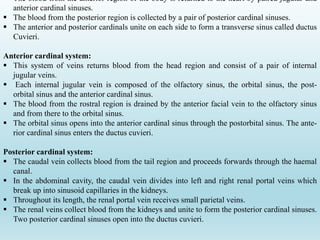
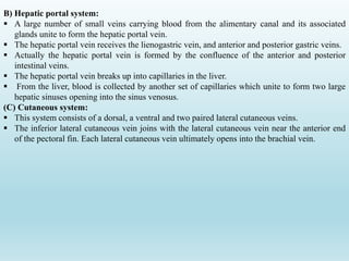
SlideShare a Scribd company logo
1 of 15
Dr. Arun Gaware
Dept. of Zoology,
Shri. Shivaji College Motala, Dist.
Buldana.
Circulatory System of Scoliodon
Circulatory system of scoliodon
The circulatory system of scoliodon consists of the circulatory fluid called blood, the heart, the
arteries and the veins.
(i) Blood:
•The blood consists of a colourless plasma and corpuscles suspended in the plasma.
• Two kinds of corpuscles are encountered the RBC (or erythrocytes) and the WBC (or
leucocytes).
•The erythrocytes are oval bodies containing a nucleus.
•Haemoglobin is present in the erythrocytes. The leucocytes are amoeboid in structure.
ii) Heart:
•It is situated mid-ventrally in head beneath the pharynx supported below by the coracoid
cartilages of the pectoral girdle.
• It lies within the pericardial cavity of two layered membranous sacs the pericardium.
•Inner layer is visral pericardium and outer layer is parietal pericardium. Between the two layers
is the pericardial cavity which is filled with paricardial fluid.
•Heart is a reddish-brown, muscular and dorso-ventrally bent, “S” shaped tube differentiated into
a series of 4 chambers; sinus venosus, auricle, ventricle and conus arteriosus.
•But only two auricles and ventricle are considered true chambers, so that heart is only two
chambers.
(a) Sinus venosus: It is triangular elastic, thin walled chamber, extending transversely
across the posterior wall of pericardium. It receives venous blood through two large
veins, called ducti cuvieri laterally one on each side and through a pair of hepatic
sinuses posteriorly. Anteriroly sinus venosum opens into auricle by a sinuatrial or sinu-
auricular aperture, guarded by a pair of membranous valves.
(b) Auricle: It lies in front of sinus venosus dorsally upon the ventricle. It is large
triangular sac moderately muscular and with walls thicker than those of sinus venosus.
Its two lateral sides projecting beyond the ventricle look like ears. It opens into ventricle
through an auriculo-ventricular aperture which is also guarded by two pocket-like
valves to prevent backward flow of blood.
(c) Ventricle: It is the most prominent and pear-shaped chamber of heart. Relatively small in
size, it has very thick muscular walls to pump the blood into all parts of the body. Chordae
tendinae are present to be attached to opposite walls to prevent ventricle from expanding beyond
its capacity.
(d) Conus arteriosus: Ventricle tapers anteriorly into a stout and muscular tube, the conus
arteriosus. Its cavity contains two transverse rows of semilunar valves to block the regurgitation
or backward flow of blood.
Each row has three valves, one dorsal and two ventro-laterals in position. Anterior valves are
larger than the posterior ones. Anteriorly conus arteriosus leave the pericardium and extends
further in the form of ventral aorta.
Working of the heart:
•The heart of Scoliodon is a venous heart containing only deoxygenated or venous blood.
•In a complete circuit of body, the blood passes through heart only once (single circulation).
Heart pumps its venous blood into the gills for aeration.
•To achieve this different parts of the heart rhythmically contract at regular intervals and in a
definite succession, first sinus venosus, then auricle, then ventricle and finally the conus.
•Each contraction called systole is followed by a relaxation called diastole.
• Different valves of the heart serve to prevent the backward flow of blood into preceding
chambers.
•The walls of heart are supplied with oxygenated blood through special coronary arteries.
(iii) Arterial System:
The arterial system of Scoliodon
is divided into two distinct
categories of arteries. The
afferent branchial arteries
arising from the ventral aorta
which bring the deoxygenated
blood to gills for oxygenation
and the efferent branchial
arteries which originate from
gills and convey the oxygenated
blood to the different parts of
the body.
(a) Afferent branchial arteries:
 The ventral aorta is situated on the ventral surface of the pharynx and extends up to the
posterior border of the hyoid arch.
 The ventral aorta divides into two branches called innominate arteries, which again bifurcates
into the first and second afferent branchial arteries.
 The third, fourth and fifth afferent arteries arise from the ventral aorta.
 Each afferent branchial artery arises from the ventral aorta by independent opening except the
anterior- most pairs which arise by a common opening.
(b) Efferent branchial arteries:
 The afferent branchial arteries break up into capillaries in the gills. From the gills the blood is
collected by efferent branchial arteries.
 There are nine pairs of efferent branchial arteries and these are equally distributed on each
side.
 The first eight arteries form a series of four complete loops around the first four gill slits.
 The ninth efferent branchial artery collects blood from the demi branch of the fifth gill pouch.
 In addition to short longitudinal connectives connecting the four loops, these are further
connected with each other by a network of longitudinal commissural vessels called the lateral
hypobranchial chain.
 From each efferent branchial loop arises an epibranchial artery. The four pairs of epibranchials
join in the mid-dorsal line to form the dorsal aorta.
Anterior arteries:
The head region gets the blood supply from the first efferent branchial artery and partly from the
proximal end of the dorsal aorta.
Arteries from the first efferent branchial (hyoidean efferent) are:
(a) The external carotid,
(b) The afferent spiracular, and
(c) The hyodean epibranchial which in turn receives a branch from dorsal aorta.
The external carotid artery originates from the first collector loop and divides into a ventral
mandibular artery giving branches to the muscles of the lower jaw and a superficial hyoid artery
which supplies the second ventral constrictor muscle, the skin and the subcutaneous tissue
beneath the hyoid arch.
Dorsal aorta and its branches:
The dorsal aorta is formed by the union of epibranchial arteries. It runs posteriorly and is situated
ventral to the vertebral column. It is continued up to the tip of the tail as the caudal artery.
Along the anteroposterior direction the following arteries have their origin from the dorsal
aorta:
1. A pair of small subclavian arteries arise from near the origin of the fourth epibranchial arteries.
The subclavian artery gets the epicoracoid artery on its way and divides into:
(i) A branchial artery to the pectoral girdle and pectoral fin,
(ii) An anterolateral artery to the body musculature and
(iii) A dorsolateral artery to the dorsal musculature.
2. A large coeliacomesenteric artery arises just behind the origin of fourth epibranchial artery. It
divides into a smaller coeliac artery and a larger anterior mesenteric artery.
3. A lienogastric artery originates posterior to the coeliacomesenteric artery and gives off:
(i) An ovarian (in females) or spermatic artery (in males),
(ii) A posterior intestinal artery,
(iii) A posterior gastric to the posterior part of the cardiac stomach and
(iv) A splenic artery to the spleen.
4. Series of paired parietal arteries emerge out behind the subclavian artery. Each parietal gives a
dorsal parietal artery and a ventral parietal artery.
The dorsal parietal artery supplies to the dorsolateral musculature, the vertebral column, the
spinal cord and the dorsal fin. The ventral parietal artery supplies to the ventral muscles and the
peritoneum. The ventral parietal gives renal branches to the kidneys.
6. A pair of iliac arteries extend to the pelvic fin as femoral arteries.
Hypobranchial chain:
•A lateral hypobranchial chain is formed by a network of slender arteries arising from the ventral
ends of the loop of the efferent branchial arteries.
•Four commissural vessels arise from the lateral hypobranchial chain which on the ventral wall of
the ventral aorta unites to form a pair of median hypo-branchials which communicate with one
another by transverse vessels.
•Posteriorly the median hypo-branchials unite to form a median coracoid artery which gives rise
to the coronary artery and a pericardial artery.
•The pericardial artery gives off the common epicoracoid artery which in turn divides into left
and right epicoracoid arteries each joining one subclavian artery.
(iv) Venous System:
The deoxygenated blood from the different
parts of the body is returned to the heart by
veins which form irregular blood sinuses
throughout their courses (Fig. 1.37). The
existence of extensive blood sinuses is a
characteristic feature of the venous system
of Scoliodon.
 The blood from the anterior region of the body is returned to the heart by paired jugular and
anterior cardinal sinuses.
 The blood from the posterior region is collected by a pair of posterior cardinal sinuses.
 The anterior and posterior cardinals unite on each side to form a transverse sinus called ductus
Cuvieri.
Anterior cardinal system:
 This system of veins returns blood from the head region and consist of a pair of internal
jugular veins.
 Each internal jugular vein is composed of the olfactory sinus, the orbital sinus, the post-
orbital sinus and the anterior cardinal sinus.
 The blood from the rostral region is drained by the anterior facial vein to the olfactory sinus
and from there to the orbital sinus.
 The orbital sinus opens into the anterior cardinal sinus through the postorbital sinus. The ante-
rior cardinal sinus enters the ductus cuvieri.
Posterior cardinal system:
 The caudal vein collects blood from the tail region and proceeds forwards through the haemal
canal.
 In the abdominal cavity, the caudal vein divides into left and right renal portal veins which
break up into sinusoid capillaries in the kidneys.
 Throughout its length, the renal portal vein receives small parietal veins.
 The renal veins collect blood from the kidneys and unite to form the posterior cardinal sinuses.
Two posterior cardinal sinuses open into the ductus cuvieri.
B) Hepatic portal system:
 A large number of small veins carrying blood from the alimentary canal and its associated
glands unite to form the hepatic portal vein.
 The hepatic portal vein receives the lienogastric vein, and anterior and posterior gastric veins.
 Actually the hepatic portal vein is formed by the confluence of the anterior and posterior
intestinal veins.
 The hepatic portal vein breaks up into capillaries in the liver.
 From the liver, blood is collected by another set of capillaries which unite to form two large
hepatic sinuses opening into the sinus venosus.
(C) Cutaneous system:
 This system consists of a dorsal, a ventral and two paired lateral cutaneous veins.
 The inferior lateral cutaneous vein joins with the lateral cutaneous vein near the anterior end
of the pectoral fin. Each lateral cutaneous vein ultimately opens into the brachial vein.
(D) Ventral system:
 This system comprises of two sets of veins—anterior ventral veins and posterior ventral
veins.
 The anterior ventral veins pour blood into the ductus cuvieri through inferior jugular
sinuses. The posterior veins discharge through the subclavian vein.
 Each inferior jugular sinus is formed by the union of the sub-mental sinus from the lower
jaw, the hyoidean sinus and the ventral veins from the gills.
 Each inferior jugular vein opens to the ductus Cuvieri. The subclavian vein also opens to
each side of the ductus cuvieri.
 Two large lateral abdominal veins are formed by a small caudal vein and two iliac veins.
 The lateral abdominal veins are connected posteriorly by a commissural vein.
 Anteriorly the lateral abdominal vein joins the brachial vein to form the subclavian vein
which in turn opens into the ductus cuvieri.
Circulatory system.pptx

More Related Content

What's hot

Comparative account of vertebrate heart
 Comparative account of vertebrate heart Comparative account of vertebrate heart
Comparative account of vertebrate heartSUTAPA DATTA
 
Canal system in porifera-different type of canal system
Canal system in porifera-different type of canal systemCanal system in porifera-different type of canal system
Canal system in porifera-different type of canal systemSoniaBajaj10
 
International Code of Zoological Nomenclature articles 1-9
International Code of Zoological Nomenclature articles 1-9International Code of Zoological Nomenclature articles 1-9
International Code of Zoological Nomenclature articles 1-9Kishor6460
 
EXRETORY SYSTEM OF PILA.pptx
EXRETORY SYSTEM OF PILA.pptxEXRETORY SYSTEM OF PILA.pptx
EXRETORY SYSTEM OF PILA.pptxfiza Iraki
 
Prawn respiratory system,Types of gills,structure of gill plates,working of ...
Prawn  respiratory system,Types of gills,structure of gill plates,working of ...Prawn  respiratory system,Types of gills,structure of gill plates,working of ...
Prawn respiratory system,Types of gills,structure of gill plates,working of ...SoniaBajaj10
 
star fish.pptx
star fish.pptxstar fish.pptx
star fish.pptxaniltuli
 
Canal system in sponges.pptx
Canal system in sponges.pptxCanal system in sponges.pptx
Canal system in sponges.pptxGeetanjaliDevdhe1
 
Phylogeny and affinities of balanoglossus
Phylogeny and affinities of balanoglossusPhylogeny and affinities of balanoglossus
Phylogeny and affinities of balanoglossusAshokkumar Bollapalli
 
External part of Body (oral & ab oral Surface) , Pedicellaria & water vascula...
External part of Body (oral & ab oral Surface) , Pedicellaria & water vascula...External part of Body (oral & ab oral Surface) , Pedicellaria & water vascula...
External part of Body (oral & ab oral Surface) , Pedicellaria & water vascula...SoniaBajaj10
 
Heart in vertebrates
Heart in vertebratesHeart in vertebrates
Heart in vertebratessunandakumre
 
Comparative anatomy of heart structure
Comparative anatomy of heart structureComparative anatomy of heart structure
Comparative anatomy of heart structureGunashreeKarunakhara
 

What's hot (20)

Comparative account of vertebrate heart
 Comparative account of vertebrate heart Comparative account of vertebrate heart
Comparative account of vertebrate heart
 
Canal system in porifera-different type of canal system
Canal system in porifera-different type of canal systemCanal system in porifera-different type of canal system
Canal system in porifera-different type of canal system
 
International Code of Zoological Nomenclature articles 1-9
International Code of Zoological Nomenclature articles 1-9International Code of Zoological Nomenclature articles 1-9
International Code of Zoological Nomenclature articles 1-9
 
Coelenterates- The Obelia
Coelenterates- The ObeliaCoelenterates- The Obelia
Coelenterates- The Obelia
 
EXRETORY SYSTEM OF PILA.pptx
EXRETORY SYSTEM OF PILA.pptxEXRETORY SYSTEM OF PILA.pptx
EXRETORY SYSTEM OF PILA.pptx
 
Algae introduction
Algae introductionAlgae introduction
Algae introduction
 
Vertebrate respiratory system
Vertebrate respiratory systemVertebrate respiratory system
Vertebrate respiratory system
 
Prawn respiratory system,Types of gills,structure of gill plates,working of ...
Prawn  respiratory system,Types of gills,structure of gill plates,working of ...Prawn  respiratory system,Types of gills,structure of gill plates,working of ...
Prawn respiratory system,Types of gills,structure of gill plates,working of ...
 
star fish.pptx
star fish.pptxstar fish.pptx
star fish.pptx
 
2. Marchantia distribution and external features
2. Marchantia distribution and external features2. Marchantia distribution and external features
2. Marchantia distribution and external features
 
Canal system in sponges.pptx
Canal system in sponges.pptxCanal system in sponges.pptx
Canal system in sponges.pptx
 
Phylogeny and affinities of balanoglossus
Phylogeny and affinities of balanoglossusPhylogeny and affinities of balanoglossus
Phylogeny and affinities of balanoglossus
 
External part of Body (oral & ab oral Surface) , Pedicellaria & water vascula...
External part of Body (oral & ab oral Surface) , Pedicellaria & water vascula...External part of Body (oral & ab oral Surface) , Pedicellaria & water vascula...
External part of Body (oral & ab oral Surface) , Pedicellaria & water vascula...
 
Brain of scoliodon
Brain of scoliodonBrain of scoliodon
Brain of scoliodon
 
Heart in vertebrates
Heart in vertebratesHeart in vertebrates
Heart in vertebrates
 
Amphioxus (Branchiostoma)
Amphioxus (Branchiostoma)Amphioxus (Branchiostoma)
Amphioxus (Branchiostoma)
 
Mollusca general characters
Mollusca general charactersMollusca general characters
Mollusca general characters
 
Comparative anatomy of heart structure
Comparative anatomy of heart structureComparative anatomy of heart structure
Comparative anatomy of heart structure
 
scoliodon.pptx
scoliodon.pptxscoliodon.pptx
scoliodon.pptx
 
Scoliodon
ScoliodonScoliodon
Scoliodon
 

Similar to Circulatory system.pptx

Arterial System-WPS Office.pptx
Arterial System-WPS Office.pptxArterial System-WPS Office.pptx
Arterial System-WPS Office.pptxHODZoology3
 
CIRCULATORY SYSTEM OF BLANO GLOSSUS.pptx
CIRCULATORY SYSTEM OF BLANO GLOSSUS.pptxCIRCULATORY SYSTEM OF BLANO GLOSSUS.pptx
CIRCULATORY SYSTEM OF BLANO GLOSSUS.pptxRajkumar716741
 
Major Arteries VS.pptx
Major Arteries VS.pptxMajor Arteries VS.pptx
Major Arteries VS.pptxGayatriHande1
 
Major Arteries AS.pptx
Major Arteries AS.pptxMajor Arteries AS.pptx
Major Arteries AS.pptxGayatriHande1
 
Comparative study of Blood vessels of vertebrates
Comparative study of Blood vessels of vertebratesComparative study of Blood vessels of vertebrates
Comparative study of Blood vessels of vertebratesVinod Bhattarai
 
Blood-circulation.pdf
Blood-circulation.pdfBlood-circulation.pdf
Blood-circulation.pdfZahidSubhani3
 
Blood circulatory system of earthworm
Blood circulatory system of earthwormBlood circulatory system of earthworm
Blood circulatory system of earthwormSoniaBajaj10
 
Circulatory system of earthworm
Circulatory system of earthwormCirculatory system of earthworm
Circulatory system of earthwormSSMV2016
 
Circulatory system of earthworm
Circulatory system of earthwormCirculatory system of earthworm
Circulatory system of earthwormSoniaBajaj10
 
circulatory system mammals.ppt
circulatory system mammals.pptcirculatory system mammals.ppt
circulatory system mammals.pptssuser880f82
 
Circulatory anes 16.pptx
Circulatory anes 16.pptxCirculatory anes 16.pptx
Circulatory anes 16.pptxWasihun Aragie
 
INTRODUCTION TO CVS PHYSIOLOGY-BSCN.pptx
INTRODUCTION TO CVS PHYSIOLOGY-BSCN.pptxINTRODUCTION TO CVS PHYSIOLOGY-BSCN.pptx
INTRODUCTION TO CVS PHYSIOLOGY-BSCN.pptxJamesMwaura15
 
CARDIOVASCULAR SYSTEM.pptx
CARDIOVASCULAR SYSTEM.pptxCARDIOVASCULAR SYSTEM.pptx
CARDIOVASCULAR SYSTEM.pptxSweety gupta
 
Cardiovascular system of goat
Cardiovascular system of goatCardiovascular system of goat
Cardiovascular system of goatDr. Waqas Nawaz
 
Johny's A & P Blood Circulation
Johny's A & P Blood CirculationJohny's A & P Blood Circulation
Johny's A & P Blood CirculationJohny Kutty Joseph
 
‏لقطة شاشة ٢٠٢٣-١١-١٢ في ١١.٤٥.٤٨ ص.pdf
‏لقطة شاشة ٢٠٢٣-١١-١٢ في ١١.٤٥.٤٨ ص.pdf‏لقطة شاشة ٢٠٢٣-١١-١٢ في ١١.٤٥.٤٨ ص.pdf
‏لقطة شاشة ٢٠٢٣-١١-١٢ في ١١.٤٥.٤٨ ص.pdfahmad2100799
 
MAJOR ARTERIES & VEINS.pptx
MAJOR ARTERIES & VEINS.pptxMAJOR ARTERIES & VEINS.pptx
MAJOR ARTERIES & VEINS.pptxDr Musadiq
 

Similar to Circulatory system.pptx (20)

Arterial System-WPS Office.pptx
Arterial System-WPS Office.pptxArterial System-WPS Office.pptx
Arterial System-WPS Office.pptx
 
CIRCULATORY SYSTEM OF BLANO GLOSSUS.pptx
CIRCULATORY SYSTEM OF BLANO GLOSSUS.pptxCIRCULATORY SYSTEM OF BLANO GLOSSUS.pptx
CIRCULATORY SYSTEM OF BLANO GLOSSUS.pptx
 
Major Arteries VS.pptx
Major Arteries VS.pptxMajor Arteries VS.pptx
Major Arteries VS.pptx
 
Major Arteries AS.pptx
Major Arteries AS.pptxMajor Arteries AS.pptx
Major Arteries AS.pptx
 
Comparative study of Blood vessels of vertebrates
Comparative study of Blood vessels of vertebratesComparative study of Blood vessels of vertebrates
Comparative study of Blood vessels of vertebrates
 
C
CC
C
 
Blood-circulation.pdf
Blood-circulation.pdfBlood-circulation.pdf
Blood-circulation.pdf
 
Blood circulatory system of earthworm
Blood circulatory system of earthwormBlood circulatory system of earthworm
Blood circulatory system of earthworm
 
Circulatory system of earthworm
Circulatory system of earthwormCirculatory system of earthworm
Circulatory system of earthworm
 
Circulatory system of earthworm
Circulatory system of earthwormCirculatory system of earthworm
Circulatory system of earthworm
 
Circulatory system of earthworm
Circulatory system of earthwormCirculatory system of earthworm
Circulatory system of earthworm
 
Heart in vetrebrates
Heart in vetrebratesHeart in vetrebrates
Heart in vetrebrates
 
circulatory system mammals.ppt
circulatory system mammals.pptcirculatory system mammals.ppt
circulatory system mammals.ppt
 
Circulatory anes 16.pptx
Circulatory anes 16.pptxCirculatory anes 16.pptx
Circulatory anes 16.pptx
 
INTRODUCTION TO CVS PHYSIOLOGY-BSCN.pptx
INTRODUCTION TO CVS PHYSIOLOGY-BSCN.pptxINTRODUCTION TO CVS PHYSIOLOGY-BSCN.pptx
INTRODUCTION TO CVS PHYSIOLOGY-BSCN.pptx
 
CARDIOVASCULAR SYSTEM.pptx
CARDIOVASCULAR SYSTEM.pptxCARDIOVASCULAR SYSTEM.pptx
CARDIOVASCULAR SYSTEM.pptx
 
Cardiovascular system of goat
Cardiovascular system of goatCardiovascular system of goat
Cardiovascular system of goat
 
Johny's A & P Blood Circulation
Johny's A & P Blood CirculationJohny's A & P Blood Circulation
Johny's A & P Blood Circulation
 
‏لقطة شاشة ٢٠٢٣-١١-١٢ في ١١.٤٥.٤٨ ص.pdf
‏لقطة شاشة ٢٠٢٣-١١-١٢ في ١١.٤٥.٤٨ ص.pdf‏لقطة شاشة ٢٠٢٣-١١-١٢ في ١١.٤٥.٤٨ ص.pdf
‏لقطة شاشة ٢٠٢٣-١١-١٢ في ١١.٤٥.٤٨ ص.pdf
 
MAJOR ARTERIES & VEINS.pptx
MAJOR ARTERIES & VEINS.pptxMAJOR ARTERIES & VEINS.pptx
MAJOR ARTERIES & VEINS.pptx
 

More from Dr. Arun Gaware, Dept. Of Zoology, Shri Shivaji Arts, Commerce and Science College, Motala, Buldhana (8)

nerve physiology 2..pptx
nerve physiology 2..pptxnerve physiology 2..pptx
nerve physiology 2..pptx
 
Dr. Morey Madam RC.pptx
Dr. Morey Madam RC.pptxDr. Morey Madam RC.pptx
Dr. Morey Madam RC.pptx
 
Scoliodon Morphology.pptx
Scoliodon Morphology.pptxScoliodon Morphology.pptx
Scoliodon Morphology.pptx
 
Chordata ppt.ppt
Chordata ppt.pptChordata ppt.ppt
Chordata ppt.ppt
 
Excretory organs in Amphioxus.pptx
Excretory organs in Amphioxus.pptxExcretory organs in Amphioxus.pptx
Excretory organs in Amphioxus.pptx
 
Reptilia.pptx
Reptilia.pptxReptilia.pptx
Reptilia.pptx
 
Amphibia.pptx
Amphibia.pptxAmphibia.pptx
Amphibia.pptx
 
Amphioxus.pptx
Amphioxus.pptxAmphioxus.pptx
Amphioxus.pptx
 

Recently uploaded

Recombinant DNA technology (Immunological screening)
Recombinant DNA technology (Immunological screening)Recombinant DNA technology (Immunological screening)
Recombinant DNA technology (Immunological screening)PraveenaKalaiselvan1
 
Artificial Intelligence In Microbiology by Dr. Prince C P
Artificial Intelligence In Microbiology by Dr. Prince C PArtificial Intelligence In Microbiology by Dr. Prince C P
Artificial Intelligence In Microbiology by Dr. Prince C PPRINCE C P
 
Physiochemical properties of nanomaterials and its nanotoxicity.pptx
Physiochemical properties of nanomaterials and its nanotoxicity.pptxPhysiochemical properties of nanomaterials and its nanotoxicity.pptx
Physiochemical properties of nanomaterials and its nanotoxicity.pptxAArockiyaNisha
 
Analytical Profile of Coleus Forskohlii | Forskolin .pptx
Analytical Profile of Coleus Forskohlii | Forskolin .pptxAnalytical Profile of Coleus Forskohlii | Forskolin .pptx
Analytical Profile of Coleus Forskohlii | Forskolin .pptxSwapnil Therkar
 
Isotopic evidence of long-lived volcanism on Io
Isotopic evidence of long-lived volcanism on IoIsotopic evidence of long-lived volcanism on Io
Isotopic evidence of long-lived volcanism on IoSérgio Sacani
 
Analytical Profile of Coleus Forskohlii | Forskolin .pdf
Analytical Profile of Coleus Forskohlii | Forskolin .pdfAnalytical Profile of Coleus Forskohlii | Forskolin .pdf
Analytical Profile of Coleus Forskohlii | Forskolin .pdfSwapnil Therkar
 
Discovery of an Accretion Streamer and a Slow Wide-angle Outflow around FUOri...
Discovery of an Accretion Streamer and a Slow Wide-angle Outflow around FUOri...Discovery of an Accretion Streamer and a Slow Wide-angle Outflow around FUOri...
Discovery of an Accretion Streamer and a Slow Wide-angle Outflow around FUOri...Sérgio Sacani
 
Orientation, design and principles of polyhouse
Orientation, design and principles of polyhouseOrientation, design and principles of polyhouse
Orientation, design and principles of polyhousejana861314
 
Unlocking the Potential: Deep dive into ocean of Ceramic Magnets.pptx
Unlocking  the Potential: Deep dive into ocean of Ceramic Magnets.pptxUnlocking  the Potential: Deep dive into ocean of Ceramic Magnets.pptx
Unlocking the Potential: Deep dive into ocean of Ceramic Magnets.pptxanandsmhk
 
Spermiogenesis or Spermateleosis or metamorphosis of spermatid
Spermiogenesis or Spermateleosis or metamorphosis of spermatidSpermiogenesis or Spermateleosis or metamorphosis of spermatid
Spermiogenesis or Spermateleosis or metamorphosis of spermatidSarthak Sekhar Mondal
 
Behavioral Disorder: Schizophrenia & it's Case Study.pdf
Behavioral Disorder: Schizophrenia & it's Case Study.pdfBehavioral Disorder: Schizophrenia & it's Case Study.pdf
Behavioral Disorder: Schizophrenia & it's Case Study.pdfSELF-EXPLANATORY
 
zoogeography of pakistan.pptx fauna of Pakistan
zoogeography of pakistan.pptx fauna of Pakistanzoogeography of pakistan.pptx fauna of Pakistan
zoogeography of pakistan.pptx fauna of Pakistanzohaibmir069
 
Disentangling the origin of chemical differences using GHOST
Disentangling the origin of chemical differences using GHOSTDisentangling the origin of chemical differences using GHOST
Disentangling the origin of chemical differences using GHOSTSérgio Sacani
 
VIRUSES structure and classification ppt by Dr.Prince C P
VIRUSES structure and classification ppt by Dr.Prince C PVIRUSES structure and classification ppt by Dr.Prince C P
VIRUSES structure and classification ppt by Dr.Prince C PPRINCE C P
 
Boyles law module in the grade 10 science
Boyles law module in the grade 10 scienceBoyles law module in the grade 10 science
Boyles law module in the grade 10 sciencefloriejanemacaya1
 
Grafana in space: Monitoring Japan's SLIM moon lander in real time
Grafana in space: Monitoring Japan's SLIM moon lander  in real timeGrafana in space: Monitoring Japan's SLIM moon lander  in real time
Grafana in space: Monitoring Japan's SLIM moon lander in real timeSatoshi NAKAHIRA
 
Traditional Agroforestry System in India- Shifting Cultivation, Taungya, Home...
Traditional Agroforestry System in India- Shifting Cultivation, Taungya, Home...Traditional Agroforestry System in India- Shifting Cultivation, Taungya, Home...
Traditional Agroforestry System in India- Shifting Cultivation, Taungya, Home...jana861314
 
Bentham & Hooker's Classification. along with the merits and demerits of the ...
Bentham & Hooker's Classification. along with the merits and demerits of the ...Bentham & Hooker's Classification. along with the merits and demerits of the ...
Bentham & Hooker's Classification. along with the merits and demerits of the ...Nistarini College, Purulia (W.B) India
 
Is RISC-V ready for HPC workload? Maybe?
Is RISC-V ready for HPC workload? Maybe?Is RISC-V ready for HPC workload? Maybe?
Is RISC-V ready for HPC workload? Maybe?Patrick Diehl
 
NAVSEA PEO USC - Unmanned & Small Combatants 26Oct23.pdf
NAVSEA PEO USC - Unmanned & Small Combatants 26Oct23.pdfNAVSEA PEO USC - Unmanned & Small Combatants 26Oct23.pdf
NAVSEA PEO USC - Unmanned & Small Combatants 26Oct23.pdfWadeK3
 

Recently uploaded (20)

Recombinant DNA technology (Immunological screening)
Recombinant DNA technology (Immunological screening)Recombinant DNA technology (Immunological screening)
Recombinant DNA technology (Immunological screening)
 
Artificial Intelligence In Microbiology by Dr. Prince C P
Artificial Intelligence In Microbiology by Dr. Prince C PArtificial Intelligence In Microbiology by Dr. Prince C P
Artificial Intelligence In Microbiology by Dr. Prince C P
 
Physiochemical properties of nanomaterials and its nanotoxicity.pptx
Physiochemical properties of nanomaterials and its nanotoxicity.pptxPhysiochemical properties of nanomaterials and its nanotoxicity.pptx
Physiochemical properties of nanomaterials and its nanotoxicity.pptx
 
Analytical Profile of Coleus Forskohlii | Forskolin .pptx
Analytical Profile of Coleus Forskohlii | Forskolin .pptxAnalytical Profile of Coleus Forskohlii | Forskolin .pptx
Analytical Profile of Coleus Forskohlii | Forskolin .pptx
 
Isotopic evidence of long-lived volcanism on Io
Isotopic evidence of long-lived volcanism on IoIsotopic evidence of long-lived volcanism on Io
Isotopic evidence of long-lived volcanism on Io
 
Analytical Profile of Coleus Forskohlii | Forskolin .pdf
Analytical Profile of Coleus Forskohlii | Forskolin .pdfAnalytical Profile of Coleus Forskohlii | Forskolin .pdf
Analytical Profile of Coleus Forskohlii | Forskolin .pdf
 
Discovery of an Accretion Streamer and a Slow Wide-angle Outflow around FUOri...
Discovery of an Accretion Streamer and a Slow Wide-angle Outflow around FUOri...Discovery of an Accretion Streamer and a Slow Wide-angle Outflow around FUOri...
Discovery of an Accretion Streamer and a Slow Wide-angle Outflow around FUOri...
 
Orientation, design and principles of polyhouse
Orientation, design and principles of polyhouseOrientation, design and principles of polyhouse
Orientation, design and principles of polyhouse
 
Unlocking the Potential: Deep dive into ocean of Ceramic Magnets.pptx
Unlocking  the Potential: Deep dive into ocean of Ceramic Magnets.pptxUnlocking  the Potential: Deep dive into ocean of Ceramic Magnets.pptx
Unlocking the Potential: Deep dive into ocean of Ceramic Magnets.pptx
 
Spermiogenesis or Spermateleosis or metamorphosis of spermatid
Spermiogenesis or Spermateleosis or metamorphosis of spermatidSpermiogenesis or Spermateleosis or metamorphosis of spermatid
Spermiogenesis or Spermateleosis or metamorphosis of spermatid
 
Behavioral Disorder: Schizophrenia & it's Case Study.pdf
Behavioral Disorder: Schizophrenia & it's Case Study.pdfBehavioral Disorder: Schizophrenia & it's Case Study.pdf
Behavioral Disorder: Schizophrenia & it's Case Study.pdf
 
zoogeography of pakistan.pptx fauna of Pakistan
zoogeography of pakistan.pptx fauna of Pakistanzoogeography of pakistan.pptx fauna of Pakistan
zoogeography of pakistan.pptx fauna of Pakistan
 
Disentangling the origin of chemical differences using GHOST
Disentangling the origin of chemical differences using GHOSTDisentangling the origin of chemical differences using GHOST
Disentangling the origin of chemical differences using GHOST
 
VIRUSES structure and classification ppt by Dr.Prince C P
VIRUSES structure and classification ppt by Dr.Prince C PVIRUSES structure and classification ppt by Dr.Prince C P
VIRUSES structure and classification ppt by Dr.Prince C P
 
Boyles law module in the grade 10 science
Boyles law module in the grade 10 scienceBoyles law module in the grade 10 science
Boyles law module in the grade 10 science
 
Grafana in space: Monitoring Japan's SLIM moon lander in real time
Grafana in space: Monitoring Japan's SLIM moon lander  in real timeGrafana in space: Monitoring Japan's SLIM moon lander  in real time
Grafana in space: Monitoring Japan's SLIM moon lander in real time
 
Traditional Agroforestry System in India- Shifting Cultivation, Taungya, Home...
Traditional Agroforestry System in India- Shifting Cultivation, Taungya, Home...Traditional Agroforestry System in India- Shifting Cultivation, Taungya, Home...
Traditional Agroforestry System in India- Shifting Cultivation, Taungya, Home...
 
Bentham & Hooker's Classification. along with the merits and demerits of the ...
Bentham & Hooker's Classification. along with the merits and demerits of the ...Bentham & Hooker's Classification. along with the merits and demerits of the ...
Bentham & Hooker's Classification. along with the merits and demerits of the ...
 
Is RISC-V ready for HPC workload? Maybe?
Is RISC-V ready for HPC workload? Maybe?Is RISC-V ready for HPC workload? Maybe?
Is RISC-V ready for HPC workload? Maybe?
 
NAVSEA PEO USC - Unmanned & Small Combatants 26Oct23.pdf
NAVSEA PEO USC - Unmanned & Small Combatants 26Oct23.pdfNAVSEA PEO USC - Unmanned & Small Combatants 26Oct23.pdf
NAVSEA PEO USC - Unmanned & Small Combatants 26Oct23.pdf
 

Circulatory system.pptx

  • 1. Dr. Arun Gaware Dept. of Zoology, Shri. Shivaji College Motala, Dist. Buldana. Circulatory System of Scoliodon
  • 3. Circulatory system of scoliodon The circulatory system of scoliodon consists of the circulatory fluid called blood, the heart, the arteries and the veins. (i) Blood: •The blood consists of a colourless plasma and corpuscles suspended in the plasma. • Two kinds of corpuscles are encountered the RBC (or erythrocytes) and the WBC (or leucocytes). •The erythrocytes are oval bodies containing a nucleus. •Haemoglobin is present in the erythrocytes. The leucocytes are amoeboid in structure. ii) Heart: •It is situated mid-ventrally in head beneath the pharynx supported below by the coracoid cartilages of the pectoral girdle. • It lies within the pericardial cavity of two layered membranous sacs the pericardium. •Inner layer is visral pericardium and outer layer is parietal pericardium. Between the two layers is the pericardial cavity which is filled with paricardial fluid. •Heart is a reddish-brown, muscular and dorso-ventrally bent, “S” shaped tube differentiated into a series of 4 chambers; sinus venosus, auricle, ventricle and conus arteriosus. •But only two auricles and ventricle are considered true chambers, so that heart is only two chambers.
  • 4. (a) Sinus venosus: It is triangular elastic, thin walled chamber, extending transversely across the posterior wall of pericardium. It receives venous blood through two large veins, called ducti cuvieri laterally one on each side and through a pair of hepatic sinuses posteriorly. Anteriroly sinus venosum opens into auricle by a sinuatrial or sinu- auricular aperture, guarded by a pair of membranous valves. (b) Auricle: It lies in front of sinus venosus dorsally upon the ventricle. It is large triangular sac moderately muscular and with walls thicker than those of sinus venosus. Its two lateral sides projecting beyond the ventricle look like ears. It opens into ventricle through an auriculo-ventricular aperture which is also guarded by two pocket-like valves to prevent backward flow of blood.
  • 5. (c) Ventricle: It is the most prominent and pear-shaped chamber of heart. Relatively small in size, it has very thick muscular walls to pump the blood into all parts of the body. Chordae tendinae are present to be attached to opposite walls to prevent ventricle from expanding beyond its capacity. (d) Conus arteriosus: Ventricle tapers anteriorly into a stout and muscular tube, the conus arteriosus. Its cavity contains two transverse rows of semilunar valves to block the regurgitation or backward flow of blood. Each row has three valves, one dorsal and two ventro-laterals in position. Anterior valves are larger than the posterior ones. Anteriorly conus arteriosus leave the pericardium and extends further in the form of ventral aorta. Working of the heart: •The heart of Scoliodon is a venous heart containing only deoxygenated or venous blood. •In a complete circuit of body, the blood passes through heart only once (single circulation). Heart pumps its venous blood into the gills for aeration. •To achieve this different parts of the heart rhythmically contract at regular intervals and in a definite succession, first sinus venosus, then auricle, then ventricle and finally the conus. •Each contraction called systole is followed by a relaxation called diastole. • Different valves of the heart serve to prevent the backward flow of blood into preceding chambers. •The walls of heart are supplied with oxygenated blood through special coronary arteries.
  • 6. (iii) Arterial System: The arterial system of Scoliodon is divided into two distinct categories of arteries. The afferent branchial arteries arising from the ventral aorta which bring the deoxygenated blood to gills for oxygenation and the efferent branchial arteries which originate from gills and convey the oxygenated blood to the different parts of the body.
  • 7. (a) Afferent branchial arteries:  The ventral aorta is situated on the ventral surface of the pharynx and extends up to the posterior border of the hyoid arch.  The ventral aorta divides into two branches called innominate arteries, which again bifurcates into the first and second afferent branchial arteries.  The third, fourth and fifth afferent arteries arise from the ventral aorta.  Each afferent branchial artery arises from the ventral aorta by independent opening except the anterior- most pairs which arise by a common opening. (b) Efferent branchial arteries:  The afferent branchial arteries break up into capillaries in the gills. From the gills the blood is collected by efferent branchial arteries.  There are nine pairs of efferent branchial arteries and these are equally distributed on each side.  The first eight arteries form a series of four complete loops around the first four gill slits.  The ninth efferent branchial artery collects blood from the demi branch of the fifth gill pouch.  In addition to short longitudinal connectives connecting the four loops, these are further connected with each other by a network of longitudinal commissural vessels called the lateral hypobranchial chain.  From each efferent branchial loop arises an epibranchial artery. The four pairs of epibranchials join in the mid-dorsal line to form the dorsal aorta.
  • 8. Anterior arteries: The head region gets the blood supply from the first efferent branchial artery and partly from the proximal end of the dorsal aorta. Arteries from the first efferent branchial (hyoidean efferent) are: (a) The external carotid, (b) The afferent spiracular, and (c) The hyodean epibranchial which in turn receives a branch from dorsal aorta. The external carotid artery originates from the first collector loop and divides into a ventral mandibular artery giving branches to the muscles of the lower jaw and a superficial hyoid artery which supplies the second ventral constrictor muscle, the skin and the subcutaneous tissue beneath the hyoid arch. Dorsal aorta and its branches: The dorsal aorta is formed by the union of epibranchial arteries. It runs posteriorly and is situated ventral to the vertebral column. It is continued up to the tip of the tail as the caudal artery.
  • 9. Along the anteroposterior direction the following arteries have their origin from the dorsal aorta: 1. A pair of small subclavian arteries arise from near the origin of the fourth epibranchial arteries. The subclavian artery gets the epicoracoid artery on its way and divides into: (i) A branchial artery to the pectoral girdle and pectoral fin, (ii) An anterolateral artery to the body musculature and (iii) A dorsolateral artery to the dorsal musculature. 2. A large coeliacomesenteric artery arises just behind the origin of fourth epibranchial artery. It divides into a smaller coeliac artery and a larger anterior mesenteric artery. 3. A lienogastric artery originates posterior to the coeliacomesenteric artery and gives off: (i) An ovarian (in females) or spermatic artery (in males), (ii) A posterior intestinal artery, (iii) A posterior gastric to the posterior part of the cardiac stomach and (iv) A splenic artery to the spleen. 4. Series of paired parietal arteries emerge out behind the subclavian artery. Each parietal gives a dorsal parietal artery and a ventral parietal artery. The dorsal parietal artery supplies to the dorsolateral musculature, the vertebral column, the spinal cord and the dorsal fin. The ventral parietal artery supplies to the ventral muscles and the peritoneum. The ventral parietal gives renal branches to the kidneys. 6. A pair of iliac arteries extend to the pelvic fin as femoral arteries.
  • 10. Hypobranchial chain: •A lateral hypobranchial chain is formed by a network of slender arteries arising from the ventral ends of the loop of the efferent branchial arteries. •Four commissural vessels arise from the lateral hypobranchial chain which on the ventral wall of the ventral aorta unites to form a pair of median hypo-branchials which communicate with one another by transverse vessels. •Posteriorly the median hypo-branchials unite to form a median coracoid artery which gives rise to the coronary artery and a pericardial artery. •The pericardial artery gives off the common epicoracoid artery which in turn divides into left and right epicoracoid arteries each joining one subclavian artery.
  • 11. (iv) Venous System: The deoxygenated blood from the different parts of the body is returned to the heart by veins which form irregular blood sinuses throughout their courses (Fig. 1.37). The existence of extensive blood sinuses is a characteristic feature of the venous system of Scoliodon.
  • 12.  The blood from the anterior region of the body is returned to the heart by paired jugular and anterior cardinal sinuses.  The blood from the posterior region is collected by a pair of posterior cardinal sinuses.  The anterior and posterior cardinals unite on each side to form a transverse sinus called ductus Cuvieri. Anterior cardinal system:  This system of veins returns blood from the head region and consist of a pair of internal jugular veins.  Each internal jugular vein is composed of the olfactory sinus, the orbital sinus, the post- orbital sinus and the anterior cardinal sinus.  The blood from the rostral region is drained by the anterior facial vein to the olfactory sinus and from there to the orbital sinus.  The orbital sinus opens into the anterior cardinal sinus through the postorbital sinus. The ante- rior cardinal sinus enters the ductus cuvieri. Posterior cardinal system:  The caudal vein collects blood from the tail region and proceeds forwards through the haemal canal.  In the abdominal cavity, the caudal vein divides into left and right renal portal veins which break up into sinusoid capillaries in the kidneys.  Throughout its length, the renal portal vein receives small parietal veins.  The renal veins collect blood from the kidneys and unite to form the posterior cardinal sinuses. Two posterior cardinal sinuses open into the ductus cuvieri.
  • 13. B) Hepatic portal system:  A large number of small veins carrying blood from the alimentary canal and its associated glands unite to form the hepatic portal vein.  The hepatic portal vein receives the lienogastric vein, and anterior and posterior gastric veins.  Actually the hepatic portal vein is formed by the confluence of the anterior and posterior intestinal veins.  The hepatic portal vein breaks up into capillaries in the liver.  From the liver, blood is collected by another set of capillaries which unite to form two large hepatic sinuses opening into the sinus venosus. (C) Cutaneous system:  This system consists of a dorsal, a ventral and two paired lateral cutaneous veins.  The inferior lateral cutaneous vein joins with the lateral cutaneous vein near the anterior end of the pectoral fin. Each lateral cutaneous vein ultimately opens into the brachial vein.
  • 14. (D) Ventral system:  This system comprises of two sets of veins—anterior ventral veins and posterior ventral veins.  The anterior ventral veins pour blood into the ductus cuvieri through inferior jugular sinuses. The posterior veins discharge through the subclavian vein.  Each inferior jugular sinus is formed by the union of the sub-mental sinus from the lower jaw, the hyoidean sinus and the ventral veins from the gills.  Each inferior jugular vein opens to the ductus Cuvieri. The subclavian vein also opens to each side of the ductus cuvieri.  Two large lateral abdominal veins are formed by a small caudal vein and two iliac veins.  The lateral abdominal veins are connected posteriorly by a commissural vein.  Anteriorly the lateral abdominal vein joins the brachial vein to form the subclavian vein which in turn opens into the ductus cuvieri.