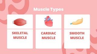
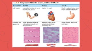
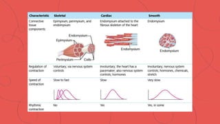
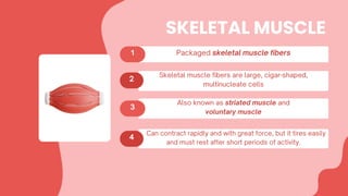
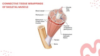
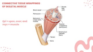
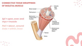
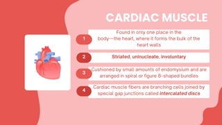
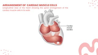
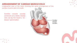
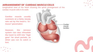
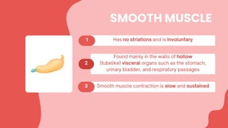
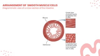
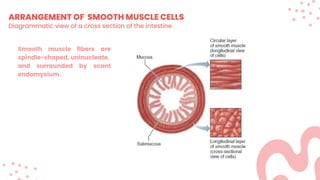
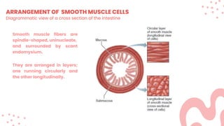
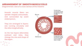
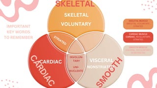
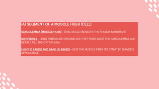
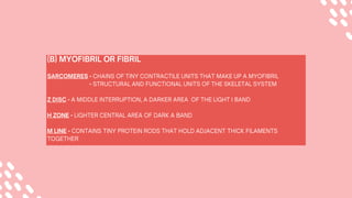
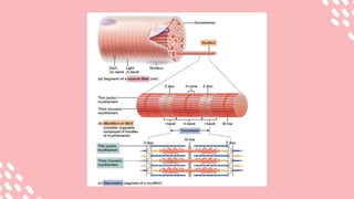
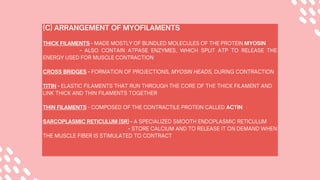
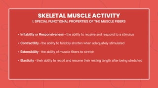
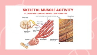
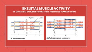
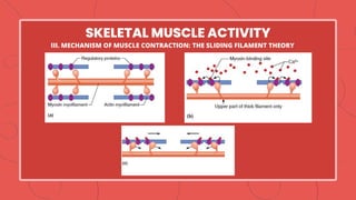
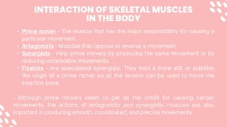
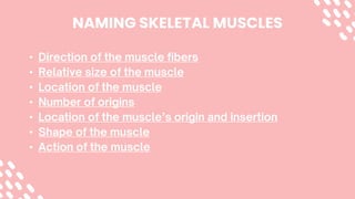
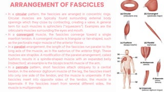
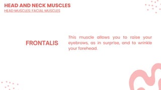
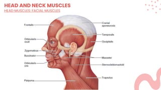
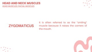
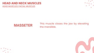
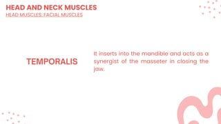
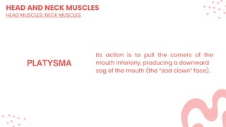
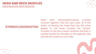
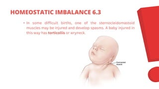
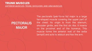
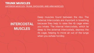
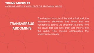
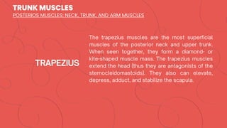
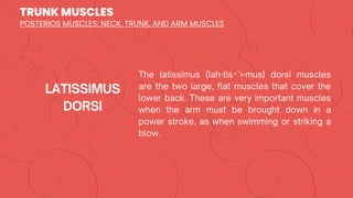
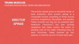
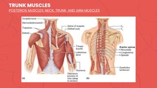
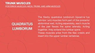
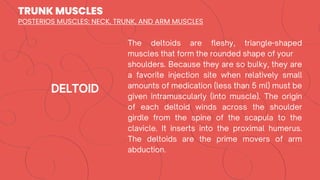
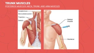
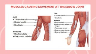
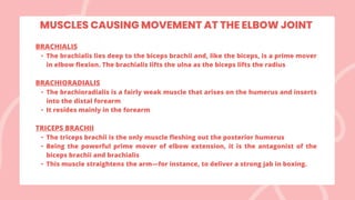
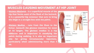
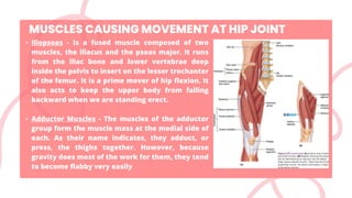
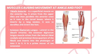
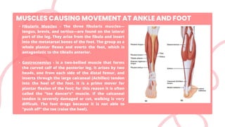
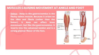
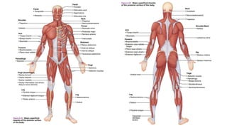
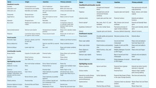
13. ARRANGEMENT OF CARDIAC MUSCLE CELLS
Longitudinal view of the heart showing the spiral arrangement of the
cardiac muscle cells in its walls
14. ARRANGEMENT OF CARDIAC MUSCLE CELLS
Cardiac muscle usually
contracts at a fairly steady
rate set by the heart’s “in-
house” pacemaker.
Longitudinal view of the heart showing the spiral arrangement of the
cardiac muscle cells in its walls
15. ARRANGEMENT OF CARDIAC MUSCLE CELLS
Cardiac muscle usually
contracts at a fairly steady
rate set by the heart’s “in-
house” pacemaker.
However, the nervous
system can also stimulate
the heart to shift into “high
gear” for short periods, as
when you run to catch a
bus.
Longitudinal view of the heart showing the spiral arrangement of the
cardiac muscle cells in its walls
17. ARRANGEMENT OF SMOOTH MUSCLE CELLS
Diagrammatic view of a cross section of the intestine
18. ARRANGEMENT OF SMOOTH MUSCLE CELLS
Diagrammatic view of a cross section of the intestine
Smooth muscle fibers are
spindle-shaped, uninucleate,
and surrounded by scant
endomysium.
19. ARRANGEMENT OF SMOOTH MUSCLE CELLS
Diagrammatic view of a cross section of the intestine
Smooth muscle fibers are
spindle-shaped, uninucleate,
and surrounded by scant
endomysium.
They are arranged in layers;
one running circularly and
the other longitudinally.
20. ARRANGEMENT OF SMOOTH MUSCLE CELLS
Diagrammatic view of a cross section of the intestine
Smooth muscle fibers are
spindle-shaped, uninucleate,
and surrounded by scant
endomysium.
They are arranged in layers;
one running circularly and
the other longitudinally.
As the two layers alternately
contract and relax, they
change the size and shape of
the organ.
33. • ALS or amyotrophic lateral sclerosis (also called Lou Gehrig’s
disease), motor neurons degenerate over time, resulting in
paralysis that gradually worsens.
• Common characteristics include malfunctioning mitochondria,
inflammation, and the generation of free radicals that damage
DNA and tissue much like intense UV light.
• The prognosis for patients with ALS is generally death within
three to five years because the breathing muscles will eventually
be affected, resulting in suffocation
HOMEOSTATIC IMBALANCE 6.1
39. CONTRACTION OF A SKELETAL
MUSCLE AS A WHOLE
In skeletal muscles, the “all-or-none” law of muscle physiology applies to the muscle fiber, not
to the whole muscle. It states that a muscle fiber will contract to its fullest extent when it is
stimulated adequately; it never partially contracts.
GRADED RESPONSES
• The whole muscle reacts to stimuli with graded responses, or different degrees of
shortening, which generate different amounts of force
• Graded responses —different degrees of skeletal muscle shortening
Graded muscle contractions can be produced two ways:
• By changing the frequency of muscle stimulation
• By changing the number of muscle cells being stimulated at one time
49. • If the nerve supply to a muscle is destroyed (as in an accident),
the muscle is no longer stimulated in this manner, and it loses
tone. Soon after, it becomes flaccid (fla˘′sid), or soft and flabby,
and begins to atrophy (waste away). This is called flaccid
paralysis.
• A condition that increases muscle tone until the muscle is no
longer controllable, example, the disease tetanus, which is
caused by a bacterial toxin. This is called spastic paralysis.
HOMEOSTATIC IMBALANCE 6.2
53. THE FIVE GOLDEN RULES OF MUSCLE ACTIVITY
• With a few exceptions, all skeletal muscles
cross at atleast one joint.
• Typically, the bulk of a skeletal muscle lies
proximal to the joint crossed.
• All skeletal muscles have at least two
attachments: the origin and insertion.
• Skeletal muscles can only pull; they never push
• During contraction, a skeletal muscle insertion
moves toward the origin.
76. HEAD AND NECK MUSCLES
HEAD MUSCLES: FACIAL MUSCLES
It flattens the cheek (as in whistling or
blowing a trumpet). It is also listed as a
chewing muscle because it compresses
the cheek to hold food between the teeth
during chewing.
84. HEAD AND NECK MUSCLES
HEAD MUSCLES: NECK MUSCLES
Its action is to pull the corners of the
mouth inferiorly, producing a downward
sag of the mouth (the “sad clown” face).
86. HEAD AND NECK MUSCLES
HEAD MUSCLES: NECK MUSCLES
When both sternocleidomastoid muscles
contract together, they flex your neck. (It is this
action of bowing the head that has led some
people to call these muscles the “prayer”
muscles.) If just one muscle contracts, the face is
rotated toward the shoulder on the opposite side
and tilts the head to its own side.
87. • In some difficult births, one of the sternocleidomastoid
muscles may be injured and develop spasms. A baby injured in
this way has torticollis or wryneck.
HOMEOSTATIC IMBALANCE 6.3
113. The upper limb muscles fall into three groups:
(1) The first group includes muscles that arise from the shoulder girdle and
cross the shoulder joint to insert into the humerus; We have already
considered these muscles, which move the arm—they are the pectoralis
major, latissimus dorsi, and deltoid.
(2) The second group causes movement at the elbow joint. These muscles
enclose the humerus and insert on the forearm bones.
(3) The third group of upper limb muscles includes the muscles of the forearm,
which insert on the hand bones and cause their movement. The muscles of this
last group are thin and spindle-shaped, and there are many of them.
For example, the flexor carpi and flexor digitorum muscles, found on the
anterior aspect of the forearm, flex the wrist and fingers, respectively
115. All anterior muscles of the humerus cause elbow flexion. In order of
decreasing strength these are the brachialis, biceps brachii, and
brachioradialis
BICEPS BRACHII
• The biceps brachii is the most familiar muscle of the arm because it
bulges when you flex your elbow
• This muscle is the powerful prime mover for flexion of the forearm and
acts to supinate the forearm
BRACHIALIS
• The brachialis lies deep to the biceps brachii and, like the biceps, is a
prime mover in elbow flexion. The brachialis lifts the ulna as the biceps
lifts the radius
MUSCLES CAUSING MOVEMENT AT THE ELBOW JOINT
117. BRACHIALIS
• The brachialis lies deep to the biceps brachii and, like the biceps, is a prime mover
in elbow flexion. The brachialis lifts the ulna as the biceps lifts the radius
BRACHIORADIALIS
• The brachioradialis is a fairly weak muscle that arises on the humerus and inserts
into the distal forearm
• It resides mainly in the forearm
TRICEPS BRACHII
• The triceps brachii is the only muscle fleshing out the posterior humerus
• Being the powerful prime mover of elbow extension, it is the antagonist of the
biceps brachii and brachialis
• This muscle straightens the arm—for instance, to deliver a strong jab in boxing.
MUSCLES CAUSING MOVEMENT AT THE ELBOW JOINT
120. • Gluteus Maximus - is a superficial muscle of the
hip that forms most of the flesh of the buttock.
It is a powerful hip extensor that acts to bring
the thigh in a straight line with the pelvis.
• Gluteus Medius - runs from the ilium to the
femur, beneath the gluteus maximus for most
of its length. The gluteus medius is a hip
abductor and is important in steadying the
pelvis during walking. It is also an important
site for giving intramuscular injections,
particularly when administering more than 5
ml.
MUSCLES CAUSING MOVEMENT AT HIP JOINT
121. • Iliopsoas - is a fused muscle composed of two
muscles, the iliacus and the psoas major. It runs
from the iliac bone and lower vertebrae deep
inside the pelvis to insert on the lesser trochanter
of the femur. It is a prime mover of hip flexion. It
also acts to keep the upper body from falling
backward when we are standing erect.
• Adductor Muscles - The muscles of the adductor
group form the muscle mass at the medial side of
each. As their name indicates, they adduct, or
press, the thighs together. However, because
gravity does most of the work for them, they tend
to become flabby very easily
MUSCLES CAUSING MOVEMENT AT HIP JOINT
122. • Tibialis Anterior - is a superficial muscle on
the anterior leg. It arises from the upper
tibia and then parallels the anterior crest
as it runs to the tarsal bones, where it
inserts by a long tendon. It acts to
dorsiflex and invert the foot.
• Extensor Digitorum Longus - Lateral to the
tibialis anterior, the extensor digitorum
longus muscle arises from the lateral tibial
condyle and proximal three-quarters of the
fibula and inserts into the phalanges of
toes 2 to 5. It is a prime mover of toe
extension
MUSCLES CAUSING MOVEMENT AT ANKLE AND FOOT
123. • Fibularis Muscles - The three fibularis muscles—
longus, brevis, and tertius—are found on the lateral
part of the leg. They arise from the fibula and insert
into the metatarsal bones of the foot. The group as a
whole plantar flexes and everts the foot, which is
antagonistic to the tibialis anterior.
• Gastrocnemius - is a two-bellied muscle that forms
the curved calf of the posterior leg. It arises by two
heads, one from each side of the distal femur, and
inserts through the large calcaneal (Achilles) tendon
into the heel of the foot. It is a prime mover for
plantar flexion of the foot; for this reason it is often
called the “toe dancer’s” muscle. If the calcaneal
tendon is severely damaged or cut, walking is very
difficult. The foot drags because it is not able to
“push off” the toe (raise the heel).
MUSCLES CAUSING MOVEMENT AT ANKLE AND FOOT
124. • Soleus - Deep to the gastrocnemius is the
fleshy soleus muscle. Because it arises on
the tibia and fibula (rather than the
femur), it does not affect knee
movement, but like the gastrocnemius, it
inserts into the calcaneal tendon and is a
strong plantar flexor of the foot.
MUSCLES CAUSING MOVEMENT AT ANKLE AND FOOT
128. • Muscular dystrophy is a group of inherited muscle-destroying
diseases that affect specific muscle groups. The muscles appear to
enlarge because of fat and connective tissue deposits, but the muscle
fibers degenerate and atrophy.
• Duchenne’s muscular dystrophy, the most common and serious form
of muscular dystrophy. Expressed almost exclusively in boys. This
tragic disease is usually diagnosed between the ages of 2 and 7 years.
• The disease progresses from the extremities upward, finally affecting
the head and chest muscles. Children with this disease rarely live
beyond their early twenties and generally die of respiratory failure.
• The diseased muscle fibers lack a protein (called dystrophin) that
helps maintain the sarcolemma— a cure is still elusive.
HOMEOSTATIC IMBALANCE 6.4
129. • Myasthenia gravis, a rare autoimmune disease
characterized by drooping upper eyelids, difficulty in
swallowing and talking, and generalized muscle weakness
and fatigability that can affect muscles during adulthood.
• The disease involves a shortage of acetylcholine receptors
at neuromuscular junctions caused by antibodies specific
for acetylcholine receptors.
• Death usually occurs when the respiratory muscles can no
longer function, which leads to respiratory failure.
HOMEOSTATIC IMBALANCE 6.5