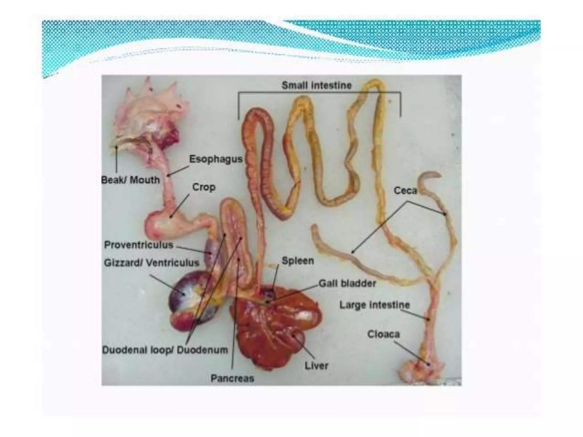
The avian digestive system begins with the beak and mouth, where food is ingested but not chewed. The esophagus transports food to the crop for temporary storage. The proventriculus is the true stomach where digestion begins through enzymes. The gizzard uses strong muscles to grind food like teeth would. The small intestine continues digestion and nutrient absorption. The caeca and large intestine absorb remaining nutrients and water before waste exits through the cloaca. Accessory glands like the salivary glands, pancreas, and liver secrete enzymes and fluids to further break down food into absorbable nutrients.
2. Introduction
The digestive system of any animal is important in converting
the food the animal eats into the nutrients its body needs for
growth, maintenance, and production (such as egg
production).
An animal's body breaks down food through both mechanical
and chemical means.
In many animals, mechanical action involves chewing;
however, because birds do not have teeth, their bodies use
other mechanical action.
Chemical action includes the release of digestive enzymes and
fluids from various parts of the digestive system.
After being released from food during digestion, nutrients are
absorbed and distributed throughout the animal's body.
3. Parts of a Chicken Digestive Tract
The chicken has a typical avian digestive system. In chickens, the digestive
tract (also referred to as the gastrointestinal tract or GI tract) begins at the
mouth, includes several important organs, and ends at the cloaca.
Organs
Mouth
Pharynx
Esophagus/gullet
Crop
Proventriculus
Gizzard
Small intestine
Caeca
Large intestine
Cloaca
Vent
4.
5. Beak/Mouth
As with most birds, a chicken obtains feed by using its
beak.
Food picked up by the beak enters the mouth. Chickens
do not have teeth, so they cannot chew their food.
However, the mouth contains glands that secrete saliva,
which wets the feed to make it easier to swallow.
Also, the saliva contains enzymes, such as amylase, that
start the digestion process.
The chicken uses its tongue to push the feed to the back
of the mouth to be swallowed.
6. Esophagus
The esophagus is a flexible tube that connects the
mouth with the rest of the digestive tract.
It carries food from the mouth to the crop and from
the crop to the proventriculus.
7. Crop
The crop is an out-pocketing of the esophagus and is
located just outside the body cavity in the neck
region .
Swallowed feed and water are stored in the crop
until they are passed to the rest of the digestive tract.
When the crop is empty or nearly empty, it sends
hunger signals to the brain so that the chicken will
eat more.
8. Although the digestive enzymes secreted in the mouth
began the digestion process, very little digestion takes
place in the crop—it is simply a temporary storage pouch.
The crop evolved for birds that are typically hunted by
other animals but need to move to the open to find feed.
These birds can consume relatively large amounts of
food quickly and then move to a more secure location to
digest that food.
Occasionally, the crop becomes impacted, or backed up.
This problem—called crop impaction, crop binding, or
pendulous crop—can occur when a chicken goes a long
time without feed and then eats too much too quickly
when feed is available again.
9. Crop impaction also can occur when a chicken free-
ranges on a pasture of tough, fibrous vegetation or
eats long pieces of string.
With crop impaction, even if a chicken continues to
eat, the feed cannot pass the impacted crop.
The swollen crop also can block the windpipe,
causing the chicken to suffocate.
10. Proventriculus
The esophagus continues past the crop, connecting the
crop to the proventriculus.
The proventriculus (also known as the true stomach) is
the glandular stomach where digestion primarily begins.
Hydrochloric acid and digestive enzymes, such as pepsin,
are added to the feed here and begin to break it down
more significantly than the enzymes secreted by the
salivary glands.
At this point, however, the food has not yet been
ground—this organ is called the proventriculus because
its location in the digestive tract is before the ventriculus,
where food is ground
11. Ventriculus/ Gizzard
The ventriculus, or gizzard, is a part of the digestive
tract of birds, reptiles, earthworms, and fish.
Often referred to as the mechanical stomach, the
gizzard is made up of two sets of strong muscles that
act as the bird's teeth and has a thick lining that
protects those muscles.
Consumed feed and the digestive juices from the
salivary glands and proventriculus pass into the
gizzard for grinding, mixing, and mashing.
12. When allowed to free-range, chickens typically eat
small stones.
The acidic environment in the proventriculus softens
the stones, and then the strong muscles of the
gizzard grind them into tiny pieces.
The stones remain in the gizzard until they are
ground into pieces small enough to pass to the rest of
the digestive tract.
13. When a chicken eats a small, sharp object, such as a
tack or staple, the object is likely to get stuck in the
gizzard.
Because of the strong grinding motion of the
gizzard's muscles, such sharp objects can put holes in
the gizzard wall.
Chickens with damaged gizzards grow thin and
eventually die.
Preventing this situation is a good reason to keep a
poultry house free of nails, glass shards, bits of wire,
and so on.
14. Small Intestine
The small intestine is made up of the duodenum (also referred
to as the duodenal loop) and the lower small intestine.
The remainder of the digestion occurs in the duodenum, and
the released nutrients are absorbed mainly in the lower small
intestine.
The duodenum receives digestive enzymes and bicarbonate
(to counter the hydrochloric acid from the proventriculus)
from the pancreas and bile from the liver (via the gall
bladder).
The digestive juices produced by the pancreas are involved
primarily in protein digestion. Bile is a detergent that is
important in the digestion of lipids and the absorption of fat-
soluble vitamins (A, D, E, and K).
15. The lower small intestine is composed of two parts,
the jejunum and the ileum.
The Meckel's diverticulum marks the end of the
jejunum and the start of the ileum .
The Meckel's diverticulum is formed during a
chicken's embryonic stage.
In the egg, the yolk sac supplies the nutrients needed
for the embryo to develop and grow. Right before
hatch, the yolk sac is taken into the navel cavity of
the embryo. The residual tiny sac is the Meckel's
diverticulum.
16. Caeca
The caeca are two blind pouches located where the small
and large intestines join.
Some of the water remaining in the digested material is
reabsorbed here.
Another important function of the caeca is the
fermentation of any remaining coarse materials.
During this fermentation, the caeca produce several fatty
acids as well as the eight B vitamins (thiamine,
riboflavin, niacin, pantothenic acid, pyridoxine, biotin,
folic acid, and vitamin B12).
Because the caeca are located so close to the end of the
digestive tract, however, few of the produced nutrients
are absorbed and available to the chicken.
17. Large Intestine (Colon)
Despite the name, the large intestine is actually
shorter than the small intestine.
The large intestine is where the last of the water
reabsorption occurs.
18. Cloaca
In the cloaca, the digestive wastes mix with wastes from
the urinary system (urates).
Chickens usually void fecal material as digestive waste
with uric acid crystals on the outer surface that is,
chickens do not urinate.
The color and texture of chicken faecal material can
indicate the health status of the chicken's digestive tract:
the white, pasty material coating chicken fecal material is
uric acid, the avian form of urine, and is normal.
The reproductive tract also exits through this area. When
a hen lays an egg, the vagina folds over to allow the egg to
leave through the cloaca opening without coming into
contact with feces or urine.
19. Accessory Digestive Glands
There are three accessory digestive glands which play a
vital role in the process of digestion
1. Salivary Glands
2. Pancreas
3. Liver
20. Salivary Glands
It is responsible for production of saliva.
Its secretions ranges from 7 to 30 ml per day.
The salivary glands are:
1. Maxillary – in the roof of the mouth.
2. Palatine – on either side of the nasal opening in the roof of the mouth.
3. Apheno-pteryoid glands – in the roof of the pharynx on each side of the
common opening for the eustachian tubes.
4. Anterior sub-mandible glands – in the angle formed by the union of the
upper and lower beaks or mandibles.
5. Posterior sub-mandibular glands.
6. Lingual glands – in the tongue.
7. Crico-arytenoid glands – around the glottis.
8. A small gland in the angle of the mouth.
21. The saliva has following functions:
1. Lubrication of the feed.
2. Digestion, it contains salivary amylase which is
responsible for carbohydrates digestion.
3. Acts as a buffer, it contains bicarbonate and other
salts.
4. Helps tasting the feed.
5. Protects the mucous membrane and keeps it moist.
6. Helps regulate the body temperature.
7. Contain enzyme known as muramidase which is
bacteriocidal in nature and thus it produces the local
immunity.
22. Accessory Digestive Gland: Pancreas
Pancreas produces a pancreatic juice.
Its pH is 6.9
It is released in the distal end of the loop of duodenum.
Pancreatic juice contains four kinds of enzymes;
1. Proteolytic Enzymes
2. Lipolytic Enzyme
3. Carbohydrate splitting Enzymes
4. Nucleolytic Enzymes
23. There are five different kinds of proteolytic enzymes
1. Trypsinogen
2. Chymotrypsinogen A
3. Chymotrypsinogen B
4. Procarboxy peptidase A
5. Procarboxy peptidase B
These enzymes are responsible for the break down of
protein molecules into simpler units.
24. Lipolytic Enzyme
There are three type of lipolytic enzymes which are
produced by the pancreas;
1. Phospholipase, lipid breakdown.
2. Pancreatic lipase, lipid breakdown.
3. Cholesterol esterase, esterification of cholesterol.
25. Carbohydrate splitting Enzymes
These consist of:
1. Pancreatic amylase, acts on the starch and converts it
into simpler units.
2. Invertase, acts on the sucrose and convert it into simpler
sugar.
27. Liver
The largest glandular organ in the body. Aids in the metabolism of
carbohydrates, fats and proteins.
Liver is a bilobed structure and it performs the following functions
1. Detoxification
2. Store of vitamins and carbohydrates, carbohydrates are stored in
the form of glycogen.
3. Formation plasma protein like albumin and globulin.
4. It activates and inactivates the protein and peptide hormones.
5. Liver is a site for the destruction of old RBCs.
6. Formation of bile, which is responsible for the emulsification of
the fat.