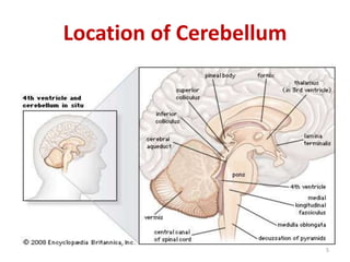
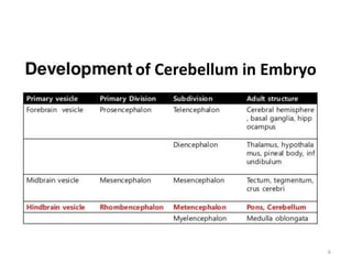
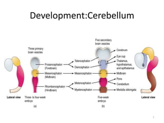
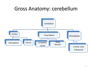
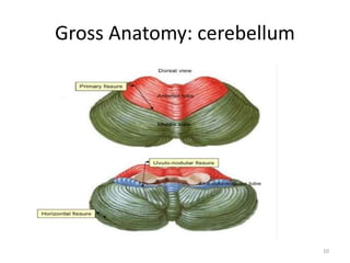
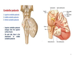
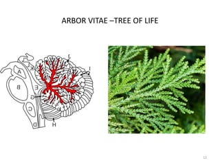
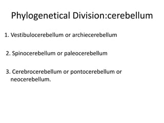
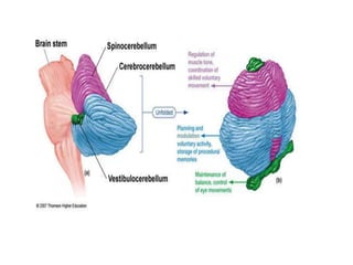
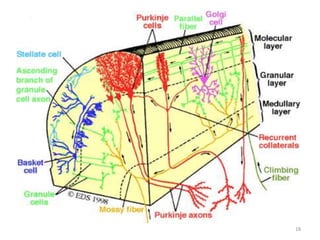
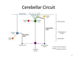
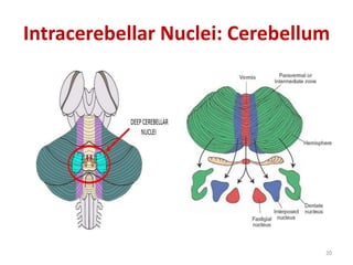
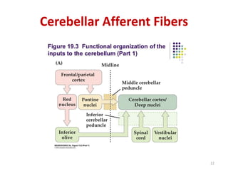
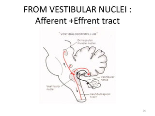
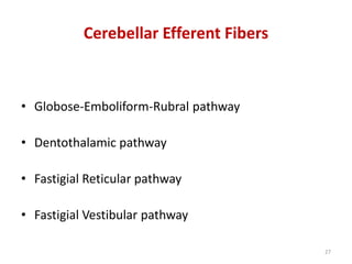
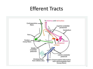
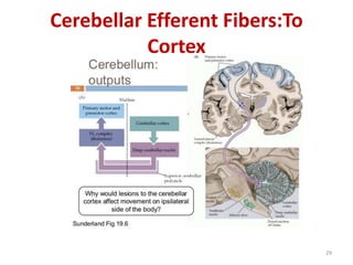
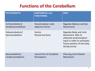
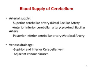
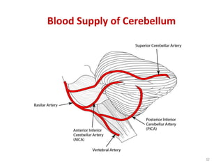
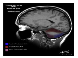
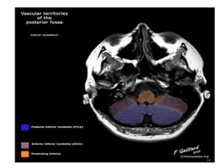
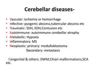
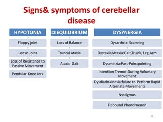
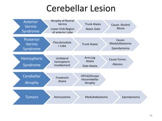
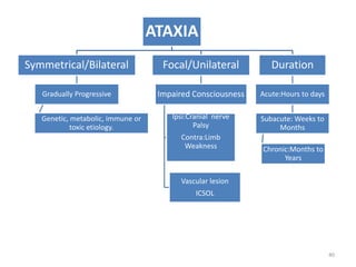
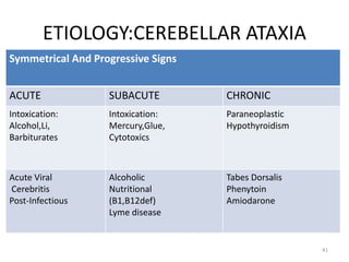
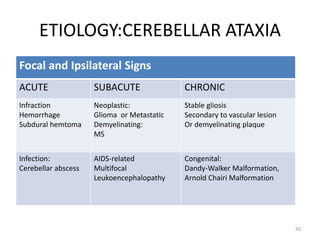
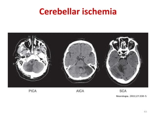
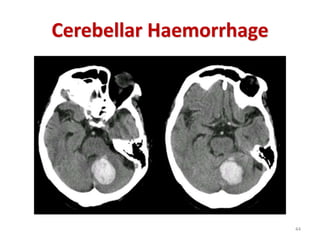
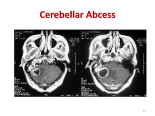
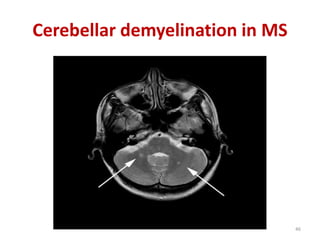
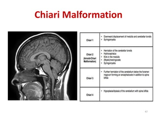
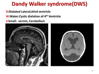
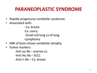
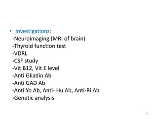
DR.TAREQ ESTEAK
Resident(PhaseA)
Neurology,NINS
1
CEREBELLUM
(THE LITTLE BRAIN)
Gross appearance of the cerebellum
Posterior view Inferior view
2
Radiographic anatomy: CT
3
Radiographic anatomy: MRI
SAGITAL SECTION AXIAL SECTION
4
Location of Cerebellum
5
of Cerebellum in Embryo
6
Development:Cerebellum
7
Gross Anatomy of Cerebellum
• Cerebellum consists of two cerebeller
hemispheres joined by a narrow median vermis.
• Three bundles of nerve fibers connect the
cerebellum to the brain stem -
- Superior (midbrain),
- Middle(pons) &
- Inferior cerebellar peduncle (medulla )
8
Gross Anatomy: cerebellum
Cerebellum
Surface
Hemisphere Vermis
Grey Matter
Cerebellar
cortex
Deep
Neuclei
WhiteMatter
1.Arbor vitae
2.Peduncle
9
Gross Anatomy: cerebellum
10
11
ARBOR VITAE –TREE OF LIFE
12
1. Vestibulocerebellum or archiecerebellum
2. Spinocerebellum or paleocerebellum
3. Cerebrocerebellum or pontocerebellum or
neocerebellum.
Phylogenetical Division:cerebellum
1. Cortex of vermis: influences movements of the long
axis of the body – neck, shoulders, thorax, abdomen,
hips.
2. Intermediate zone: (Just lateral to vermis) controls
distal parts of the limbs hands and feet.
3. Lateral zone: planning of sequential movements of
conscious assessment of movement errors.
Functional Division: Cerebellum
Functional Division
Structure of the Grey Matter: Cortex
• Layers of cortex:
1.External molecular layer
2.Middle granular layer.
3.Internal Medullary layer
17
18
Cerebellar Circuit
19
Intracerebellar Nuclei: Cerebellum
20
Cerebellar Cortical Mechanism
• Purkinje cells form the center of a functional unit of
cerebellar cortex.
• Climbing fibers-terminal fibers of olivo-cerebellar
tract. Excitatory effect on Purkinje cells.
• Mossy fibers-terminal fibers of all other cerebellar
afferent tracts. Diffuse excitatory effect on Purkinje
cells.
• Stellate, basket and Golgi cells- inhibitory
interneurons. Influence the degree of Purkinje cell
excitation by climbing and mossy fibers.
21
Cerebellar Afferent Fibers
22
FROM SPINAL CORD:
Afferent tracts
1. Anterior Spinocerebellar tract
2. Posterior Spinocerebellar tract
3.Cuneo-cerebellar tract.
23
24
25
FROM VESTIBULAR NUCLEI :
Afferent +Effrent tract
26
Cerebellar Efferent Fibers
• Globose-Emboliform-Rubral pathway
• Dentothalamic pathway
• Fastigial Reticular pathway
• Fastigial Vestibular pathway
27
Efferent Tracts
28
Cerebellar Efferent Fibers:To
Cortex
29
PHYLOGENETIC ANATOMICAL and
FUNCTIONAL
ROLE
Archicerebellum/
Vestibulocerebellum
Floculonodular Lobe
Adjacent Vermis
Regulate Balance and Eye
Movements
Paleocerebellum/
Spinocerebellum
Vermis
Paravermal Zone
Regulate Body and Limb
Movement. Able to
elaborate proprioceptive
Input in order to anticipate
future position of the body
during course
Neocerebellum/
Cerebrocerebellum
Lateral Part of Cerebellar
Hemisphere
Planning and Initiation
Movement.
Functions of the Cerebellum
30
• Arterial supply:
-Superior cerebellar artery<Distal Bacillar Artery
-Anterior inferior cerebellar artery<proximal Bacillar
Artery
-Posterior inferior cerebellar artery<Vetebral Artery
• Venous drainage:
-Supirior and Inferior Cerebellar vein
-Adjacent venous sinuses.
Blood Supply of Cerebellum
31
Blood Supply of Cerebellum
32
33
34
APPLIED PART OF CEREBELLUM
35
Cerebellar diseases-
• Vascular: ischemia or hemorrhage
• Infective: pyogenic abscess,tubercular abscess etc
• Traumatic: SDH, EDH,Contusion etc
• Autoimmune: autoimmune cerebellar atrophy
• Metabolic: Hypoxia
• Inflammatory: MS
• Neoplastic: primary: medulloblastoma
Secondary: metastasis
Congenital & others: DWM,Chiari malformations,SCA
etc.
HYPOTONIA
Floppy joint
Loose Joint
Loss of Resistance to
Passive Movement
Pendular Knee Jerk
DIEQUILIBRIUM
Loss of Balance
Truncal Ataxia
Ataxic Gait
DYSYNERGIA
Dysarthria: Scanning
Dystaxia/Ataxia:Gait,Trunk, Leg,Arm
Dysmetria:Past-Pointpointing
Intention Tremor:During Voluntary
Movement
Dysdiadokinesia:faiure to Perform Rapid
Alternate Movements
Nystigmus
Rebound Phenomenon
Signs& symptoms of cerebellar
disease
37
Cerebellar Lesion
38
Anterior
Vermis
Syndrome
Atrophy of Rostral
Vermis
Lower limb Region
of anterior Lobe
Trunk Ataxia
Ataxic Gate
Cause: Alcohol
Abuse
Posterior
Vermis
Syndrome
Floculonodula
r Lobe
Trunk Ataxia
Cause:
Meduloblastoma
Ependymoma
Hemispheric
Syndrome
Unilateral
hemispheric
Involbement
Arm,Leg
Ataxia
Gate Ataxia
Cause:Tumor,
Abscess
Cerebellar
Atrophy
Friedreich
Ataxia
OPCA(Olivopo
ntocerebellar
Atrophy
Tumors Astrocytoma Medulloblastoma Ependymoma
APPROACH TO THE PATIENT WITH ATAXIA:
39
ATAXIA
Symmetrical/Bilateral
Gradually Progressive
Genetic, metabolic, immune or
toxic etiology.
Focal/Unilateral
Impaired Consciousness
Ipsi:Cranial nerve
Palsy
Contra:Limb
Weakness
Vascular lesion
ICSOL
Duration
Acute:Hours to days
Subacute: Weeks to
Months
Chronic:Months to
Years
40
ETIOLOGY:CEREBELLAR ATAXIA
Symmetrical And Progressive Signs
ACUTE SUBACUTE CHRONIC
Intoxication:
Alcohol,Li,
Barbiturates
Intoxication:
Mercury,Glue,
Cytotoxics
Paraneoplastic
Hypothyroidism
Acute Viral
Cerebritis
Post-Infectious
Alcoholic
Nutritional
(B1,B12def)
Lyme disease
Tabes Dorsalis
Phenytoin
Amiodarone
41
ETIOLOGY:CEREBELLAR ATAXIA
Focal and Ipsilateral Signs
ACUTE SUBACUTE CHRONIC
Infraction
Hemorrhage
Subdural hemtoma
Neoplastic:
Glioma or Metastatic
Demyelinating:
MS
Stable gliosis
Secondary to vascular lesion
Or demyelinating plaque
Infection:
Cerebellar abscess
AIDS-related
Multifocal
Leukoencephalopathy
Congenital:
Dandy-Walker Malformation,
Arnold Chairi Malformation
42
Cerebellar ischemia
43
Cerebellar Haemorrhage
44
Cerebellar Abcess
45
Cerebellar demyelination in MS
46
Chiari Malformation
47
Dandy Walker syndrome(DWS)
D:Dialated Lateral,third ventricle
W:Water:Cystic dialation of 4th Ventricle
S:Small: vermis, Cerebellum
48
• These are – Autosomal dominant
- Autosomal recessive
-Mitochondrial inheritence
• Clinical manifestation dominated by cerebellar
signs but there are changes in
-basal ganglia –spinal cord – Optic nerve-
Retina – Peripheral nerves
• Rarely dementia is present.
INHERITED ATAXIAS:
49
• Begins in adult life.
• Occurs in each generation of pedigree.
• SCA type 1 to SCA type 28, episodic ataxia
type 1&2.
• Clinical phenotypes overlaps.
• Genetic analysis is the gold standard for
diagnosis and classification.
AUTOSOMAL DOMINANT ATAXIAS
50
• Begins in childhood or early adulthood.
• Parental consanguinity of marriage is more
likely but not essential.
• Examples:
Friedreich’s ataxia.
Ataxia with isolated Vit-E deficiency.
AUTOSOMAL RECESSIVE ATAXIAS
51
• Mutation in maternal mitochondrial DNA
• E.g.-
 MERRF syndrome ( Myoclonic epilepsy with
ragged red fibres ).
 MELAS ( Mitochondrial myopathy,
encephalopathy, lactic acidosis & stroke like
episodes ).
 Kearns-Sayre syndrome.
MITOCHONDRIAL ATAXIAS
52
• Findings- reflexes normal but knee and ankles
are lost, planter bilateral extensor.
• Mild dementia
• Sphincter disturbance
• Summary: Ataxia
Ophthalmoparesis
Pyramidal and extrapyramidal
features.
SCA 1
53
SCA-1
54
SCA1 (OPCA)
MRI findings:
-Atrophy of cerebellum
-Marked shrinkage of ventral
pons
-Disappearance of Olivary
eminence of medulla.
55
• Most common hereditary ataxia
• Presents before the age of 25yrs
• Primary sites of pathology – Spinal cord, dorsal root
ganglia, peripheral nerve and cerebellum
• Neurological- nystagmus, optic atrophy, deep tendon
reflex absent, planter b/l extensor.
• Skeletal deformity- scoliosis, pes cavus
• Presentation: Progressive staggering gait, frequent
fall and titubation.
FRIEDRIECH’S ATAXIA:
56
57
Friedreichs Ataxia
• METASTASIS:
In adults metastasis is the most common
tumor.
• Primary sites:
-Bronchus - Breast -Thyroid
-Stomach -Kidney -Prostate
-Testes -Melanoma
CEREBELLAR TUMORS
58
A)Hemangioblastoma:
-Commonest tumor in adult
-More in male
-Benign tumor of vascular origin
-Association: Polycythemia
PRIMARY CEREBELLAR TUMOR:
59
Haemangioblastoma
60
B) Medulloblastoma:
-Primitive neuroectodermal tumor
-Occurs in childhood
-Peak age of onset is 5yrs of age
-Highly malignant tumor
-Arise in vermis and extend into 4th ventricle
-Presentation- truncal ataxia, raised ICP, visual loss.
- Treatment: Surgery, radiotherapy.
61
Medulloblastoma
62
C)Cerebellar Astrocytoma:
-It is low grade in nature in contrast to
astrocytoma of cerebral hemisphere.
D) Ependymoma:
-Arises from ependymal cells
E) CP angle tumor:
- Acoustic neuroma - Meningioma
63
Astrocytoma
64
C-P angle tumor:
• Vestibular schwannoma
• Meningioma
• Metastasis
• Neurofibroma
• Epidermoid
• Rapidly progressive cerebellar syndrome.
• Associated with-
- Ca. breast
-Ca. ovary
-Small cell lung ca of lung
-Lymphoma
• MRI of brain shows cerebellar atrophy.
• Tumor markers:
-Anti yo Ab – ovarian ca
-Anti Hu Ab – SCLC
-Anti ri Ab – Ca. breast
PARANEOPLASTIC SYNDROME
66
• Investigations:
-Neuroimaging (MRI of brain)
-Thyroid function test
-VDRL
-CSF study
-Vit B12, Vit E level
-Anti Gliadin Ab
-Anti GAD Ab
-Anti Yo Ab, Anti- Hu Ab, Anti-Ri Ab
-Genetic analysis
67
Thank you
68

Cerebellum

  • 1.
  • 2.
    Gross appearance ofthe cerebellum Posterior view Inferior view 2
  • 3.
  • 4.
    Radiographic anatomy: MRI SAGITALSECTION AXIAL SECTION 4
  • 5.
  • 6.
  • 7.
  • 8.
    Gross Anatomy ofCerebellum • Cerebellum consists of two cerebeller hemispheres joined by a narrow median vermis. • Three bundles of nerve fibers connect the cerebellum to the brain stem - - Superior (midbrain), - Middle(pons) & - Inferior cerebellar peduncle (medulla ) 8
  • 9.
    Gross Anatomy: cerebellum Cerebellum Surface HemisphereVermis Grey Matter Cerebellar cortex Deep Neuclei WhiteMatter 1.Arbor vitae 2.Peduncle 9
  • 10.
  • 11.
  • 12.
  • 13.
    1. Vestibulocerebellum orarchiecerebellum 2. Spinocerebellum or paleocerebellum 3. Cerebrocerebellum or pontocerebellum or neocerebellum. Phylogenetical Division:cerebellum
  • 15.
    1. Cortex ofvermis: influences movements of the long axis of the body – neck, shoulders, thorax, abdomen, hips. 2. Intermediate zone: (Just lateral to vermis) controls distal parts of the limbs hands and feet. 3. Lateral zone: planning of sequential movements of conscious assessment of movement errors. Functional Division: Cerebellum
  • 16.
  • 17.
    Structure of theGrey Matter: Cortex • Layers of cortex: 1.External molecular layer 2.Middle granular layer. 3.Internal Medullary layer 17
  • 18.
  • 19.
  • 20.
  • 21.
    Cerebellar Cortical Mechanism •Purkinje cells form the center of a functional unit of cerebellar cortex. • Climbing fibers-terminal fibers of olivo-cerebellar tract. Excitatory effect on Purkinje cells. • Mossy fibers-terminal fibers of all other cerebellar afferent tracts. Diffuse excitatory effect on Purkinje cells. • Stellate, basket and Golgi cells- inhibitory interneurons. Influence the degree of Purkinje cell excitation by climbing and mossy fibers. 21
  • 22.
  • 23.
    FROM SPINAL CORD: Afferenttracts 1. Anterior Spinocerebellar tract 2. Posterior Spinocerebellar tract 3.Cuneo-cerebellar tract. 23
  • 24.
  • 25.
  • 26.
    FROM VESTIBULAR NUCLEI: Afferent +Effrent tract 26
  • 27.
    Cerebellar Efferent Fibers •Globose-Emboliform-Rubral pathway • Dentothalamic pathway • Fastigial Reticular pathway • Fastigial Vestibular pathway 27
  • 28.
  • 29.
  • 30.
    PHYLOGENETIC ANATOMICAL and FUNCTIONAL ROLE Archicerebellum/ Vestibulocerebellum FloculonodularLobe Adjacent Vermis Regulate Balance and Eye Movements Paleocerebellum/ Spinocerebellum Vermis Paravermal Zone Regulate Body and Limb Movement. Able to elaborate proprioceptive Input in order to anticipate future position of the body during course Neocerebellum/ Cerebrocerebellum Lateral Part of Cerebellar Hemisphere Planning and Initiation Movement. Functions of the Cerebellum 30
  • 31.
    • Arterial supply: -Superiorcerebellar artery<Distal Bacillar Artery -Anterior inferior cerebellar artery<proximal Bacillar Artery -Posterior inferior cerebellar artery<Vetebral Artery • Venous drainage: -Supirior and Inferior Cerebellar vein -Adjacent venous sinuses. Blood Supply of Cerebellum 31
  • 32.
    Blood Supply ofCerebellum 32
  • 33.
  • 34.
  • 35.
    APPLIED PART OFCEREBELLUM 35
  • 36.
    Cerebellar diseases- • Vascular:ischemia or hemorrhage • Infective: pyogenic abscess,tubercular abscess etc • Traumatic: SDH, EDH,Contusion etc • Autoimmune: autoimmune cerebellar atrophy • Metabolic: Hypoxia • Inflammatory: MS • Neoplastic: primary: medulloblastoma Secondary: metastasis Congenital & others: DWM,Chiari malformations,SCA etc.
  • 37.
    HYPOTONIA Floppy joint Loose Joint Lossof Resistance to Passive Movement Pendular Knee Jerk DIEQUILIBRIUM Loss of Balance Truncal Ataxia Ataxic Gait DYSYNERGIA Dysarthria: Scanning Dystaxia/Ataxia:Gait,Trunk, Leg,Arm Dysmetria:Past-Pointpointing Intention Tremor:During Voluntary Movement Dysdiadokinesia:faiure to Perform Rapid Alternate Movements Nystigmus Rebound Phenomenon Signs& symptoms of cerebellar disease 37
  • 38.
    Cerebellar Lesion 38 Anterior Vermis Syndrome Atrophy ofRostral Vermis Lower limb Region of anterior Lobe Trunk Ataxia Ataxic Gate Cause: Alcohol Abuse Posterior Vermis Syndrome Floculonodula r Lobe Trunk Ataxia Cause: Meduloblastoma Ependymoma Hemispheric Syndrome Unilateral hemispheric Involbement Arm,Leg Ataxia Gate Ataxia Cause:Tumor, Abscess Cerebellar Atrophy Friedreich Ataxia OPCA(Olivopo ntocerebellar Atrophy Tumors Astrocytoma Medulloblastoma Ependymoma
  • 39.
    APPROACH TO THEPATIENT WITH ATAXIA: 39
  • 40.
    ATAXIA Symmetrical/Bilateral Gradually Progressive Genetic, metabolic,immune or toxic etiology. Focal/Unilateral Impaired Consciousness Ipsi:Cranial nerve Palsy Contra:Limb Weakness Vascular lesion ICSOL Duration Acute:Hours to days Subacute: Weeks to Months Chronic:Months to Years 40
  • 41.
    ETIOLOGY:CEREBELLAR ATAXIA Symmetrical AndProgressive Signs ACUTE SUBACUTE CHRONIC Intoxication: Alcohol,Li, Barbiturates Intoxication: Mercury,Glue, Cytotoxics Paraneoplastic Hypothyroidism Acute Viral Cerebritis Post-Infectious Alcoholic Nutritional (B1,B12def) Lyme disease Tabes Dorsalis Phenytoin Amiodarone 41
  • 42.
    ETIOLOGY:CEREBELLAR ATAXIA Focal andIpsilateral Signs ACUTE SUBACUTE CHRONIC Infraction Hemorrhage Subdural hemtoma Neoplastic: Glioma or Metastatic Demyelinating: MS Stable gliosis Secondary to vascular lesion Or demyelinating plaque Infection: Cerebellar abscess AIDS-related Multifocal Leukoencephalopathy Congenital: Dandy-Walker Malformation, Arnold Chairi Malformation 42
  • 43.
  • 44.
  • 45.
  • 46.
  • 47.
  • 48.
    Dandy Walker syndrome(DWS) D:DialatedLateral,third ventricle W:Water:Cystic dialation of 4th Ventricle S:Small: vermis, Cerebellum 48
  • 49.
    • These are– Autosomal dominant - Autosomal recessive -Mitochondrial inheritence • Clinical manifestation dominated by cerebellar signs but there are changes in -basal ganglia –spinal cord – Optic nerve- Retina – Peripheral nerves • Rarely dementia is present. INHERITED ATAXIAS: 49
  • 50.
    • Begins inadult life. • Occurs in each generation of pedigree. • SCA type 1 to SCA type 28, episodic ataxia type 1&2. • Clinical phenotypes overlaps. • Genetic analysis is the gold standard for diagnosis and classification. AUTOSOMAL DOMINANT ATAXIAS 50
  • 51.
    • Begins inchildhood or early adulthood. • Parental consanguinity of marriage is more likely but not essential. • Examples: Friedreich’s ataxia. Ataxia with isolated Vit-E deficiency. AUTOSOMAL RECESSIVE ATAXIAS 51
  • 52.
    • Mutation inmaternal mitochondrial DNA • E.g.-  MERRF syndrome ( Myoclonic epilepsy with ragged red fibres ).  MELAS ( Mitochondrial myopathy, encephalopathy, lactic acidosis & stroke like episodes ).  Kearns-Sayre syndrome. MITOCHONDRIAL ATAXIAS 52
  • 53.
    • Findings- reflexesnormal but knee and ankles are lost, planter bilateral extensor. • Mild dementia • Sphincter disturbance • Summary: Ataxia Ophthalmoparesis Pyramidal and extrapyramidal features. SCA 1 53
  • 54.
  • 55.
    SCA1 (OPCA) MRI findings: -Atrophyof cerebellum -Marked shrinkage of ventral pons -Disappearance of Olivary eminence of medulla. 55
  • 56.
    • Most commonhereditary ataxia • Presents before the age of 25yrs • Primary sites of pathology – Spinal cord, dorsal root ganglia, peripheral nerve and cerebellum • Neurological- nystagmus, optic atrophy, deep tendon reflex absent, planter b/l extensor. • Skeletal deformity- scoliosis, pes cavus • Presentation: Progressive staggering gait, frequent fall and titubation. FRIEDRIECH’S ATAXIA: 56
  • 57.
  • 58.
    • METASTASIS: In adultsmetastasis is the most common tumor. • Primary sites: -Bronchus - Breast -Thyroid -Stomach -Kidney -Prostate -Testes -Melanoma CEREBELLAR TUMORS 58
  • 59.
    A)Hemangioblastoma: -Commonest tumor inadult -More in male -Benign tumor of vascular origin -Association: Polycythemia PRIMARY CEREBELLAR TUMOR: 59
  • 60.
  • 61.
    B) Medulloblastoma: -Primitive neuroectodermaltumor -Occurs in childhood -Peak age of onset is 5yrs of age -Highly malignant tumor -Arise in vermis and extend into 4th ventricle -Presentation- truncal ataxia, raised ICP, visual loss. - Treatment: Surgery, radiotherapy. 61
  • 62.
  • 63.
    C)Cerebellar Astrocytoma: -It islow grade in nature in contrast to astrocytoma of cerebral hemisphere. D) Ependymoma: -Arises from ependymal cells E) CP angle tumor: - Acoustic neuroma - Meningioma 63
  • 64.
  • 65.
    C-P angle tumor: •Vestibular schwannoma • Meningioma • Metastasis • Neurofibroma • Epidermoid
  • 66.
    • Rapidly progressivecerebellar syndrome. • Associated with- - Ca. breast -Ca. ovary -Small cell lung ca of lung -Lymphoma • MRI of brain shows cerebellar atrophy. • Tumor markers: -Anti yo Ab – ovarian ca -Anti Hu Ab – SCLC -Anti ri Ab – Ca. breast PARANEOPLASTIC SYNDROME 66
  • 67.
    • Investigations: -Neuroimaging (MRIof brain) -Thyroid function test -VDRL -CSF study -Vit B12, Vit E level -Anti Gliadin Ab -Anti GAD Ab -Anti Yo Ab, Anti- Hu Ab, Anti-Ri Ab -Genetic analysis 67
  • 68.