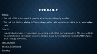
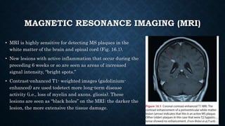
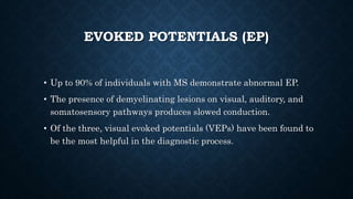
MULTIPLE SCLEROSIS
SAYALI GUJJEWAR
MPT II
INTRODUCTION
• Multiple sclerosis (MS) is an autoimmune disease characterized by
inflammation, selective demyelination, and gliosis.
• MS affects approximately 2.1 million people worldwide.
• It was first defined by Dr. Jean Charcot in 1868 by its clinical and
pathological characteristics: paralysis and the cardinal symptoms of
intention tremor, scanning speech, and nystagmus, later termed
Charcot’s triad.
• The onset of MS typically occurs between ages 20 and 40 years.
• MS is rare in children, as is the onset of symptoms in adults
older than age 50 years.
• The disease is more common in woman than in men by a ratio
of 2:1 to 3:1.
• MS affects predominantly white populations; African
Americans demonstrate approximately half the risk of
acquiring the disease. Low rates are also reported in Asians
and Native Americans.
ETIOLOGY
Family
• The risk of MS is increased in persons with an affected family member.
• The risk is 3.0% for a sibling, 5.0% for a fraternal co-twin, and rises to 25.0% for an identical co-
twin.
Genetic
• Genetic studies have revealed many interacting alleles that may contribute to MS susceptibility
with mutations in the human leukocyte antigen major histocompatibility complex (MHC) gene
most strongly correlated.
Viral infection
Vitamin D deficiency
Smoking
PATHOPHYSIOLOGY
TYPES
SYMPTOMS
DIAGNOSIS
• The diagnosis of MS is made by the neurologist based on a careful
medical history, a complete neurological examination, and supportive
laboratory tests.
• Evidence of damage must be present in at least two separate areas of
the CNS (dissemination of lesions in space) and damage must have
occurred at two separate points in time at lease 1 month apart
(dissemination of lesions in time).
LABORATORY TESTS
• Magnetic Resonance Imaging (MRI)
• Evoked Potentials (EP)
• Lumbar Puncture (LP) With Cerebrospinal Fluid (CSF) Analysis
MAGNETIC RESONANCE IMAGING (MRI)
• MRI is highly sensitive for detecting MS plaques in the
white matter of the brain and spinal cord (Fig. 16.1).
• New lesions with active inflammation that occur during the
preceding 6 weeks or so are seen as areas of increased
signal intensity, “bright spots.”
• Contrast-enhanced T1- weighted images (gadolinium-
enhanced) are used todetect more long-term disease
activity (i.e., loss of myelin and axons, gliosis). These
lesions are seen as “black holes” on the MRI; the darker the
lesion, the more extensive the tissue damage.
EVOKED POTENTIALS (EP)
• Up to 90% of individuals with MS demonstrate abnormal EP.
• The presence of demyelinating lesions on visual, auditory, and
somatosensory pathways produces slowed conduction.
• Of the three, visual evoked potentials (VEPs) have been found to
be the most helpful in the diagnostic process.
LUMBAR PUNCTURE (LP) WITH
CEREBROSPINAL FLUID (CSF) ANALYSIS
• Patients with MS show elevated total immunoglobulin (IgG) in
CSF and the presence of oligoclonal IgG bands (seen in 90% to 95%
of patients) in response to inflammatory demyelinating lesions.
• Patients with PPMS have higher levels of immunoglobulins in
spinal fluid than patients with RRMS.
MEDICAL MANAGEMENT
• Immunosupression (ATCH)- initial phase
• Steroidal therapy- maintenance
• Beta-Interferon – treatment of relapses
• Immuno Globulin g - prevention of relapse
Multiple sclerosis

Multiple sclerosis

  • 1.
  • 2.
    INTRODUCTION • Multiple sclerosis(MS) is an autoimmune disease characterized by inflammation, selective demyelination, and gliosis. • MS affects approximately 2.1 million people worldwide. • It was first defined by Dr. Jean Charcot in 1868 by its clinical and pathological characteristics: paralysis and the cardinal symptoms of intention tremor, scanning speech, and nystagmus, later termed Charcot’s triad.
  • 3.
    • The onsetof MS typically occurs between ages 20 and 40 years. • MS is rare in children, as is the onset of symptoms in adults older than age 50 years. • The disease is more common in woman than in men by a ratio of 2:1 to 3:1. • MS affects predominantly white populations; African Americans demonstrate approximately half the risk of acquiring the disease. Low rates are also reported in Asians and Native Americans.
  • 4.
    ETIOLOGY Family • The riskof MS is increased in persons with an affected family member. • The risk is 3.0% for a sibling, 5.0% for a fraternal co-twin, and rises to 25.0% for an identical co- twin. Genetic • Genetic studies have revealed many interacting alleles that may contribute to MS susceptibility with mutations in the human leukocyte antigen major histocompatibility complex (MHC) gene most strongly correlated. Viral infection Vitamin D deficiency Smoking
  • 5.
  • 6.
  • 8.
  • 10.
    DIAGNOSIS • The diagnosisof MS is made by the neurologist based on a careful medical history, a complete neurological examination, and supportive laboratory tests. • Evidence of damage must be present in at least two separate areas of the CNS (dissemination of lesions in space) and damage must have occurred at two separate points in time at lease 1 month apart (dissemination of lesions in time).
  • 12.
    LABORATORY TESTS • MagneticResonance Imaging (MRI) • Evoked Potentials (EP) • Lumbar Puncture (LP) With Cerebrospinal Fluid (CSF) Analysis
  • 13.
    MAGNETIC RESONANCE IMAGING(MRI) • MRI is highly sensitive for detecting MS plaques in the white matter of the brain and spinal cord (Fig. 16.1). • New lesions with active inflammation that occur during the preceding 6 weeks or so are seen as areas of increased signal intensity, “bright spots.” • Contrast-enhanced T1- weighted images (gadolinium- enhanced) are used todetect more long-term disease activity (i.e., loss of myelin and axons, gliosis). These lesions are seen as “black holes” on the MRI; the darker the lesion, the more extensive the tissue damage.
  • 14.
    EVOKED POTENTIALS (EP) •Up to 90% of individuals with MS demonstrate abnormal EP. • The presence of demyelinating lesions on visual, auditory, and somatosensory pathways produces slowed conduction. • Of the three, visual evoked potentials (VEPs) have been found to be the most helpful in the diagnostic process.
  • 15.
    LUMBAR PUNCTURE (LP)WITH CEREBROSPINAL FLUID (CSF) ANALYSIS • Patients with MS show elevated total immunoglobulin (IgG) in CSF and the presence of oligoclonal IgG bands (seen in 90% to 95% of patients) in response to inflammatory demyelinating lesions. • Patients with PPMS have higher levels of immunoglobulins in spinal fluid than patients with RRMS.
  • 16.
    MEDICAL MANAGEMENT • Immunosupression(ATCH)- initial phase • Steroidal therapy- maintenance • Beta-Interferon – treatment of relapses • Immuno Globulin g - prevention of relapse