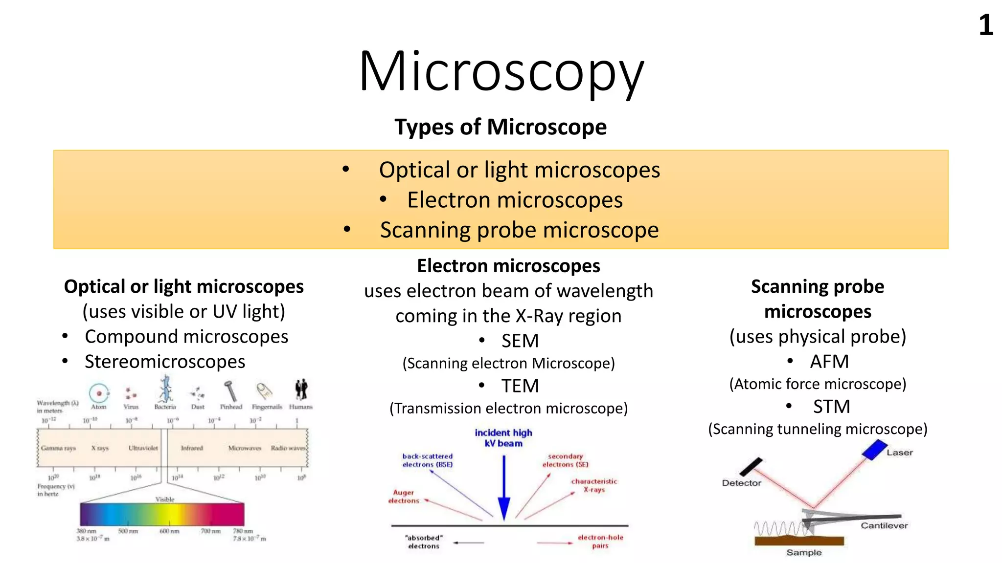
1) The document describes different types of microscopes including optical, electron, and scanning probe microscopes.
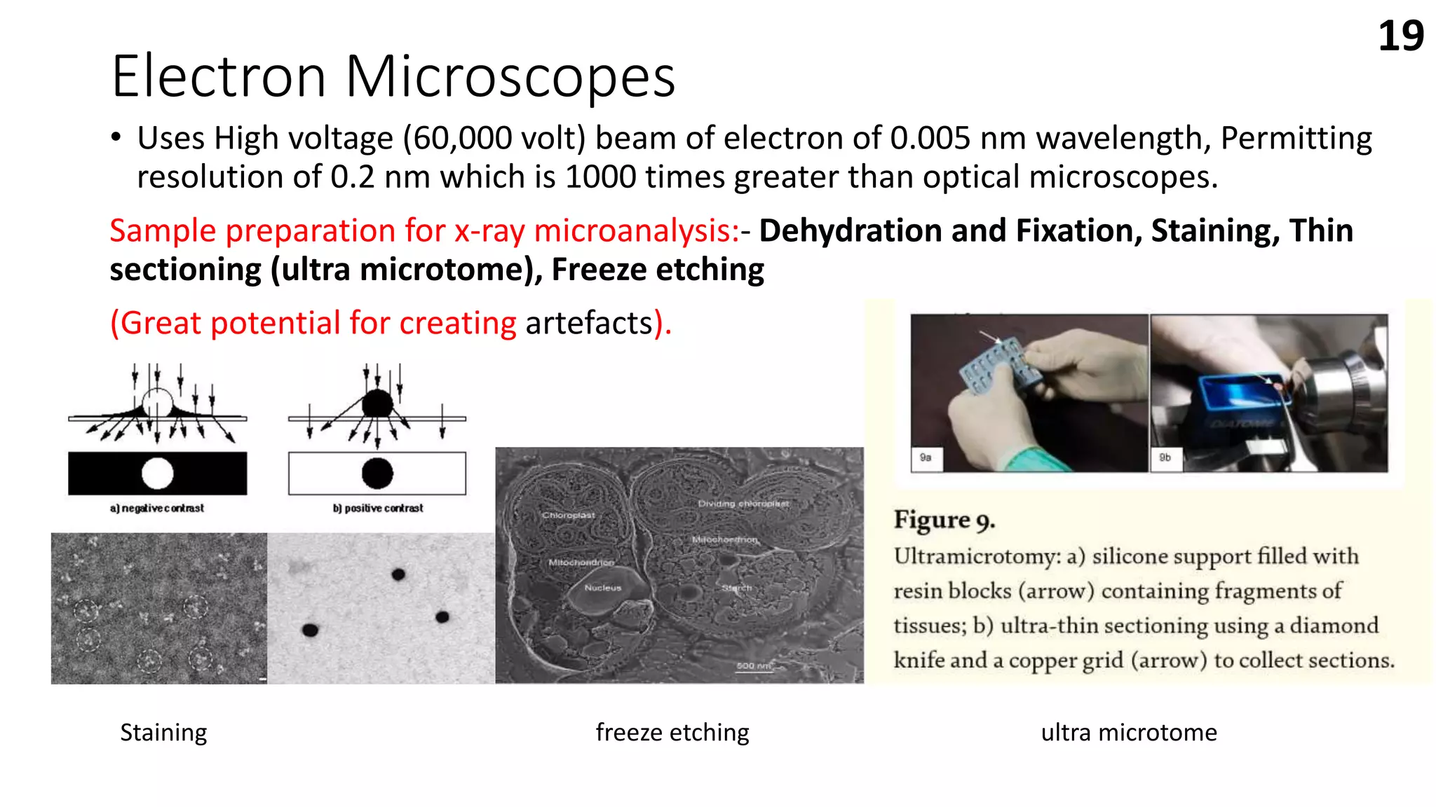
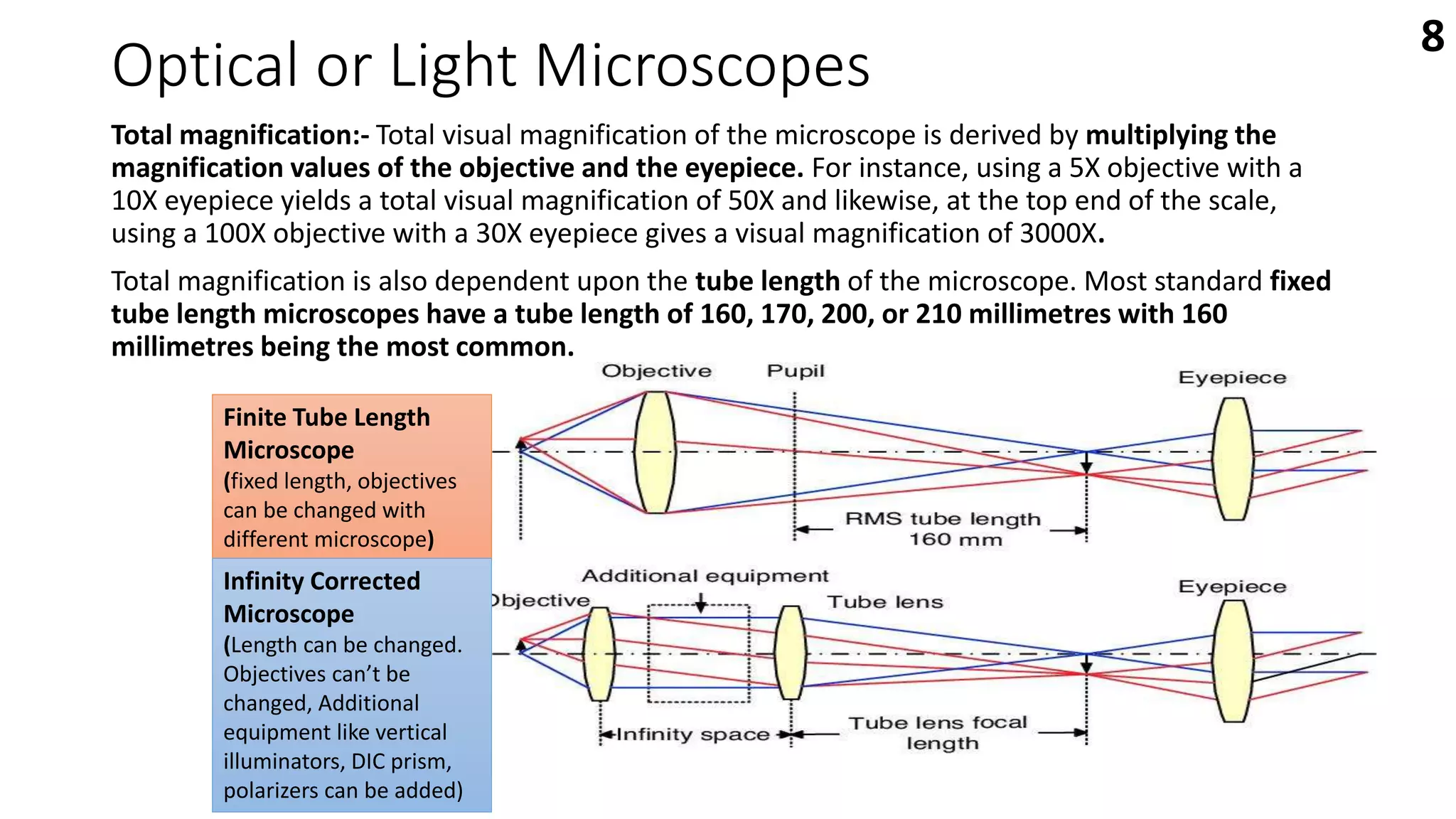
2) Optical microscopes use visible light and lenses to magnify images up to 2000x, while electron microscopes use electron beams for higher magnification up to 2 millionx and better resolution.
3) Scanning probe microscopes like atomic force microscopes and scanning tunneling microscopes use physical probes to generate high resolution images of surface topography at the atomic scale without using light or electron beams.
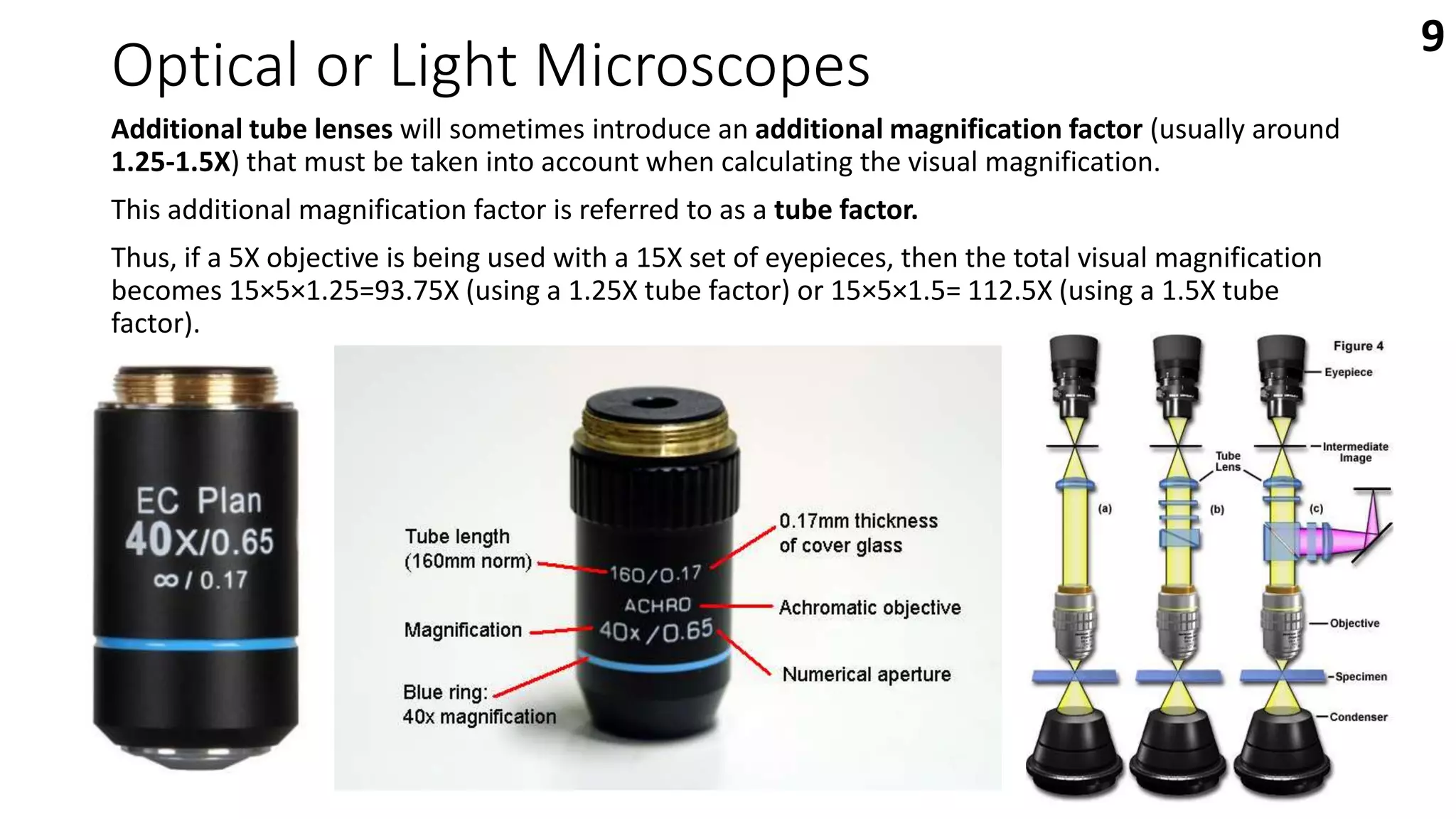
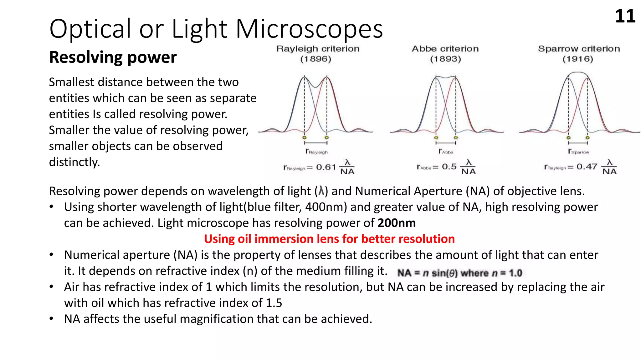
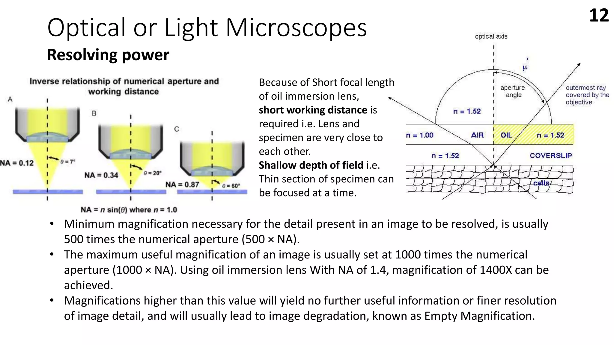
this presentation deals with the introduction of some of the commonly used optical microscopes in forensic labs; compound microscope, stereoscopic microscope, comparison microscope, fluorescence microscope and polarized microscope.
Types of Microscopes with their applications - Microbiologynote.com
https://microbiologynote.com/types-of-microscopes-with-their-applications/
Youtube Lecture Video:
https://www.youtube.com/watch?v=nuJZtXohFFQ&ab_channel=MicrobiologyNote
during this ppt of microscopes we will be able to know
INTRODUCTION
DEFINITION
HISTORICAL BACKGROUND
VARIABLES USED IN MICROSCOPY
VARIOUS TYPES OF MICROSCOPES
COMPOUND MICROSCOPE - Structure and Function
USE OF MICROSCOPE
CARE OF MICROSCOPE
defintion
A microscope (Greek: micron = small and scopos = aim)
MICROSCOPE - An instrument for viewing objects that are too small to be seen by the naked or unaided eye
MICROSCOPY - The science of investigating small objects using such an instrument is called microscopy
microscope (1).pdf this is a project for for botany majorarpitakhairwar123
Name - Arpita khairwar
Class - B.sc 1st year
Subject - Botany Major
College - Govt. Jayashankar Trivedi College Balaghat
Guided by - Dr. Pratima bisen
Submitted by - Arpita khairwar
While this ppt given by Dr pratima mam this ppt is a educational institution. My ppt is about microscope
Deep Behavioral Phenotyping in Systems Neuroscience for Functional Atlasing a...Ana Luísa Pinho
Functional Magnetic Resonance Imaging (fMRI) provides means to characterize brain activations in response to behavior. However, cognitive neuroscience has been limited to group-level effects referring to the performance of specific tasks. To obtain the functional profile of elementary cognitive mechanisms, the combination of brain responses to many tasks is required. Yet, to date, both structural atlases and parcellation-based activations do not fully account for cognitive function and still present several limitations. Further, they do not adapt overall to individual characteristics. In this talk, I will give an account of deep-behavioral phenotyping strategies, namely data-driven methods in large task-fMRI datasets, to optimize functional brain-data collection and improve inference of effects-of-interest related to mental processes. Key to this approach is the employment of fast multi-functional paradigms rich on features that can be well parametrized and, consequently, facilitate the creation of psycho-physiological constructs to be modelled with imaging data. Particular emphasis will be given to music stimuli when studying high-order cognitive mechanisms, due to their ecological nature and quality to enable complex behavior compounded by discrete entities. I will also discuss how deep-behavioral phenotyping and individualized models applied to neuroimaging data can better account for the subject-specific organization of domain-general cognitive systems in the human brain. Finally, the accumulation of functional brain signatures brings the possibility to clarify relationships among tasks and create a univocal link between brain systems and mental functions through: (1) the development of ontologies proposing an organization of cognitive processes; and (2) brain-network taxonomies describing functional specialization. To this end, tools to improve commensurability in cognitive science are necessary, such as public repositories, ontology-based platforms and automated meta-analysis tools. I will thus discuss some brain-atlasing resources currently under development, and their applicability in cognitive as well as clinical neuroscience.
Professional air quality monitoring systems provide immediate, on-site data for analysis, compliance, and decision-making.
Monitor common gases, weather parameters, particulates.
Richard's entangled aventures in wonderlandRichard Gill
Since the loophole-free Bell experiments of 2020 and the Nobel prizes in physics of 2022, critics of Bell's work have retreated to the fortress of super-determinism. Now, super-determinism is a derogatory word - it just means "determinism". Palmer, Hance and Hossenfelder argue that quantum mechanics and determinism are not incompatible, using a sophisticated mathematical construction based on a subtle thinning of allowed states and measurements in quantum mechanics, such that what is left appears to make Bell's argument fail, without altering the empirical predictions of quantum mechanics. I think however that it is a smoke screen, and the slogan "lost in math" comes to my mind. I will discuss some other recent disproofs of Bell's theorem using the language of causality based on causal graphs. Causal thinking is also central to law and justice. I will mention surprising connections to my work on serial killer nurse cases, in particular the Dutch case of Lucia de Berk and the current UK case of Lucy Letby.
THE IMPORTANCE OF MARTIAN ATMOSPHERE SAMPLE RETURN.Sérgio Sacani
The return of a sample of near-surface atmosphere from Mars would facilitate answers to several first-order science questions surrounding the formation and evolution of the planet. One of the important aspects of terrestrial planet formation in general is the role that primary atmospheres played in influencing the chemistry and structure of the planets and their antecedents. Studies of the martian atmosphere can be used to investigate the role of a primary atmosphere in its history. Atmosphere samples would also inform our understanding of the near-surface chemistry of the planet, and ultimately the prospects for life. High-precision isotopic analyses of constituent gases are needed to address these questions, requiring that the analyses are made on returned samples rather than in situ.
This pdf is about the Schizophrenia.
For more details visit on YouTube; @SELF-EXPLANATORY;
https://www.youtube.com/channel/UCAiarMZDNhe1A3Rnpr_WkzA/videos
Thanks...!
Seminar of U.V. Spectroscopy by SAMIR PANDASAMIR PANDA
Spectroscopy is a branch of science dealing the study of interaction of electromagnetic radiation with matter.
Ultraviolet-visible spectroscopy refers to absorption spectroscopy or reflect spectroscopy in the UV-VIS spectral region.
Ultraviolet-visible spectroscopy is an analytical method that can measure the amount of light received by the analyte.
Multi-source connectivity as the driver of solar wind variability in the heli...Sérgio Sacani
The ambient solar wind that flls the heliosphere originates from multiple
sources in the solar corona and is highly structured. It is often described
as high-speed, relatively homogeneous, plasma streams from coronal
holes and slow-speed, highly variable, streams whose source regions are
under debate. A key goal of ESA/NASA’s Solar Orbiter mission is to identify
solar wind sources and understand what drives the complexity seen in the
heliosphere. By combining magnetic feld modelling and spectroscopic
techniques with high-resolution observations and measurements, we show
that the solar wind variability detected in situ by Solar Orbiter in March
2022 is driven by spatio-temporal changes in the magnetic connectivity to
multiple sources in the solar atmosphere. The magnetic feld footpoints
connected to the spacecraft moved from the boundaries of a coronal hole
to one active region (12961) and then across to another region (12957). This
is refected in the in situ measurements, which show the transition from fast
to highly Alfvénic then to slow solar wind that is disrupted by the arrival of
a coronal mass ejection. Our results describe solar wind variability at 0.5 au
but are applicable to near-Earth observatories.
(May 29th, 2024) Advancements in Intravital Microscopy- Insights for Preclini...Scintica Instrumentation
Intravital microscopy (IVM) is a powerful tool utilized to study cellular behavior over time and space in vivo. Much of our understanding of cell biology has been accomplished using various in vitro and ex vivo methods; however, these studies do not necessarily reflect the natural dynamics of biological processes. Unlike traditional cell culture or fixed tissue imaging, IVM allows for the ultra-fast high-resolution imaging of cellular processes over time and space and were studied in its natural environment. Real-time visualization of biological processes in the context of an intact organism helps maintain physiological relevance and provide insights into the progression of disease, response to treatments or developmental processes.
In this webinar we give an overview of advanced applications of the IVM system in preclinical research. IVIM technology is a provider of all-in-one intravital microscopy systems and solutions optimized for in vivo imaging of live animal models at sub-micron resolution. The system’s unique features and user-friendly software enables researchers to probe fast dynamic biological processes such as immune cell tracking, cell-cell interaction as well as vascularization and tumor metastasis with exceptional detail. This webinar will also give an overview of IVM being utilized in drug development, offering a view into the intricate interaction between drugs/nanoparticles and tissues in vivo and allows for the evaluation of therapeutic intervention in a variety of tissues and organs. This interdisciplinary collaboration continues to drive the advancements of novel therapeutic strategies.
Richard's aventures in two entangled wonderlandsRichard Gill
Since the loophole-free Bell experiments of 2020 and the Nobel prizes in physics of 2022, critics of Bell's work have retreated to the fortress of super-determinism. Now, super-determinism is a derogatory word - it just means "determinism". Palmer, Hance and Hossenfelder argue that quantum mechanics and determinism are not incompatible, using a sophisticated mathematical construction based on a subtle thinning of allowed states and measurements in quantum mechanics, such that what is left appears to make Bell's argument fail, without altering the empirical predictions of quantum mechanics. I think however that it is a smoke screen, and the slogan "lost in math" comes to my mind. I will discuss some other recent disproofs of Bell's theorem using the language of causality based on causal graphs. Causal thinking is also central to law and justice. I will mention surprising connections to my work on serial killer nurse cases, in particular the Dutch case of Lucia de Berk and the current UK case of Lucy Letby.
What is greenhouse gasses and how many gasses are there to affect the Earth.moosaasad1975
What are greenhouse gasses how they affect the earth and its environment what is the future of the environment and earth how the weather and the climate effects.
1. Microscopy
Types of Microscope
Optical or light microscopes
(uses visible or UV light)
• Compound microscopes
• Stereomicroscopes
• Optical or light microscopes
• Electron microscopes
• Scanning probe microscope
Electron microscopes
uses electron beam of wavelength
coming in the X-Ray region
• SEM
(Scanning electron Microscope)
• TEM
(Transmission electron microscope)
Scanning probe
microscopes
(uses physical probe)
• AFM
(Atomic force microscope)
• STM
(Scanning tunneling microscope)
1
2. Optical or Light Microscopes
• Uses system of lenses and properties of light transmission like Refraction,
Diffraction, Reflection, Absorption and Fluorescence. to generate magnified
image of a small object.
Refraction
Diffraction
2
Fluorescence
3. Optical or Light Microscopes
• The past Centuries has witnessed an enormous growth in the application of optical
microscopy for micron and submicron level investigations in a wide variety of
disciplines.
• But early microscopists (late 18th century) were Facing some difficulties in some of the
aspects like image distortion, blurred images, and poor lens design.
Problems with the image quality!!
• Distortion of microscopic lenses (Spherical and chromatic aberration).
• Poor Magnification and resolution.
• Poor Contrast of the image.
3
4. Optical or Light Microscopes
Distortion of microscopic lenses
• Distorted images occur when not all the light from parallel rays passing through
lens focus at the same focal point.
• This distortion based on the shape of lenses is called spherical aberration.
• Lights of differing wavelength will also focus at different focal point. This is known
as chromatic aberration.
Aspherical aberration
Corrected
Achromatic aberration
Corrected
4
5. Optical or Light Microscopes
In 1830, Joseph Jackson Lister made massive strides in correcting a phenomenon called spherical
aberration. Lister built a device by placing weak lenses at precise distances from each other.
5
6. Optical or Light Microscopes
Magnification
• Visible or UV light is Refracted by a series of lenses to achieve magnification.
Using two convex lens systems i.e. Eyepiece or Ocular lens and objective lens in
combination, the image formed is magnified.
Objective lens :- The first lens of a microscope, closest to
the object is called the objective.
Light passes through the specimen and into the objective
which then projects a real, inverted, and magnified image
of the specimen to a fixed plane within the microscope
that is termed the intermediate image plane.
The intermediate image plane is usually located about 10
millimetres below the top of the microscope body tube.
• Objectives typically have magnifying powers that
range from 4X (or 5X), 10X, 20X, 40X (or 50X), and
100X.
6
7. Optical or Light Microscopes
Eyepiece (ocular lens) :- The eyepiece or ocular, which fits into the
microscope body tube at the upper end, is the farthest optical
component from the specimen. Ocular lens further magnifies the
primary real image projected by the objective. The eye of the observer
sees this secondarily magnified image as if it were at a distance of 10
inches (25 centimetres) from the eye.
Their magnification factors vary between 5X and 30X with the most
commonly used eyepieces having a value of 10X-15X.
7
8. Optical or Light Microscopes
Total magnification:- Total visual magnification of the microscope is derived by multiplying the
magnification values of the objective and the eyepiece. For instance, using a 5X objective with a
10X eyepiece yields a total visual magnification of 50X and likewise, at the top end of the scale,
using a 100X objective with a 30X eyepiece gives a visual magnification of 3000X.
Total magnification is also dependent upon the tube length of the microscope. Most standard fixed
tube length microscopes have a tube length of 160, 170, 200, or 210 millimetres with 160
millimetres being the most common.
Finite Tube Length
Microscope
(fixed length, objectives
can be changed with
different microscope)
Infinity Corrected
Microscope
(Length can be changed.
Objectives can’t be
changed, Additional
equipment like vertical
illuminators, DIC prism,
polarizers can be added)
8
9. Optical or Light Microscopes
Additional tube lenses will sometimes introduce an additional magnification factor (usually around
1.25-1.5X) that must be taken into account when calculating the visual magnification.
This additional magnification factor is referred to as a tube factor.
Thus, if a 5X objective is being used with a 15X set of eyepieces, then the total visual magnification
becomes 15×5×1.25=93.75X (using a 1.25X tube factor) or 15×5×1.5= 112.5X (using a 1.5X tube
factor).
9
10. Optical or Light Microscopes
Resolving power
• Magnifying an image by using microscope is useful
only when the details can be preserved accurately
and observed.
Rayleigh’s criterion:- diffraction of light
Limit of resolution:- smallest linear or angular
distance between the two objects at which they
appear separated(just resolved)
Resolving power is the reciprocal of the limit of
resolution.
10
11. Optical or Light Microscopes
Resolving power
Smallest distance between the two
entities which can be seen as separate
entities Is called resolving power.
Smaller the value of resolving power,
smaller objects can be observed
distinctly.
Resolving power depends on wavelength of light (λ) and Numerical Aperture (NA) of objective lens.
• Using shorter wavelength of light(blue filter, 400nm) and greater value of NA, high resolving power
can be achieved. Light microscope has resolving power of 200nm
Using oil immersion lens for better resolution
• Numerical aperture (NA) is the property of lenses that describes the amount of light that can enter
it. It depends on refractive index (n) of the medium filling it.
• Air has refractive index of 1 which limits the resolution, but NA can be increased by replacing the air
with oil which has refractive index of 1.5
• NA affects the useful magnification that can be achieved.
11
12. Optical or Light Microscopes
Resolving power
• Minimum magnification necessary for the detail present in an image to be resolved, is usually
500 times the numerical aperture (500 × NA).
• The maximum useful magnification of an image is usually set at 1000 times the numerical
aperture (1000 × NA). Using oil immersion lens With NA of 1.4, magnification of 1400X can be
achieved.
• Magnifications higher than this value will yield no further useful information or finer resolution
of image detail, and will usually lead to image degradation, known as Empty Magnification.
Because of Short focal length
of oil immersion lens,
short working distance is
required i.e. Lens and
specimen are very close to
each other.
Shallow depth of field i.e.
Thin section of specimen can
be focused at a time.
12
13. Optical or Light Microscopes
Contrast of the image
• To achieve better contrast, two methods are available ,i.e. Staining and light
illumination.
• Light illumination can be either Reflected OR Transmitted
Transmitted light illumination
(light passes through sample)
• Bright Field
• Differential interface contrast(DIC)
• Dark Field
• Phase contrast
• Polarization
Reflected light Illumination
(Light reflected back from the sample)
• Fluorescence
• Confocal
• Multiphoton
• TIRF(total internal reflection
fluorescence)
• Super resolution
13
14. Optical or Light Microscopes
Contrast of the image
• Bright field
Used for fixed
stained samples.
• Differential interference
contrast
• Unstained, transparent
sample appears three
dimensional
• Dark field
Combining light
waves that are
out of phase
Blocking out of the central light rays that
ordinarily pass through or around the
specimen and allowing only oblique rays to
illuminate the specimen.
14
15. Optical or Light Microscopes
Contrast of the image
• Phase
contrast
Separation of Direct light or undeviated
light from diffracted light
• Polarization
Birefringence :- an incident ray of light is
split into two rays, called an ordinary ray and
an extraordinary ray, which are polarized
15
16. Optical or Light Microscopes
Contrast of the image
• Fluorescence
• Illuminated at one
wavelength and
observed at different
wavelengths.
Flurescein
Isothiocyanate
• Excitation filters
• Barrier filters
• Confocal
Scanning
• Beam of light from laser is
focused and scanned
through 2 mirrors.
• Eliminated the diffracted
light that blur the image.
• Do not form 2D image
16
17. Optical or Light Microscopes
1) Compound Microscope
• Designed with compound lens system, loaded with
several objective lens(5× to 100×) and eyepieces
(generally 10x).
Upright Microscope Inverted Microscope
• Objectives are above the
sample stage.
• For Fixed samples such as
cells and tissues section.
• Objectives are below the
sample stage.
• For live cell imaging.
• Sterile working conditions.
17
18. Optical or Light Microscopes
2) Stereomicroscopes
• Comparatively low power as compared to compound microscopes.(usually below
100×) objective is [1× to 2×] and eyepiece is 10×
• They can have Fixed magnification system or
Zoom magnification system.
• Working distance is much longer.
• Also known as dissecting microscope
18
19. • Uses High voltage (60,000 volt) beam of electron of 0.005 nm wavelength, Permitting
resolution of 0.2 nm which is 1000 times greater than optical microscopes.
Sample preparation for x-ray microanalysis:- Dehydration and Fixation, Staining, Thin
sectioning (ultra microtome), Freeze etching
(Great potential for creating artefacts).
Electron Microscopes
Staining freeze etching ultra microtome
19
20. Electron Microscopes
1) TEM:- Transmission Electron Microscope
Electromagnetic
lens
• Used to view thin specimens through
which electrons can pass generating a
projection image.
• Hot Tungsten Filament in the electron
gun provides electron beam.
• Vacuum system is maintained to prevent
collision with electron.
20
21. Electron Microscopes
2) SEM:- Scanning Electron Microscope
• Primary electron beam knocks electron out of
specimen. This secondary electron are
transmitted to detector.
• More intense signal is developed.
• Gives detailed topography of specimen surface.
21
22. Scanning probe microscope
• Images formation using a physical probe that scans the specimen surface.
• Resolution varies somewhat from technique to technique(atomic resolution)
• This family of techniques can be called “piezoelectric techniques”.
• The data are typically obtained as a two-dimensional grid of data points,
visualized in false colour as a computer image.
• The nature of an SPM probe tip depends entirely on the type of SPM being used.
• The apex of the probe defines the resolution of the microscope, the sharper the
probe the better the resolution
“contact mode” or “tapping mode.” Contact mode
maintains a constant force between the cantilever
tip of the microscope and the sample surface(fast
production of the image). Tapping mode involves
oscillation of the cantilever, so the tip intermittently
comes into contact with the sample surface(useful
for softer samples).
22
23. Scanning probe microscope
1) AFM:-Atomic Force Microscope
• measuring intermolecular forces and
sees atoms by using probed surfaces of
the specimen in nanoscale.
• Takes the image of the surface topography of
the sample by scanning the cantilever over a
section of interest.
23
24. Scanning probe microscope
2) STM:- Scanning Tunneling Microscope
24
• Used to obtain ultra-high resolution
images at the atomic scale, without
using light or electron beams.
• Quantum mechanical process (electron
tunneling),i.e. Electrons travel a barrier
(in this case, a tiny gap between the tip
and surface)
• No physical contact
• This tunneling current is highly sensitive
and can be detected.
• Image can be seen in Armstrong.
• Used in material science, measures
electron density gradient.