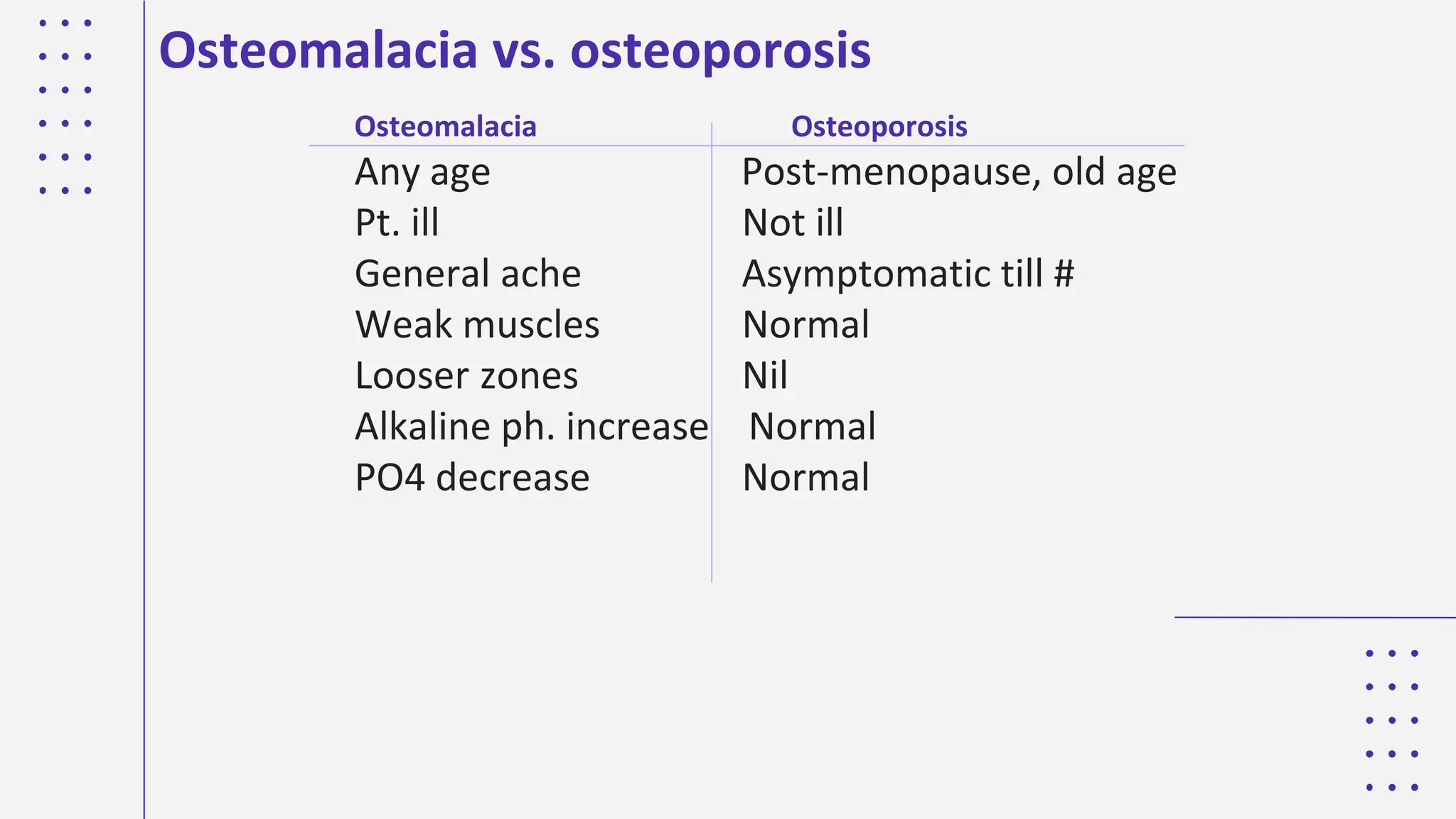
A PRESENTATION BY:
EKEOHA MICHAEL TOCHUKWU
UJ/2016/MD/0011
AUDU VICTORY
UJ/2016/MD/0014
METABOLIC
BONE
DISEASE
IMAGING MODALITIES
03
OUTLINE
INTRODUCTION
01
CLASSIFICATION METABOLIC BONE
DISEASES
02
CONCLUSIONS
04
INTRODUCTION
INTRODUCTION
ANATOMY OF BONE
• Cortical bone: the external part of each bone consists of
dense skeletal tissue known as cortical (compact) bone,
which contributes to most of the skeleton’s mechanical
strength.
• Trabecular bone: within the vertebrae and the ends of long
bones, the internal space is filled with a fine network of bone
tissue called trabecular (cancellous) bone.
This is in intimate contact with the bone marrow and is
largely responsible for the skeleton’s metabolic role as a
reservoir for body calcium. In addition, trabecular elements
are thought to contribute to the ability of vertebrae to
withstand compressive forces, with loss of these contributing
to the vertebral collapse seen in osteoporosis.
INTRODUCTION
FUNCTION OF BONE
Bone has two main functions:
1. To provide an endoskeleton
During growth, bone formation and resorption are regulated as
part of the modelling process that results in the micro- and macro
architecture of the adult skeleton.
• Modelling: involves resorption secondary to bone formation
• Remodeling: consists of repeated cycles of bone resorption
followed by formation, at discrete sites throughout the skeleton
2. To act as a reservoir for body calcium
Many essential intracellular processes are critically dependent on
the concentration of ionized extracellular calcium, powerful
homoeostatic mechanisms preserve the concentration of
extracellular calcium by using skeletal calcium stores, this can
ultimately lead to a significant loss of calcium from bone.
INTRODUCTION
Metabolic bone diseases are a diverse group of
diseases that result in abnormalities of:
(a) bone mass
(b) structure mineral homeostasis
(c) bone turnover
(d) growth
They lead to an altered serum calcium concentration
and/or skeletal failure.
CLASSIFICATION OF METABOLIC BONE DISEASES
1. TOO LITTLE CALCIFIED BONE
A. Too little bone formed
◆too little matrix—osteoporosis
◆ too little calcification of matrix– osteomalacia or rickets
B. Too much bone resorbed—Hyperparathyroidism
2. TOO MUCH CALCIFIED BONE
A. Too much bone formed
◆too much matrix— Poisonings, Excessive stress, Repair
B. Too little bone resorbed- Osteosclerosis
GENERAL IMAGING TECHNIQUES
Plain radiography
Metabolic bone disease can be associated with adverse effects
on the skeleton that can be readily recognized on plain
radiographs. Although changes such as low bone density are
non-specific, specific changes pointing to a particular diagnosis
such as Looser’s zones in osteomalacia may be evident.
GENERAL IMAGING TECHNIQUES
Isotope bone scan
Metabolic bone diseases can be associated with local areas of
increased technetium uptake, especially if there are associated
fractures. Widespread changes are most commonly a result of
skeletal secondary malignant deposits but also occur in
osteomalacia and Paget’s disease of bone.
Computed Tomography
GENERAL IMAGING TECHNIQUES
Bone densitometry
A number of techniques have been developed to quantify the
amount of bone mineral present at a given skeletal site, from
which other values such as bone mineral density can be derived.
They largely consist of dual energy absorptiometry (DXA) and
ultrasound-based approaches to assess fracture risk when
preventative therapy is being considered. DXA measures bone
mineral density (BMD), which is bone mineral content partially
corrected for size, either centrally (spinal BMD) or peripherally
(forearm or heel BMD). Quantitative ultrasound analyses
transmission of high-frequency sound through bone at the
calcaneus, phalanges and other skeletal sites.
OSTEOPOROSIS
01
Osteoporosis is defined as a condition characterized
by diminished but otherwise normal bone. An
osteoporotic state may arise either when bone
formation is inadequate or when bone resorption
exceeds bone formation. Osteoporosis may be a
local phenomenon (as in disuse osteoporosis) or a
generalized condition. The imaging features depend
to some degree on the rate at which osteoporosis
develops.
OSTEOPOROSIS
Risk Factors in Post menopausal Osteoporosis
● Race
● Hereditary
● Body build
● Early menopause
● Smoking/ alcohol intake/ drug abuse
Clinical Features of Osteoporosis
● Osteoporosis is a Silent disease
● Osteoporosis is Serious due to possible complications :mainly
fractures
● Osteoporosis does not cause pain usually
● Osteoporosis causes gradual increase in dorsal kyphosis
● Osteoporosis leads to loss of height
● Osteoporosis is not osteoarthritis; but the two conditions may co-
exist
Osteoporosis: Primary and Secondary
● Primary Osteoporosis :
Post menopausal
Senile
Post menopausal Osteoporosis
● Due to rapid decline in estrogen level
● This results in increased osteoclastic activity
● Normal bone loss usually 0.3% per year
● Post menopausal bone loss 3% per year
Osteoporotic Fractures
 They are Pathological fractures
 Most common is osteoporotic compression fracture ( OVC #s )
 Vertebral micro fractures occur unnoticed (dull ache)
 Most serious is hip fractures
 Also common is wrist fractures ( Colles fracture )
Secondary Osteoporosis
 Drug induced : steroids, alcohol, smoking, phenytoin,heparin
 Hyperparathyroidism, hyperthyroidism, Cushing's syndrome, gonadal
disorders, malabsorption, mal nutrition
 Chronic diseases : RA, renal failure, tuberculosis
 Malignancy : multiple myeloma, leukemia, metastasis
Disuse Osteoporosis
● Occurs locally adjacent to immobilized bone or joint
● May be generalized in in bed ridden patients
● Awareness of and attempts for prevention are helpful
Prevention of Osteoporosis
● Prevention of osteoporosis should start from childhood
● Healthy diet, adequate sunshine, regular exercise, avoidance of
smoking or alcohol, caution in steroid use
● At some time in the past there was a recommendation of HRT
(Hormone replacement Therapy ) for post menopausal women ? And
men; but now this is discontinued
IMAGING
IMAGING
IMAGING
IMAGING
IMAGING
Management of Osteoporosis
● Drugs
● Exercise
● Management of fractures
 Estrogen has a definite therapeutic effect and was used extensively
as HRT but cannot be recommended now due to serious possible
side effects
 Adequate intake of calcium and vitamin D is mandatory
 Drugs which inhibit osteoclast activities : e.g. Bisphosphonates like
sodium alendronate FOSAMAX , BONVIVA
 Drugs which enhance osteoblast activities : bone stimulating
agents like PROTELOS, FORTEO
Drug Therapy in Osteoporosis
• Resistive exercises
• Weight bearing exercises
• Exercise should be intelligent to
avoid injury which may lead to
fracture
Exercise in Osteoporosis
Rickets and
Osteomalacia
02
Rickets is the interruption of orderly development
and mineralization of growth plates. Osteomalacia
is inadequate or abnormal mineralization of osteoid
in cortical and trabecular bone. Prior to growth
plate fusion, rickets and osteomalacia coexist
Rickets and Osteomalacia
RICKETS
Rickets results from deficiency of vitamin D in a
child before epiphyseal closure.
Age= 4-18 months
Patients presents with tetany or convulsion in the
first 6 months of life due to low calcium level.
Later, they present with swollen tender joints.
RADIOLOGICAL FEATURES
● Changes are maximal at site of bone growth, best seen in
the knees, wrists, and ankles.
● The features are due to non-calcification of osteoid and the
effect of stress on the weakened bone.
● The changes in the long bones include:
a. The earliest radiological sign is loss of zone of provisional
calcification
b. Indistinctness of the metaphyseal margins with fraying
c. Splaying and cupping of the metaphyseal margins
d. Indistinctness of the cortical margins
e. Soft tissue swelling of the metaphysis due to pilling up of
osteoid
f. Generalized decrease in bone density
g. Delay in bone maturation and epiphyseal fusion
h. Incomplete fractures- loosers zone may occur
i. Appendicular skeletal deformities
1. genu varum
2. genu valgus
3. wind swept deformity
RADIOLOGICAL FEATURES
● Thoracic changes;
a. Kyphosis
b. Scoliosis
c. Pigeon chest
d. Harrisons sulcus
e. Rickety rosary
RADIOLOGICAL FEATURES
● Skull
a. Frontal bossing
b. Basilar invagination
c. Loss of lamina dura
d. Tam-o'-shanter skull (thickening of
the skull vault with basilar
invagination)
MNEMONIC: "RICKETS"
● Reaction of periosteum may occur
● Indistinct cortex
● Coarse trabeculation
● Knees + wrists + ankles mainly
affected
● Epiphyseal plates widened +
irregular
● Tremendous metaphysis (fraying,
splaying, cupping)
● Spur (metaphyseal)
RADIOLOGICAL FEATURES
Osteomalacia vs. osteoporosis
Osteomalacia Osteoporosis
Any age Post-menopause, old age
Pt. ill Not ill
General ache Asymptomatic till #
Weak muscles Normal
Looser zones Nil
Alkaline ph. increase Normal
PO4 decrease Normal
HYPOPHOSPHATAEMIA
03
Hypophosphatasia
Hypophosphatasia is a rare genetic disorder caused by mutations in the gene
that encodes tissue-nonspecific alkaline phosphatase, resulting in
accumulation of pyrophosphate, an inhibitor of bone mineralization.
The skeletal findings of hypophosphatasia resemble those of rickets and
osteomalacia.
The clinical spectrum of disease varies widely, and it can be roughly
categorized into the following four clinical phenotypes of decreasing severity:
1. perinatal
2. Infantile
3. Childhood
4. Adult
In the perinatal form, mineralization can be remarkably poor, with entire
segments of the spine not depicted (absent) on radiographs. In the infantile
and childhood forms, there can be craniosynostosis; and in the childhood
form, characteristic “tongues” of lucency extend from the growth plate to the
metaphysis.
Hypophosphatasia
Hypophosphatasia
Hyperparathyroidism
04
Hyperparathyroidism
Hyperparathyroidism is a pathologic state of elevated parathyroid hormone
concentrations, which causes increased bone resorption.
Primary hyperparathyroidism is a state of autonomous parathyroid hormone
secretion by the parathyroid glands and lack of feedback inhibition by serum
calcium. Primary hyperparathyroidism is usually caused by a parathyroid
adenoma.
Secondary hyperparathyroidism is more common than primary
hyperparathyroidism and is a response to low serum calcium levels. The most
common cause is chronic renal failure, in which chronically elevated serum
phosphate levels depress the serum calcium level, which leads to
compensatory hyperplasia of the chief cells of the parathyroid gland
Hyperparathyroidism
● Excessive PTH secretion : primary, secondary or tertiary
● Leads to increased bone resorption , sub periosteal erosions, osteitis
manifested by fibrous replacement of bone
● Significant feature is hypercalcemia
● In severe cases : osteitis fibrosa cystica and formation of Brown
tumours
Radiological changes in Hyperparathyroidism
● Generalized decrease in bone density
● Sub-periosteal bone resorption ( scalloping of metacarpals and
phalanges )
● Brown tumors
● Chondrocalcinosis ( wrist, knee, shoulder )
Hyperparathyroidism
Hyperparathyroidism
Hyperparathyroidism
Hyperparathyroidism
Hyperparathyroidism
Management of Hyperparathyroidism
● By management of the cause :
● Primary hyperparathyroidism due to neoplasm ( adenoma or
carcinoma ) by excision
● Secondary hyperparathyroidism by correcting the cause of
hypocalcemia
● Tertiary hyperparathyroidism by excision of hyperactive
● ( autonomous )nodule
● Extreme care should be applied after surgery to avoid hypocalcemia
due hungry bones syndrome
Renal Osteodystrophy
05
Renal Osteodystrophy
Renal osteodystrophy refers to the complex of
findings observed in the setting of chronic renal
insufficiency.
These include the findings of osteomalacia (and rickets in children) and
secondary hyperparathyroidism. In any given patient, the findings of one or
the other may predominate.
In patients with chronic renal insufficiency, radiographs may show a diffuse
increase in bone radiodensity, a finding that is seen more often in the axial
skeleton, which has more trabecular bone than cortical bone.
Despite the increased radiodensity, the bone is structurally weak and
prone to stress fractures.
Renal Osteodystrophy
Renal osteodystrophy in a 60-year-old man after he
underwent renal transplantation for
glomerulonephritis. Anteroposterior radiograph of
the lumbar spine shows diffusely increased
radiodensity of the depicted bones.
Renal Osteodystrophy
Paget disease(Osteitis Deformans).
06
Paget disease (Osteitis Deformans).
● It is a disorder characterized by episodes of localized, increased
osteoclastic activity & bone resorption, followed by exuberant bone
formation. The end result of these intermittent processes is skeletal
deformity.
● Uncommon under 40 years, male < female.
● Usually sporadic but some cases are familial with abnormality on
chromosome 18.
Pathogenesis of Paget disease.
○ Paget disease is inflammatory in nature. Current evidence suggests that
Paget disease is of infectious etiology, depending on the following
facts.
I. Paramyxovirus like particles & antigens have been identified within
osteoclasts obtained from patients with Paget disease.
II. More recently other virus agents are isolated from the lesion of Paget
disease which is Measles virus.
● These viral agents are thought to be play a role in pathogenesis of Paget
disease by activating osteoclastic activity.
Morphology of Paget disease.
Gross. Either solitary or multifocal lesions.
Microscopic.
Usually pass in phases.
1. Primary (osteolytic) phase. There is bone resorption by osteoclasts.
2. Osteoclasts & Osteoblasts proliferation. (Mixed phase).
3. Osteosclerotic bone formation phase. Which is characterized by cessation
of osteoclastic activity & continuing osteoblastic activity, that result in
formation of WOVEN BONE (weak bone & is subject to fracture &
deformity).
Complications of paget disease.
1. Bone deformity (enlargement of head, kyphosis, scoliosis…).
2. Backache is due to involvement of vertebrae (vertebral fractures & nerve
root compression).
3. Chalk stick fracture of long bones.
4. Osteosarcoma in les than 1% of cases(usually multicentric & poor
prognosis malignancy).
5. High cardiac output failure in extensive cases.
Paget disease / Gross
References
● 1. Skowrońska-Jóźwiak E, Lorenc RS. Metabolic bone disease in
children: etiology and treatment options. Treat Endocrinol
2006;5(5):297–318.
● 2. Guglielmi G, Muscarella S, Bazzocchi A. Integrated imaging
approach to osteoporosis: state-of-the-art review and update.
RadioGraphics 2011;31(5):1343–1364.
● 3. O’Neill TW, Felsenberg D, Varlow J, Cooper C, Kanis JA,
Silman AJ. The prevalence of vertebral deformity in European
men and women: the European Vertebral Osteoporosis Study. J
Bone Miner Res 1996;11(7):1010–1018.
● 4. Looker AC, Orwoll ES, Johnston CC Jr, et al. Prevalence of low
femoral bone density in older U.S. adults from NHANES III. J
Bone Miner Res 1997;12(11):1761–1768.
RESEARCH
References
● 5. Kanis J, on behalf of the World Health Organization Scientific
Group. Assessment of osteoporosis at the primary health care
level: technical report. Sheffield, United Kingdom: World Health
Organization Collaborating Centre for Metabolic Bones
Diseases, University of Sheffield, 2007. University of Sheffield
website. https://www.shef.ac.uk/FRAX/pdfs/
WHO_Technical_Report.pdf. Accessed November 12, 2015.
● 6. Johnell O, Kanis JA. An estimate of the worldwide prevalence
and disability associated with osteoporotic fractures.
Osteoporos Int 2006;17(12):1726–1733
RESEARCH
CONCLUSIONS
Metabolic bone diseases are a
heterogeneous group of disorders that
diffusely affect the bones. Understanding
the underlying mechanism of the diseases
helps the radiologist to also understand
the radiographic findings and to make a
correct diagnosis.

THE SCRIBES SLIDE-MET BONE DISEASE, management.pdf

  • 1.
    A PRESENTATION BY: EKEOHAMICHAEL TOCHUKWU UJ/2016/MD/0011 AUDU VICTORY UJ/2016/MD/0014 METABOLIC BONE DISEASE
  • 2.
  • 3.
  • 4.
    INTRODUCTION ANATOMY OF BONE •Cortical bone: the external part of each bone consists of dense skeletal tissue known as cortical (compact) bone, which contributes to most of the skeleton’s mechanical strength. • Trabecular bone: within the vertebrae and the ends of long bones, the internal space is filled with a fine network of bone tissue called trabecular (cancellous) bone. This is in intimate contact with the bone marrow and is largely responsible for the skeleton’s metabolic role as a reservoir for body calcium. In addition, trabecular elements are thought to contribute to the ability of vertebrae to withstand compressive forces, with loss of these contributing to the vertebral collapse seen in osteoporosis.
  • 5.
    INTRODUCTION FUNCTION OF BONE Bonehas two main functions: 1. To provide an endoskeleton During growth, bone formation and resorption are regulated as part of the modelling process that results in the micro- and macro architecture of the adult skeleton. • Modelling: involves resorption secondary to bone formation • Remodeling: consists of repeated cycles of bone resorption followed by formation, at discrete sites throughout the skeleton 2. To act as a reservoir for body calcium Many essential intracellular processes are critically dependent on the concentration of ionized extracellular calcium, powerful homoeostatic mechanisms preserve the concentration of extracellular calcium by using skeletal calcium stores, this can ultimately lead to a significant loss of calcium from bone.
  • 6.
    INTRODUCTION Metabolic bone diseasesare a diverse group of diseases that result in abnormalities of: (a) bone mass (b) structure mineral homeostasis (c) bone turnover (d) growth They lead to an altered serum calcium concentration and/or skeletal failure.
  • 7.
    CLASSIFICATION OF METABOLICBONE DISEASES 1. TOO LITTLE CALCIFIED BONE A. Too little bone formed ◆too little matrix—osteoporosis ◆ too little calcification of matrix– osteomalacia or rickets B. Too much bone resorbed—Hyperparathyroidism 2. TOO MUCH CALCIFIED BONE A. Too much bone formed ◆too much matrix— Poisonings, Excessive stress, Repair B. Too little bone resorbed- Osteosclerosis
  • 8.
    GENERAL IMAGING TECHNIQUES Plainradiography Metabolic bone disease can be associated with adverse effects on the skeleton that can be readily recognized on plain radiographs. Although changes such as low bone density are non-specific, specific changes pointing to a particular diagnosis such as Looser’s zones in osteomalacia may be evident.
  • 9.
    GENERAL IMAGING TECHNIQUES Isotopebone scan Metabolic bone diseases can be associated with local areas of increased technetium uptake, especially if there are associated fractures. Widespread changes are most commonly a result of skeletal secondary malignant deposits but also occur in osteomalacia and Paget’s disease of bone. Computed Tomography
  • 10.
    GENERAL IMAGING TECHNIQUES Bonedensitometry A number of techniques have been developed to quantify the amount of bone mineral present at a given skeletal site, from which other values such as bone mineral density can be derived. They largely consist of dual energy absorptiometry (DXA) and ultrasound-based approaches to assess fracture risk when preventative therapy is being considered. DXA measures bone mineral density (BMD), which is bone mineral content partially corrected for size, either centrally (spinal BMD) or peripherally (forearm or heel BMD). Quantitative ultrasound analyses transmission of high-frequency sound through bone at the calcaneus, phalanges and other skeletal sites.
  • 11.
  • 12.
    Osteoporosis is definedas a condition characterized by diminished but otherwise normal bone. An osteoporotic state may arise either when bone formation is inadequate or when bone resorption exceeds bone formation. Osteoporosis may be a local phenomenon (as in disuse osteoporosis) or a generalized condition. The imaging features depend to some degree on the rate at which osteoporosis develops. OSTEOPOROSIS
  • 13.
    Risk Factors inPost menopausal Osteoporosis ● Race ● Hereditary ● Body build ● Early menopause ● Smoking/ alcohol intake/ drug abuse
  • 14.
    Clinical Features ofOsteoporosis ● Osteoporosis is a Silent disease ● Osteoporosis is Serious due to possible complications :mainly fractures ● Osteoporosis does not cause pain usually ● Osteoporosis causes gradual increase in dorsal kyphosis ● Osteoporosis leads to loss of height ● Osteoporosis is not osteoarthritis; but the two conditions may co- exist
  • 15.
    Osteoporosis: Primary andSecondary ● Primary Osteoporosis : Post menopausal Senile
  • 16.
    Post menopausal Osteoporosis ●Due to rapid decline in estrogen level ● This results in increased osteoclastic activity ● Normal bone loss usually 0.3% per year ● Post menopausal bone loss 3% per year
  • 17.
    Osteoporotic Fractures  Theyare Pathological fractures  Most common is osteoporotic compression fracture ( OVC #s )  Vertebral micro fractures occur unnoticed (dull ache)  Most serious is hip fractures  Also common is wrist fractures ( Colles fracture )
  • 18.
    Secondary Osteoporosis  Druginduced : steroids, alcohol, smoking, phenytoin,heparin  Hyperparathyroidism, hyperthyroidism, Cushing's syndrome, gonadal disorders, malabsorption, mal nutrition  Chronic diseases : RA, renal failure, tuberculosis  Malignancy : multiple myeloma, leukemia, metastasis
  • 19.
    Disuse Osteoporosis ● Occurslocally adjacent to immobilized bone or joint ● May be generalized in in bed ridden patients ● Awareness of and attempts for prevention are helpful
  • 20.
    Prevention of Osteoporosis ●Prevention of osteoporosis should start from childhood ● Healthy diet, adequate sunshine, regular exercise, avoidance of smoking or alcohol, caution in steroid use ● At some time in the past there was a recommendation of HRT (Hormone replacement Therapy ) for post menopausal women ? And men; but now this is discontinued
  • 21.
  • 22.
  • 23.
  • 24.
  • 25.
  • 26.
    Management of Osteoporosis ●Drugs ● Exercise ● Management of fractures
  • 27.
     Estrogen hasa definite therapeutic effect and was used extensively as HRT but cannot be recommended now due to serious possible side effects  Adequate intake of calcium and vitamin D is mandatory  Drugs which inhibit osteoclast activities : e.g. Bisphosphonates like sodium alendronate FOSAMAX , BONVIVA  Drugs which enhance osteoblast activities : bone stimulating agents like PROTELOS, FORTEO Drug Therapy in Osteoporosis
  • 28.
    • Resistive exercises •Weight bearing exercises • Exercise should be intelligent to avoid injury which may lead to fracture Exercise in Osteoporosis
  • 29.
  • 30.
    Rickets is theinterruption of orderly development and mineralization of growth plates. Osteomalacia is inadequate or abnormal mineralization of osteoid in cortical and trabecular bone. Prior to growth plate fusion, rickets and osteomalacia coexist Rickets and Osteomalacia
  • 31.
  • 32.
    RICKETS Rickets results fromdeficiency of vitamin D in a child before epiphyseal closure. Age= 4-18 months Patients presents with tetany or convulsion in the first 6 months of life due to low calcium level. Later, they present with swollen tender joints.
  • 33.
    RADIOLOGICAL FEATURES ● Changesare maximal at site of bone growth, best seen in the knees, wrists, and ankles. ● The features are due to non-calcification of osteoid and the effect of stress on the weakened bone. ● The changes in the long bones include: a. The earliest radiological sign is loss of zone of provisional calcification b. Indistinctness of the metaphyseal margins with fraying c. Splaying and cupping of the metaphyseal margins d. Indistinctness of the cortical margins e. Soft tissue swelling of the metaphysis due to pilling up of osteoid f. Generalized decrease in bone density g. Delay in bone maturation and epiphyseal fusion h. Incomplete fractures- loosers zone may occur i. Appendicular skeletal deformities 1. genu varum 2. genu valgus 3. wind swept deformity
  • 34.
  • 35.
    ● Thoracic changes; a.Kyphosis b. Scoliosis c. Pigeon chest d. Harrisons sulcus e. Rickety rosary RADIOLOGICAL FEATURES ● Skull a. Frontal bossing b. Basilar invagination c. Loss of lamina dura d. Tam-o'-shanter skull (thickening of the skull vault with basilar invagination)
  • 36.
    MNEMONIC: "RICKETS" ● Reactionof periosteum may occur ● Indistinct cortex ● Coarse trabeculation ● Knees + wrists + ankles mainly affected ● Epiphyseal plates widened + irregular ● Tremendous metaphysis (fraying, splaying, cupping) ● Spur (metaphyseal) RADIOLOGICAL FEATURES
  • 37.
  • 38.
  • 39.
    Osteomalacia vs. osteoporosis OsteomalaciaOsteoporosis Any age Post-menopause, old age Pt. ill Not ill General ache Asymptomatic till # Weak muscles Normal Looser zones Nil Alkaline ph. increase Normal PO4 decrease Normal
  • 40.
  • 41.
    Hypophosphatasia Hypophosphatasia is arare genetic disorder caused by mutations in the gene that encodes tissue-nonspecific alkaline phosphatase, resulting in accumulation of pyrophosphate, an inhibitor of bone mineralization. The skeletal findings of hypophosphatasia resemble those of rickets and osteomalacia. The clinical spectrum of disease varies widely, and it can be roughly categorized into the following four clinical phenotypes of decreasing severity: 1. perinatal 2. Infantile 3. Childhood 4. Adult In the perinatal form, mineralization can be remarkably poor, with entire segments of the spine not depicted (absent) on radiographs. In the infantile and childhood forms, there can be craniosynostosis; and in the childhood form, characteristic “tongues” of lucency extend from the growth plate to the metaphysis.
  • 42.
  • 43.
  • 44.
  • 45.
    Hyperparathyroidism Hyperparathyroidism is apathologic state of elevated parathyroid hormone concentrations, which causes increased bone resorption. Primary hyperparathyroidism is a state of autonomous parathyroid hormone secretion by the parathyroid glands and lack of feedback inhibition by serum calcium. Primary hyperparathyroidism is usually caused by a parathyroid adenoma. Secondary hyperparathyroidism is more common than primary hyperparathyroidism and is a response to low serum calcium levels. The most common cause is chronic renal failure, in which chronically elevated serum phosphate levels depress the serum calcium level, which leads to compensatory hyperplasia of the chief cells of the parathyroid gland
  • 46.
    Hyperparathyroidism ● Excessive PTHsecretion : primary, secondary or tertiary ● Leads to increased bone resorption , sub periosteal erosions, osteitis manifested by fibrous replacement of bone ● Significant feature is hypercalcemia ● In severe cases : osteitis fibrosa cystica and formation of Brown tumours
  • 47.
    Radiological changes inHyperparathyroidism ● Generalized decrease in bone density ● Sub-periosteal bone resorption ( scalloping of metacarpals and phalanges ) ● Brown tumors ● Chondrocalcinosis ( wrist, knee, shoulder )
  • 48.
  • 49.
  • 50.
  • 51.
  • 52.
  • 53.
    Management of Hyperparathyroidism ●By management of the cause : ● Primary hyperparathyroidism due to neoplasm ( adenoma or carcinoma ) by excision ● Secondary hyperparathyroidism by correcting the cause of hypocalcemia ● Tertiary hyperparathyroidism by excision of hyperactive ● ( autonomous )nodule ● Extreme care should be applied after surgery to avoid hypocalcemia due hungry bones syndrome
  • 54.
  • 55.
    Renal Osteodystrophy Renal osteodystrophyrefers to the complex of findings observed in the setting of chronic renal insufficiency. These include the findings of osteomalacia (and rickets in children) and secondary hyperparathyroidism. In any given patient, the findings of one or the other may predominate. In patients with chronic renal insufficiency, radiographs may show a diffuse increase in bone radiodensity, a finding that is seen more often in the axial skeleton, which has more trabecular bone than cortical bone. Despite the increased radiodensity, the bone is structurally weak and prone to stress fractures.
  • 56.
  • 57.
    Renal Osteodystrophy Renal osteodystrophyin a 60-year-old man after he underwent renal transplantation for glomerulonephritis. Anteroposterior radiograph of the lumbar spine shows diffusely increased radiodensity of the depicted bones.
  • 58.
  • 59.
  • 60.
    Paget disease (OsteitisDeformans). ● It is a disorder characterized by episodes of localized, increased osteoclastic activity & bone resorption, followed by exuberant bone formation. The end result of these intermittent processes is skeletal deformity. ● Uncommon under 40 years, male < female. ● Usually sporadic but some cases are familial with abnormality on chromosome 18. Pathogenesis of Paget disease. ○ Paget disease is inflammatory in nature. Current evidence suggests that Paget disease is of infectious etiology, depending on the following facts. I. Paramyxovirus like particles & antigens have been identified within osteoclasts obtained from patients with Paget disease. II. More recently other virus agents are isolated from the lesion of Paget disease which is Measles virus. ● These viral agents are thought to be play a role in pathogenesis of Paget disease by activating osteoclastic activity.
  • 61.
    Morphology of Pagetdisease. Gross. Either solitary or multifocal lesions. Microscopic. Usually pass in phases. 1. Primary (osteolytic) phase. There is bone resorption by osteoclasts. 2. Osteoclasts & Osteoblasts proliferation. (Mixed phase). 3. Osteosclerotic bone formation phase. Which is characterized by cessation of osteoclastic activity & continuing osteoblastic activity, that result in formation of WOVEN BONE (weak bone & is subject to fracture & deformity).
  • 62.
    Complications of pagetdisease. 1. Bone deformity (enlargement of head, kyphosis, scoliosis…). 2. Backache is due to involvement of vertebrae (vertebral fractures & nerve root compression). 3. Chalk stick fracture of long bones. 4. Osteosarcoma in les than 1% of cases(usually multicentric & poor prognosis malignancy). 5. High cardiac output failure in extensive cases.
  • 63.
  • 64.
    References ● 1. Skowrońska-JóźwiakE, Lorenc RS. Metabolic bone disease in children: etiology and treatment options. Treat Endocrinol 2006;5(5):297–318. ● 2. Guglielmi G, Muscarella S, Bazzocchi A. Integrated imaging approach to osteoporosis: state-of-the-art review and update. RadioGraphics 2011;31(5):1343–1364. ● 3. O’Neill TW, Felsenberg D, Varlow J, Cooper C, Kanis JA, Silman AJ. The prevalence of vertebral deformity in European men and women: the European Vertebral Osteoporosis Study. J Bone Miner Res 1996;11(7):1010–1018. ● 4. Looker AC, Orwoll ES, Johnston CC Jr, et al. Prevalence of low femoral bone density in older U.S. adults from NHANES III. J Bone Miner Res 1997;12(11):1761–1768. RESEARCH
  • 65.
    References ● 5. KanisJ, on behalf of the World Health Organization Scientific Group. Assessment of osteoporosis at the primary health care level: technical report. Sheffield, United Kingdom: World Health Organization Collaborating Centre for Metabolic Bones Diseases, University of Sheffield, 2007. University of Sheffield website. https://www.shef.ac.uk/FRAX/pdfs/ WHO_Technical_Report.pdf. Accessed November 12, 2015. ● 6. Johnell O, Kanis JA. An estimate of the worldwide prevalence and disability associated with osteoporotic fractures. Osteoporos Int 2006;17(12):1726–1733 RESEARCH
  • 66.
    CONCLUSIONS Metabolic bone diseasesare a heterogeneous group of disorders that diffusely affect the bones. Understanding the underlying mechanism of the diseases helps the radiologist to also understand the radiographic findings and to make a correct diagnosis.