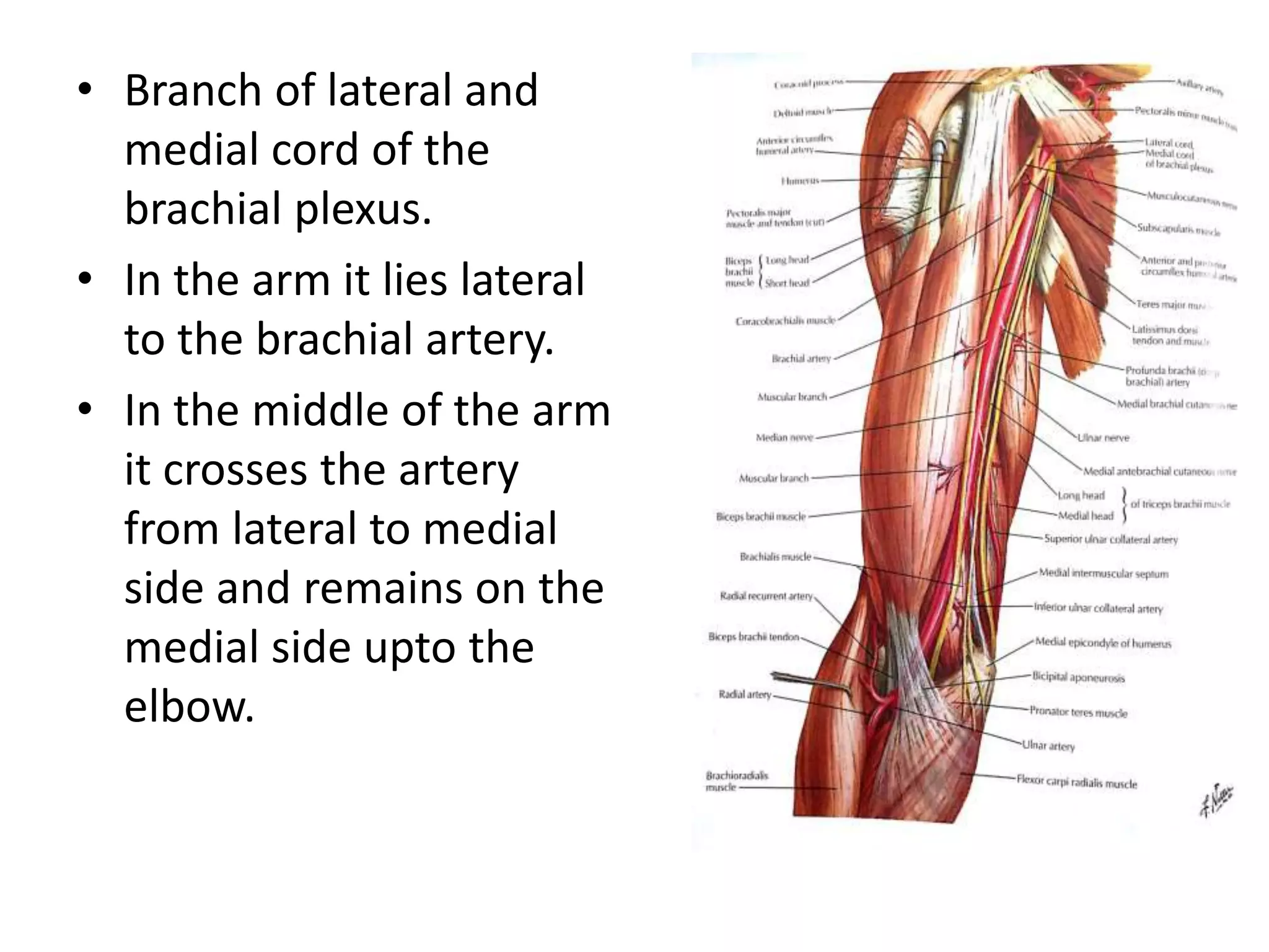
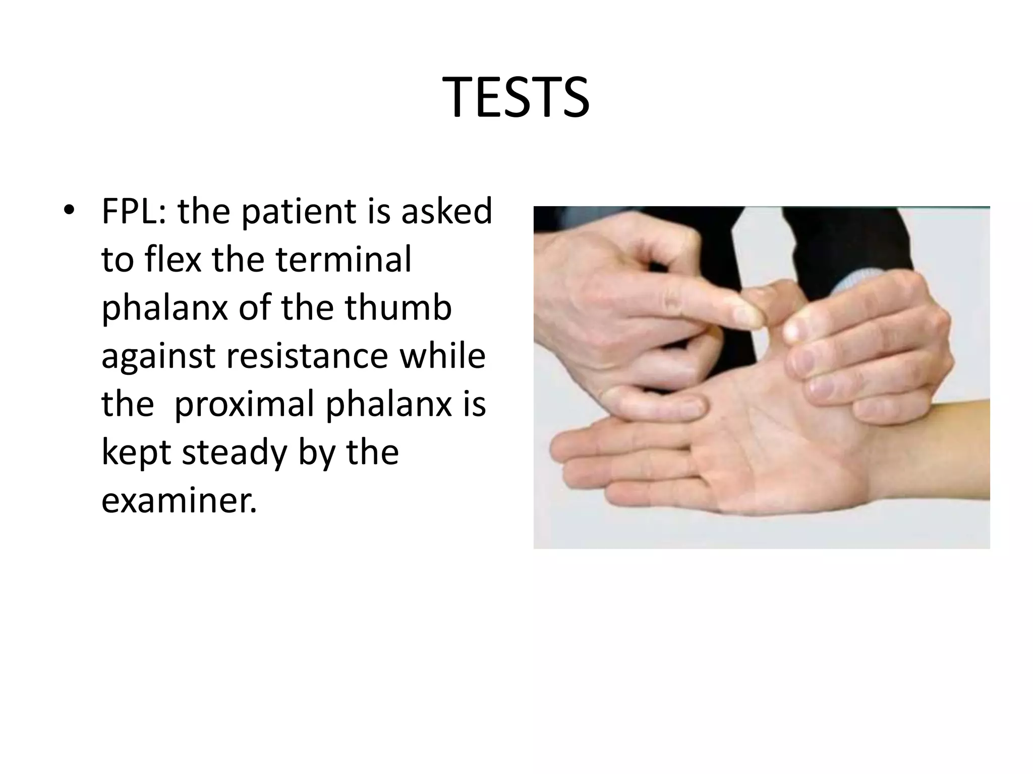
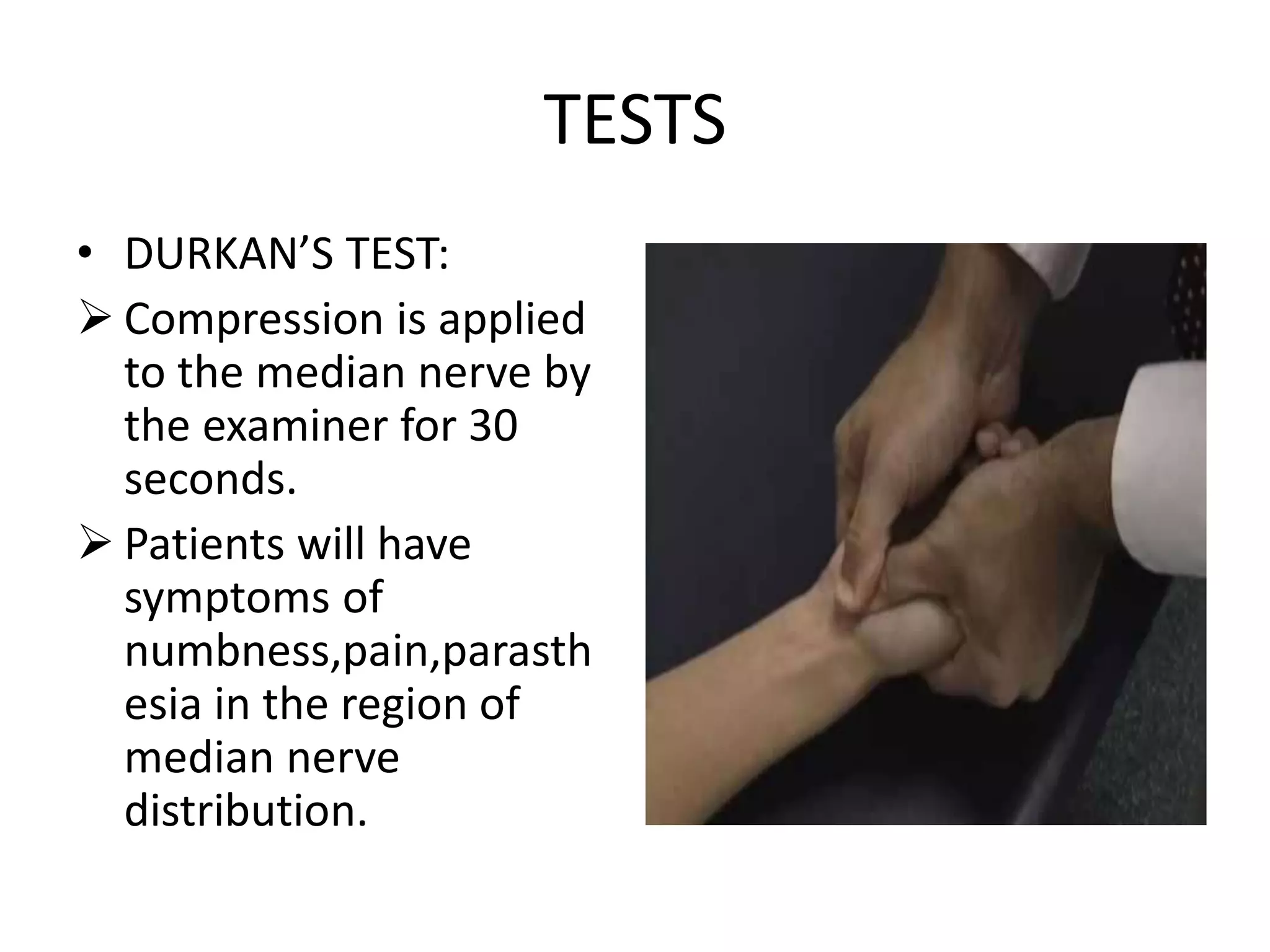
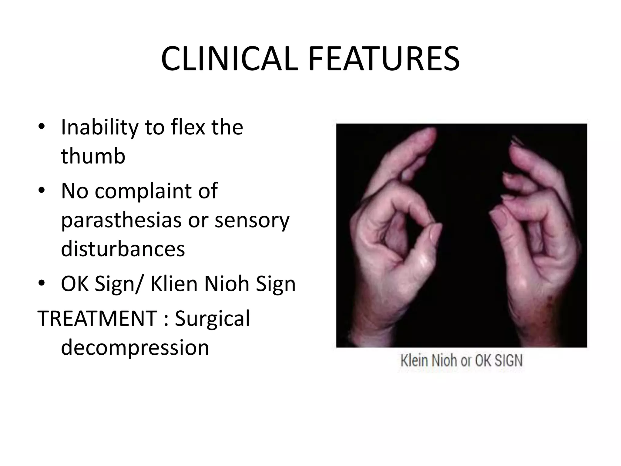
MEDIAN NERVE
Dr.HARSHA NANDINI TALASILA
M.S Ortho
• Branch of lateral and
medial cord of the
brachial plexus.
• In the arm it lies lateral
to the brachial artery.
• In the middle of the arm
it crosses the artery
from lateral to medial
side and remains on the
medial side upto the
elbow.
• In the arm: branch to the elbow joint and
vascular branch to the brachial artery.
• In the cubital fossa it lies medial to the
brachial artery.
• It enters the forearm by passing between the
2 heads of the pronator teres.
• In the forearm it passes beneath the fibrous
arch of flexor digitorum superficialis and on
the surface of flexor digitorum profundus.
• About 5cm above the flexor retinaculum it
becomes superficial and lies between the
tendons of flexor carpi radialis and FDS.
• It is overlapped by the tendon of palmaris
longus.
• Median nerve enters the hand by passing
deep to flexor retinaculum through the carpal
tunnel.
• Muscular branches to all the superficial flexors
of forearm except FCU
• Anterior interosseous nerve is given off in the
upper part of the forearm,it supplies the
flexor pollicis longus,the lateral half of FDP
and pronator quadratus
• DRUJ and wrist joints
Branches in the hand
MOTOR BRANCHES
• Abductor pollicis brevis
• Flexor pollicis brevis
• Opponens pollicis
• 1st and 2nd lumbricals
SENSORY SUPPLY
• Lateral 3½ digits on
palmar side and distal
half of dorsal surface
INJURY TO MEDIAN NERVE
• Median nerve is injured near the wrist or high up in
the fore arm.
1. High median nerve palsy
2. Low median nerve palsy
HIGH MEDIAN NERVE PALSY
• Injury proximal to the elbow.
• Stabs and gunshot wounds may damage the nerve at any level
• This will cause paralysis of all the muscles supplied by the
median nerve in the forearm and hand
LOW MEDIAN NERVE PALSY
• Injury in the distal third of the forearm
• Cuts in front of wrist or by carpal dislocation
• There will be sparing of the forearm muscles , but the muscles
of the hand will be paralysed
• There will be anaesthesia over the median nerve distribution in
the hand
• Thenar eminence is wasted and thumb
abduction and opposition are weak.
• Sensation is lost over the radial three and half
digits.
TESTS
• FPL: the patient is asked
to flex the terminal
phalanx of the thumb
against resistance while
the proximal phalanx is
kept steady by the
examiner.
OSCHNER’S CLASPING TEST
• FDS and lateral half of the
FDP: if the patient is
asked to clasp his
hand,the index finger will
remain straight,the so
called pointing index.
• This occurs because both
the finger flexors (FDS
and FDP)are paralysed
but the medial half of the
FDP makes flexion of the
others fingers possible.
• FCR: normally the
palmar flexion at the
wrist occurs at in the
long axis of the
forearm.
• In the patient with
paralysed flexor carpi
radialis,the wrist
deviates to the ulnar
side while the palmar
flexion occurs.
• ABDUCTOR POLLICIS
BREVIS: the patient is
asked to lay his hand
flat on the table with
palm facing the ceiling.
• A pen is held above the
thumb and the patient
is asked to touch the
pen with tip of his
thumb-PEN TEST
COMPRESSIVE NEUROPATHY
CARPAL TUNNEL SYNDROME
• Boundaries of carpal tunnel:
1. Medial: hook of hamate and pisiform
2. Lateral: scaphoid and trapezium
3. Roof : flexor retinaculum
• The most palmar structure in the carpal
tunnel is median nerve.
• Deep to the median nerve are present the
long flexor tendons of thumb and fingers.
• Entrapment of the median nerve at the wrist.
• CAUSES:
 Decrease in the size of the tunnel
1. Bony abnormalities of the carpal bones
2. Acromegaly .
 Increase in the contents of the tunnel.
1. Scaphoid or distal radius fractures
2. Scaphoid subluxation or lunate dislocation
3. Local tumors like ganglion,neuroma,lipoma
4. Hematoma
 Neuropathic conditions
1. Diabetes mellitus
2. Alcoholism
 Inflammatory conditions
1. Rheumatoid arthritis
2. Gout
3. Infections
 Alterations of fluid balance.
1. Pregnancy
2. Thyroid disease
3. Renal failure
4. Obesity
5. Amyloidosis
External forces.
1. Vibration
2. Direct pressure
TESTS
• DURKAN’S TEST:
 Compression is applied
to the median nerve by
the examiner for 30
seconds.
 Patients will have
symptoms of
numbness,pain,parasth
esia in the region of
median nerve
distribution.
• PHALEN’S TEST:
 Patient is asked to keep
the wrist flexed
continuously for 1
minute.
 Patient will have
tingling and numbness
in the lateral 3 and a
half fingers.
• GILIAC TEST:
Arm torniquet is inflated above systolic pressure
for 60seconds.
Tingling and numbness in the median nerve
distribution.
MANAGEMENT
1. Splinting of the wrist in neutral position
2. Corticosteroid injection into the carpal tunnel
1. SURGERY
a) Constant parasthesias
not relieving after
conservative
management
b) Thenar atrophy
c) Delayed median nerve
conduction velocity on
NCV
PRONATOR SYNDROME
• ETIOLOGY:
 Supracondylar process:
• On the anteromedial surface of the
humerus,about 5cm above the medial
epicondyle ,an aberrant spur of bone
• A ligament of struthers runs from spur to the
medial epicondyle.
• Median nerve,brachial artery and vein run
beneath this ligament.
• CLINICAL FEATURES:
Vague anterior forearm pain
Sensory disturbances over the palmar
cutaneous branch of the median nerve.
Tenderness over the course of the median
nerve in the forearm
• Tests:
PROXIMAL FOREARM COMPRESSION TEST
Firm direct pressure is applied to the forearm at
the elbow for 30 seconds.
Pain in the forearm and sensory disturbance
along the median nerve course.
TREATMENT
• NSAIDS
• ELBOW SPLINTING in 90 degree flexion and
neutral forearm rotation
ANTERIOR INTEROSSEOUS SYNDROME
• Seen in athletes resulting from a violent
muscle contracture from aggressive forearm
exercises
• This syndrome involves motor loss without
sensory involvement.
• AIN innervates: FDP,FPL,Pronator quadratus
CLINICAL FEATURES
• Inability to flex the
thumb
• No complaint of
parasthesias or sensory
disturbances
• OK Sign/ Klien Nioh Sign
TREATMENT : Surgical
decompression
TENDON TRANSFERS
• BRANDS TECHNIQUE and BUNNELL
PROCEDURE: FDS of ring finger to OPPONENS
POLLICIS
• CAMITZ PROCEDURE :PALMARIS LONGUS to
APB
• BURKHALTER TECHNIQUE: EXTENSOR INDICES
PROPRIUS to OPPONENS POLLICIS
MUSCULOCUTANEOUS NERVE
• Musculocutaneous nerve is a branch of lateral
cord of the brachial plexus.
• COURSE :
In the axilla it is present lateral to the axillary
artery.
It pierces the coracobrachialis and enters the
anterior aspect of the arm.
It runs downwards and laterally between the
biceps brachii and brachialis muscles.
It terminates as the lateral cutaneous nerve of
the forearm.
• Motor supply:
1. Biceps brachii.
2. Brachialis
3. Coracobrachialis
• Sensory supply
 Lateral cutaneous nerve of the forearm: Skin
over the lateral aspect of the forearm
• Causes of injury:
Penetrating injury
Rarely by anterior dislocation of
shoulder,fracture of humerus
• Musculocutaneous neuropathy is rare
• Possible cause due to lifting heavy weights on
the shoulder with the arm curled around the
object: CARPET CARRIER’S PALSY.
• More common entrapment is the distal
sensory nerve at the elbow between the
biceps tendon and brachialis muscle.
• Clinical features:
Decreased sensation over the lateral aspect of
the forearm.
Wasting of biceps and brachialis.
Motor loss is not significant as flexion of the
elbow is also performed by brachioradialis
TREATMENT
• REST
• Corticosteroid injection
• Massages and manual exercises to release the
fascial adhesions and any scar tissue in the
muscle which are entrapping the nerve.
• Surgical decompression if symptoms persist
more than 6months and donot respond to
conservative management.
• CAMPBELL’S OPERATIVE ORTHOPAEDICS
• NETTAR’S ATLAS OF HUMAN ANATOMY
• CHAURASIA TEXTBOOK OF ANATOMY
THANK YOU

Median nerve

  • 1.
  • 2.
    • Branch oflateral and medial cord of the brachial plexus. • In the arm it lies lateral to the brachial artery. • In the middle of the arm it crosses the artery from lateral to medial side and remains on the medial side upto the elbow.
  • 3.
    • In thearm: branch to the elbow joint and vascular branch to the brachial artery. • In the cubital fossa it lies medial to the brachial artery. • It enters the forearm by passing between the 2 heads of the pronator teres. • In the forearm it passes beneath the fibrous arch of flexor digitorum superficialis and on the surface of flexor digitorum profundus.
  • 5.
    • About 5cmabove the flexor retinaculum it becomes superficial and lies between the tendons of flexor carpi radialis and FDS. • It is overlapped by the tendon of palmaris longus. • Median nerve enters the hand by passing deep to flexor retinaculum through the carpal tunnel.
  • 6.
    • Muscular branchesto all the superficial flexors of forearm except FCU • Anterior interosseous nerve is given off in the upper part of the forearm,it supplies the flexor pollicis longus,the lateral half of FDP and pronator quadratus • DRUJ and wrist joints
  • 7.
    Branches in thehand MOTOR BRANCHES • Abductor pollicis brevis • Flexor pollicis brevis • Opponens pollicis • 1st and 2nd lumbricals SENSORY SUPPLY • Lateral 3½ digits on palmar side and distal half of dorsal surface
  • 8.
    INJURY TO MEDIANNERVE • Median nerve is injured near the wrist or high up in the fore arm. 1. High median nerve palsy 2. Low median nerve palsy
  • 9.
    HIGH MEDIAN NERVEPALSY • Injury proximal to the elbow. • Stabs and gunshot wounds may damage the nerve at any level • This will cause paralysis of all the muscles supplied by the median nerve in the forearm and hand
  • 10.
    LOW MEDIAN NERVEPALSY • Injury in the distal third of the forearm • Cuts in front of wrist or by carpal dislocation • There will be sparing of the forearm muscles , but the muscles of the hand will be paralysed • There will be anaesthesia over the median nerve distribution in the hand
  • 11.
    • Thenar eminenceis wasted and thumb abduction and opposition are weak. • Sensation is lost over the radial three and half digits.
  • 12.
    TESTS • FPL: thepatient is asked to flex the terminal phalanx of the thumb against resistance while the proximal phalanx is kept steady by the examiner.
  • 13.
    OSCHNER’S CLASPING TEST •FDS and lateral half of the FDP: if the patient is asked to clasp his hand,the index finger will remain straight,the so called pointing index. • This occurs because both the finger flexors (FDS and FDP)are paralysed but the medial half of the FDP makes flexion of the others fingers possible.
  • 14.
    • FCR: normallythe palmar flexion at the wrist occurs at in the long axis of the forearm. • In the patient with paralysed flexor carpi radialis,the wrist deviates to the ulnar side while the palmar flexion occurs.
  • 15.
    • ABDUCTOR POLLICIS BREVIS:the patient is asked to lay his hand flat on the table with palm facing the ceiling. • A pen is held above the thumb and the patient is asked to touch the pen with tip of his thumb-PEN TEST
  • 16.
  • 17.
    CARPAL TUNNEL SYNDROME •Boundaries of carpal tunnel: 1. Medial: hook of hamate and pisiform 2. Lateral: scaphoid and trapezium 3. Roof : flexor retinaculum • The most palmar structure in the carpal tunnel is median nerve. • Deep to the median nerve are present the long flexor tendons of thumb and fingers.
  • 18.
    • Entrapment ofthe median nerve at the wrist. • CAUSES:  Decrease in the size of the tunnel 1. Bony abnormalities of the carpal bones 2. Acromegaly .
  • 19.
     Increase inthe contents of the tunnel. 1. Scaphoid or distal radius fractures 2. Scaphoid subluxation or lunate dislocation 3. Local tumors like ganglion,neuroma,lipoma 4. Hematoma
  • 20.
     Neuropathic conditions 1.Diabetes mellitus 2. Alcoholism  Inflammatory conditions 1. Rheumatoid arthritis 2. Gout 3. Infections
  • 21.
     Alterations offluid balance. 1. Pregnancy 2. Thyroid disease 3. Renal failure 4. Obesity 5. Amyloidosis External forces. 1. Vibration 2. Direct pressure
  • 22.
    TESTS • DURKAN’S TEST: Compression is applied to the median nerve by the examiner for 30 seconds.  Patients will have symptoms of numbness,pain,parasth esia in the region of median nerve distribution.
  • 23.
    • PHALEN’S TEST: Patient is asked to keep the wrist flexed continuously for 1 minute.  Patient will have tingling and numbness in the lateral 3 and a half fingers.
  • 24.
    • GILIAC TEST: Armtorniquet is inflated above systolic pressure for 60seconds. Tingling and numbness in the median nerve distribution.
  • 25.
    MANAGEMENT 1. Splinting ofthe wrist in neutral position 2. Corticosteroid injection into the carpal tunnel
  • 26.
    1. SURGERY a) Constantparasthesias not relieving after conservative management b) Thenar atrophy c) Delayed median nerve conduction velocity on NCV
  • 27.
    PRONATOR SYNDROME • ETIOLOGY: Supracondylar process: • On the anteromedial surface of the humerus,about 5cm above the medial epicondyle ,an aberrant spur of bone • A ligament of struthers runs from spur to the medial epicondyle. • Median nerve,brachial artery and vein run beneath this ligament.
  • 28.
    • CLINICAL FEATURES: Vagueanterior forearm pain Sensory disturbances over the palmar cutaneous branch of the median nerve. Tenderness over the course of the median nerve in the forearm
  • 29.
    • Tests: PROXIMAL FOREARMCOMPRESSION TEST Firm direct pressure is applied to the forearm at the elbow for 30 seconds. Pain in the forearm and sensory disturbance along the median nerve course.
  • 30.
    TREATMENT • NSAIDS • ELBOWSPLINTING in 90 degree flexion and neutral forearm rotation
  • 31.
    ANTERIOR INTEROSSEOUS SYNDROME •Seen in athletes resulting from a violent muscle contracture from aggressive forearm exercises • This syndrome involves motor loss without sensory involvement. • AIN innervates: FDP,FPL,Pronator quadratus
  • 32.
    CLINICAL FEATURES • Inabilityto flex the thumb • No complaint of parasthesias or sensory disturbances • OK Sign/ Klien Nioh Sign TREATMENT : Surgical decompression
  • 33.
    TENDON TRANSFERS • BRANDSTECHNIQUE and BUNNELL PROCEDURE: FDS of ring finger to OPPONENS POLLICIS • CAMITZ PROCEDURE :PALMARIS LONGUS to APB • BURKHALTER TECHNIQUE: EXTENSOR INDICES PROPRIUS to OPPONENS POLLICIS
  • 34.
  • 35.
    • Musculocutaneous nerveis a branch of lateral cord of the brachial plexus. • COURSE : In the axilla it is present lateral to the axillary artery. It pierces the coracobrachialis and enters the anterior aspect of the arm. It runs downwards and laterally between the biceps brachii and brachialis muscles. It terminates as the lateral cutaneous nerve of the forearm.
  • 36.
    • Motor supply: 1.Biceps brachii. 2. Brachialis 3. Coracobrachialis • Sensory supply  Lateral cutaneous nerve of the forearm: Skin over the lateral aspect of the forearm
  • 37.
    • Causes ofinjury: Penetrating injury Rarely by anterior dislocation of shoulder,fracture of humerus
  • 38.
    • Musculocutaneous neuropathyis rare • Possible cause due to lifting heavy weights on the shoulder with the arm curled around the object: CARPET CARRIER’S PALSY. • More common entrapment is the distal sensory nerve at the elbow between the biceps tendon and brachialis muscle.
  • 39.
    • Clinical features: Decreasedsensation over the lateral aspect of the forearm. Wasting of biceps and brachialis. Motor loss is not significant as flexion of the elbow is also performed by brachioradialis
  • 40.
    TREATMENT • REST • Corticosteroidinjection • Massages and manual exercises to release the fascial adhesions and any scar tissue in the muscle which are entrapping the nerve. • Surgical decompression if symptoms persist more than 6months and donot respond to conservative management.
  • 41.
    • CAMPBELL’S OPERATIVEORTHOPAEDICS • NETTAR’S ATLAS OF HUMAN ANATOMY • CHAURASIA TEXTBOOK OF ANATOMY
  • 42.