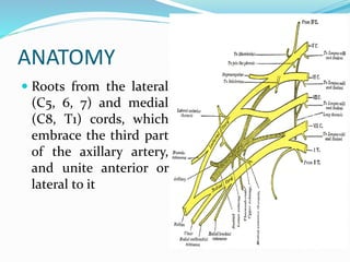
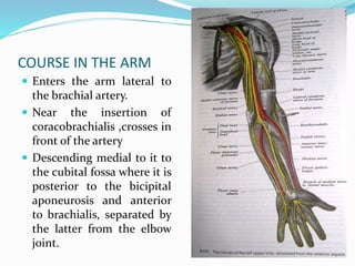
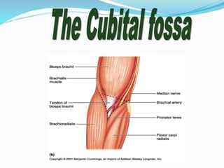
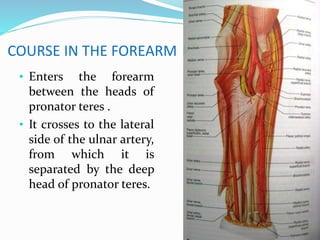
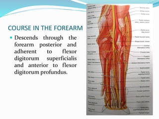
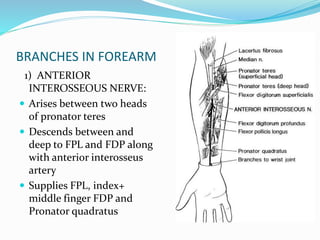
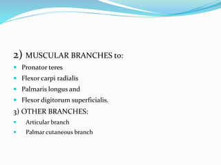
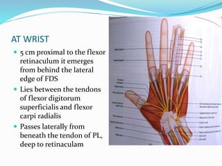
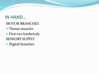
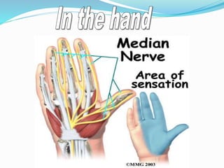
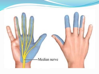
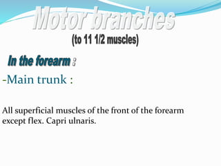
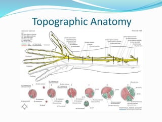
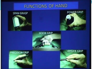
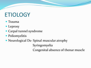
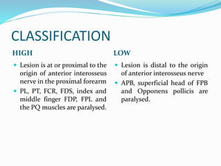
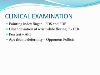
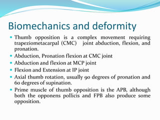
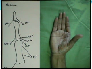
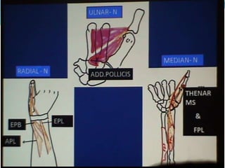
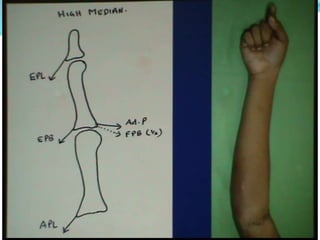
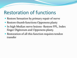
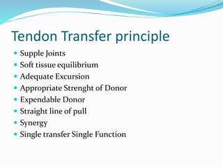
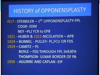
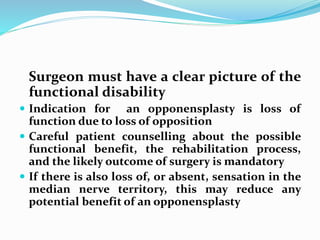
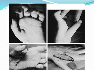
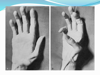
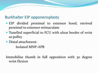
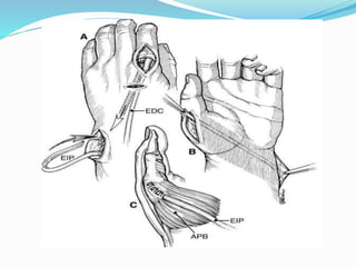
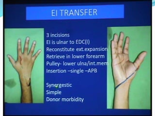
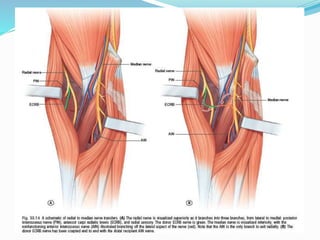
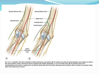
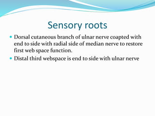
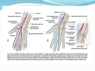
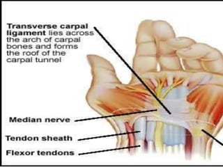
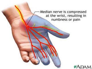
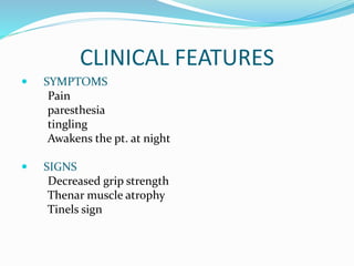
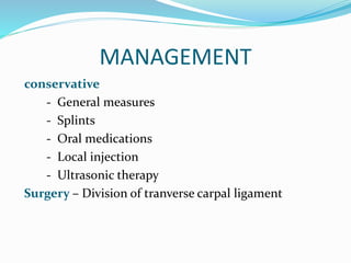
Dr. Hardik Dodia
ANATOMY
 Roots from the lateral
(C5, 6, 7) and medial
(C8, T1) cords, which
embrace the third part
of the axillary artery,
and unite anterior or
lateral to it
COURSE IN THE ARM
 Enters the arm lateral to
the brachial artery.
 Near the insertion of
coracobrachialis ,crosses in
front of the artery
 Descending medial to it to
the cubital fossa where it is
posterior to the bicipital
aponeurosis and anterior
to brachialis, separated by
the latter from the elbow
joint.
COURSE IN THE FOREARM
• Enters the forearm
between the heads of
pronator teres .
• It crosses to the lateral
side of the ulnar artery,
from which it is
separated by the deep
head of pronator teres.
COURSE IN THE FOREARM
 Descends through the
forearm posterior and
adherent to flexor
digitorum superficialis
and anterior to flexor
digitorum profundus.
BRANCHES IN FOREARM
1) ANTERIOR
INTEROSSEOUS NERVE:
 Arises between two heads
of pronator teres
 Descends between and
deep to FPL and FDP along
with anterior interosseus
artery
 Supplies FPL, index+
middle finger FDP and
Pronator quadratus
2) MUSCULAR BRANCHES to:
 Pronator teres
 Flexor carpi radialis
 Palmaris longus and
 Flexor digitorum superficialis.
3) OTHER BRANCHES:
 Articular branch
 Palmar cutaneous branch
AT WRIST
 5 cm proximal to the flexor
retinaculum it emerges
from behind the lateral
edge of FDS
 Lies between the tendons
of flexor digitorum
superficialis and flexor
carpi radialis
 Passes laterally from
beneath the tendon of PL,
deep to retinaculam
IN HAND…
MOTOR BRANCHES
 Thenar muscles
 First two lumbricals
SENSORY SUPPLY
 Digital branches
3) Branches in the hand:
1) the lat .terminal branch :
A) 3 common palmer digital branches.
B) recurrent muscular branch.
2) the med . Terminal branch :
It gives 2 common palmer brs :
-The lat. Branch.
-The med. Branch.
6) Its End
-Main trunk :
All superficial muscles of the front of the forearm
except flex. Capri ulnaris.
- Ant. Interosseous br.:
- supplies 2 ½ muscles :
All deep muscles of the front of forearm except
the med. ½ of flex. Digit. Profound.
Lat. Terminal br : 4 muscles
i.e. the 1st lumbrical m + all thenar m (except add .poll.)
Med. Terminal br.: 2nd lumbrical m
-The palmer cutaneous br.: Arises in the lower
end of forearm & descends to the hand to supply the
skin of the lat. 2/3 of the palm.
Lat. Terminal br.
Gives 3 palmer digital branches
Med. Terminal br.:
Gives 2 palmer digital brs.
-
-Main trunk
Supplies elbow & sup. Radioulnar joint .
-Ant. Interosseous
Supplies wrist & inf . Radioulnar joint
To the radial &
ulnar arteries.
So patients with
lesions
Dry scaly skin
and brittle
nails.
Topographic Anatomy
ETIOLOGY
 Trauma
 Leprosy
 Carpal tunnel syndrome
 Poliomyelitis
 Neurological Ds- Spinal muscular atrophy
Syringomyelia
Congenital absence of thenar muscle
CLASSIFICATION
HIGH LOW
 Lesion is at or proximal to the
origin of anterior interosseus
nerve in the proximal forearm
 PL, PT, FCR, FDS, index and
middle finger FDP, FPL and
the PQ muscles are paralysed.
 Lesion is distal to the origin
of anterior interosseus nerve
 APB, superficial head of FPB
and Opponens pollicis are
paralysed.
(1) Paralysis of all muscles supplied .
(2) loss of pronation of the forearm .
(3) weak flexion of the wrist .
(4) loss of the flexion & opposition of the thumb.
(1) hyper-extended thumb .
(2) adduction .
(3) flat thenar eminence .
- lat. 2/3 of the palm of the hand .
-lat. 3 ½ fingers anteriorly & their distal
halves posteriorly.
- lat. 2/3 of the palm of the hand .
-lat. 3 ½ fingers anteriorly & their distal
halves posteriorly.
-Paralysis of the 5 hand muscles supplied by the
nerve.
-The forearm muscles escape the injury as they are
supplied at elbow.
CLINICAL EXAMINATION
 Pointing index finger – FDS and FDP
 Ulnar deviation of wrist while flexing it - FCR
 Pen test – APB
 Ape thumb deformity – Opponens Pollicis
Work up
Radiographs
• X ray of arm and forearm to detect or rule out a
fracture
EMG and NCV study
Performed initially to provide a baseline, but
unless the nerve is severed, no changes will be
observed for 3-6 weeks.
• Help to locate the site of injury
• Help to monitor the nerve recovery over time.
Timing of nerve repairs
Open injuries
 Require early exploration.
 Sharp lacerations can be repaired immediately and directly.
 Wound must be relatively clean and free of gross
contamination.
 A primary repair is not recommended with injuries
secondary to a crush injury /significant soft tissue damage.
 At 3 weeks (or when the wound permits), the nerve is re-
explored, and definitive repair or graft can be performed.
 At that time, the zone of injury is apparent based on the
extent of scar formation.
Closed injuries
 In closed or blunt trauma, initial management is
expectant with close observation.
 If complete recovery is not observed within 6 weeks,

 Electrodiagnostic studies should be obtained for
baseline evaluation.
 Monthly clinical and EMG evaluation

 If motor unit potentials are seen with EMG, ►
spontaneous reinnervation is anticipated,
 Lack of clinical or electrical evidence of
reinnervation at 3 months requires operative
exploration.
Principles of nerve repair
 The results have improved with the advent of
microsurgical techniques.
1)Quantitative pre-operative assessment of motor and
sensory systems
2)Microsurgical technique including proper
magnification, instrumentation, and microsutures
3)Tension-free repair-prime object
4)When a tension-free direct repair is not technically
possible, use of an interpositional nerve graft
5)Primary repair when the conditions permit
6)Postural manuevers can’t substitute tension free repair
7)Fascicular repair when fascicular anatomy discernible
and epineural repair when it is indiscernible
8)Delay in repair for approximately 3 weeks in cases
where primary repair is not optimal (e.g, with a severe
crush, stretch, or loss of nerve tissue)
9)Early protected range of movement to allow nerve
gliding
10)Post op physiotherapy to maintain range of motion
and assist in postoperative sensory and motor re-
education and rehabilitation to maximize the clinical
outcome
Biomechanics and deformity
 Thumb opposition is a complex movement requiring
trapeziometacarpal (CMC) joint abduction, flexion, and
pronation.
 Abduction, Pronation flexion at CMC joint
 Abduction and flexion at MCP joint
 Flexion and Extension at IP joint
 Axial thumb rotation, usually 90 degrees of pronation and
60 degrees of supination.
 Prime muscle of thumb opposition is the APB, although
both the opponens pollicis and FPB also produce some
opposition.
 Considerable diversity in the pattern of innervation-
reason behind intact opposition after isolated MNP
 Superficial head of the FPB has dual median and ulnar
innervation in 30% of hands, whereas its deep head
has dual innervation in 79% of cases
 Oblique head of the adductor pollicis has dual ulnar
and median innervation in 35% of cases
 Thenar muscles sometimes do have dual nerve supply
from ulnar nerve.
 Martin Gruber Anastomosis- motor nerve
interconnection between median (or anterior
interroseus) and ulnar nerve in proximal forearm.
 17 percent of people have it.
Restoration of functions
 Restore Sensation by primary repair of nerve
 Restore thumb functions Opponens plasty
 In high Median nerve lesions- Restore FPL, Index
finger Digitorum and Opponens plasty.
 Restoration of all this function requires tendon
transfer
Tendon Transfer principle
 Supple Joints
 Soft tissue equilibrium
 Adequate Excursion
 Appropriate Strenght of Donor
 Expendable Donor
 Straight line of pull
 Synergy
 Single transfer Single Function
Surgeon must have a clear picture of the
functional disability
 Indication for an opponensplasty is loss of
function due to loss of opposition
 Careful patient counselling about the possible
functional benefit, the rehabilitation process,
and the likely outcome of surgery is mandatory
 If there is also loss of, or absent, sensation in the
median nerve territory, this may reduce any
potential benefit of an opponensplasty
Prevention and Preoperative
Treatment of Contractures
 It is always easier to prevent a soft tissue contracture
than to correct an established one
 Prevented by passive thumb abduction and opposition
exercises and by abduction splints
 Temporary internal splintage of the thumb
metacarpal in abduction with a K wire may be
indicated
 Established soft tissue contractures must be corrected
by releasing skin, fascia over Ad PL , first dorsal
interossei ,joint capsule or joint itself
Selection of motor
 Muscle selected for opponensplasty must be
expendable, and this muscle must have strength and
potential excursion similar to that of the APB and
opponens pollicis muscles
 Should have a tension fraction similar to that of the
combined APB and opponens pollicis tension (1.1 + 1.9
= 3.0) and a muscle fiber length that is at least as long
as that of the APB-3.7 cm
 Avoid tendon graft
Pulley design
 Hypothenar fibrous raphe
 Around FCU tendon
 Hooked FCU tendon
 Distal FCU strip attached to ECU
 Angle between PL and flexor retinaculam
 Window in flexor retinaculam
Four standard
opponensplasties
 Superficialis opponensplasty:
1)Royle-thompson technique
2)bunnell technique
 Burkhalter EIP opponensplasty
 Huber ADM transfer
 Camitz opponensplasty (palmaris longus)
SUPERFICIALIS opponensplasty
Superficialis tendon harvest:
Royle and thompson both divided ring finger
superficialis tendon at its middle phalnyx insertion
North and Littler recommended division proximal to
its bifurcation through window between A1 and A2
pulleys with finger fully flexed
Royle-Thompson opponensplasty:
 Ring finger FDS as motor
 Angle between flexor retinaculam & palmar
aponeurosis as pulley
 Inserted on insertion of APB
 Attachment to the thumb is then sutured while the
thumb is held in full opposition and the wrist is in
neutral for 4-6wks
Bunnell’s opponensplasty
 Ring finger FDS through FCU pulley made from distal
4 cm of FCU tendon
 Inserted with a dorsoulnar-venteroradial drill hole at
base of proximal phalanx
 Thumb immobilised in full opposition with neutral
wrist for 4-6 weeks
 DEMERITS OF SUPERFICIALIS TRANSFER:
Not useful for high MNP
Burkhalter EIP opponensplasty
 EIP divided proximal to extensor hood, retrived
proximal to extensor retinaculam
 Tunelled superficial to FCU with ulnar border of wrist
as pulley
 Distal attachment:
Isolated MNP-APB
Immobilise thumb in full opposition with 30 degree
wrist flexion
Advantage:
 Useful for both high and low MNP
 More stable, adjustable natural pulley
 Easy to perform
Disadvantage:
short fiber length of EIP
Huber’s ADM opponensplasty
 Two insertion divided along with pisiform attachment,
leaving behind FCU attachment and pedicle
 No pulley needs to be used, turned 180* to insert on APB
 As having only sufficient length, invariably remain in
adequate tension. Used mostly in patients with congenital
thenar atrophy as it gives bulk to thenar territory
 Splinted post op for 3-4 wks
DISADVANTAGE:
 Difficult to perform
 Critical blood supply
Camitz PL opponensplasty
 For carpal tunnell syndrome, done simultaneous with
decompression
 Conform position of tendon preop
 Avoid injury to palmar cutaneous branch of median
nerve
 Divide tendon with 1 cm strip of aponeurosis and
inserted on APB/ EPB/ MP joint capsule, no pulley
used
OTHER OPPONENSPLASTIES
 ECU OPPONENSPLASTY
 ECRL OPPONENSPLASTY
 EDM OPPONENSPLASTY
 COMPROMISE OPPONENSPLASTY
EPL/FPL opponensplasty
Post op management
 Thumb immobilized in opposition for 3 wks after most
opponensplasty with cast/ cyanoacrylate glue
 Wrist immobilized in neutral, as, extrinsic tendons
crosses wrist. Transfer of muscles with short
excursions, like, EIP, should be relaxed by wrist in 30*
flexion
 Early thumb & wrist movement emphasised after
3wks,except, high MNP+ UNP/profound sensory loss
like in leprosy-protect Camitz PL opponensplasty for
up to 3mth
HIGH MEDIAN NERVE PALSY
THUMB OPPOSITION:
 Pt use APL and extrinsic extensors to position their
thumb when picking up an article from flat surface
 Thumb remain in supination and extension, pt
pronate hand either by pronating forearm or internally
rotating shoulder-so, can’t use sight as substitute for
sensory loss-so, few recommend early opponensplasty
 EIP,EPL and EDM are most readily available
HIGH MEDIAN NERVE PALSY
Timing and selection of transfer:
 Prime aim is to restore thumb+ index finger flexion with
opposition
 BR,ECRL and ECU-available options, ECU may be required
for opponensplasty-all three having excursion less than
recipient
 Sensory loss is most imp single disability, some believe it
strong contraindication
 Due to poor recovery in sensory fn and opposition after
nerve repair, early transfer less recommended
 Transfer only if after max recovery, pt and surgeon believe it
as cause of debility and it will improve hand function
INDEX FINGER FLEXION:
 Only in pts who need strength on radial side of hand
 ECRL to Index Profundus transfer/side-to-
sidesuturing with other finger profundus tendons in
distal forearm
 For ECRL transfer, tension is adjusted when finger is
fully extended with wrist flexed 30* to avoid excess
tension
THUMB FLEXION:
 To achieve 30 mm excursion , BR muscle
freed in distal 2/3 forearm
 Tension set as above
 Because, BR is primarily elbow flexor, weak
thumb flexion when elbow flexed
COMPLICATIONS
Aggravation of Swan neck deformity:
 Special consideration for pts with hyperextensible
joints-
 1)transfer of ECRL to index and middle finger FDS
 2)transfer of ECRL to index and middle finger FDP
with FDP sutured to A4 pulleys
 3)transfer of ECRL to index and middle finger FDP
with DIP joint arthrodesis
CONCLUSION
 TENDON TRANSFER CAN RESTORESELECTEDMOTOR FN
AFTER HIGH AND LOW MNP, BUT THEIRABILITY
RESTRICTED BY SEVERITY OF SENSORY LOSS
 EIP/PLTRANSFER IMP FOR LOW PALSY AND
SIMULTANEOUS BR/ECRL TRANSFER FORHIGH PALSY
Nerve Transfers
Motor roots
1) Radial ECRB to AIN
2) Musculocutaneous Brachialis branch to AIN
Sensory roots
 Dorsal cutaneous branch of ulnar nerve coapted with
end to side with radial side of median nerve to restore
first web space function.
 Distal third webspace is end to side with ulnar nerve
MEDIAN NERVE COMPRESSION
NEUROPATHY
 CARPAL TUNNEL SYNDROME
 PRONATOR TERES SYNDROME
 ANTERIOR INTERROSEUS SYNDROME
It is compression of median n. as it passes through the
carpal tunnel.
(1) dislocation of one of the carpal bones.
(2) thickening of the tendons passing.
(3) myxoedema or tumour inside the carpal tunnel .
As in the injury below the elbow but there is no sensory
loss in the palm because the palmer cutaneous br . Passes
outside the carpal tunnel.
The characteristic deformity of all median n.
injuries is monkey hand.
Atrophy
CLINICAL FEATURES
 SYMPTOMS
Pain
paresthesia
tingling
Awakens the pt. at night
 SIGNS
Decreased grip strength
Thenar muscle atrophy
Tinels sign
MANAGEMENT
conservative
- General measures
- Splints
- Oral medications
- Local injection
- Ultrasonic therapy
Surgery – Division of tranverse carpal ligament
PRONATOR SYNDROME
 Ligament of Struthers. (Most common)
 Anamolous bony spur in supracondylar process.
 Bicipital aponeurosis.
 Symptomatic treatment
 Correction of cause
Anterior interosseus syndrome
 Accesory head of FPL
 Fibrous arch of FDS
 Aberrant palmaris profundus
 Fascial bands of deep head of pronator teres
 Symptomatic treatment
 Correction of the cause
.

Median nerve injuries

  • 1.
  • 2.
    ANATOMY  Roots fromthe lateral (C5, 6, 7) and medial (C8, T1) cords, which embrace the third part of the axillary artery, and unite anterior or lateral to it
  • 3.
    COURSE IN THEARM  Enters the arm lateral to the brachial artery.  Near the insertion of coracobrachialis ,crosses in front of the artery  Descending medial to it to the cubital fossa where it is posterior to the bicipital aponeurosis and anterior to brachialis, separated by the latter from the elbow joint.
  • 5.
    COURSE IN THEFOREARM • Enters the forearm between the heads of pronator teres . • It crosses to the lateral side of the ulnar artery, from which it is separated by the deep head of pronator teres.
  • 6.
    COURSE IN THEFOREARM  Descends through the forearm posterior and adherent to flexor digitorum superficialis and anterior to flexor digitorum profundus.
  • 7.
    BRANCHES IN FOREARM 1)ANTERIOR INTEROSSEOUS NERVE:  Arises between two heads of pronator teres  Descends between and deep to FPL and FDP along with anterior interosseus artery  Supplies FPL, index+ middle finger FDP and Pronator quadratus
  • 8.
    2) MUSCULAR BRANCHESto:  Pronator teres  Flexor carpi radialis  Palmaris longus and  Flexor digitorum superficialis. 3) OTHER BRANCHES:  Articular branch  Palmar cutaneous branch
  • 9.
    AT WRIST  5cm proximal to the flexor retinaculum it emerges from behind the lateral edge of FDS  Lies between the tendons of flexor digitorum superficialis and flexor carpi radialis  Passes laterally from beneath the tendon of PL, deep to retinaculam
  • 10.
    IN HAND… MOTOR BRANCHES Thenar muscles  First two lumbricals SENSORY SUPPLY  Digital branches
  • 11.
    3) Branches inthe hand: 1) the lat .terminal branch : A) 3 common palmer digital branches. B) recurrent muscular branch. 2) the med . Terminal branch : It gives 2 common palmer brs : -The lat. Branch. -The med. Branch.
  • 14.
  • 15.
    -Main trunk : Allsuperficial muscles of the front of the forearm except flex. Capri ulnaris.
  • 16.
    - Ant. Interosseousbr.: - supplies 2 ½ muscles : All deep muscles of the front of forearm except the med. ½ of flex. Digit. Profound. Lat. Terminal br : 4 muscles i.e. the 1st lumbrical m + all thenar m (except add .poll.) Med. Terminal br.: 2nd lumbrical m
  • 17.
    -The palmer cutaneousbr.: Arises in the lower end of forearm & descends to the hand to supply the skin of the lat. 2/3 of the palm. Lat. Terminal br. Gives 3 palmer digital branches Med. Terminal br.: Gives 2 palmer digital brs.
  • 18.
    - -Main trunk Supplies elbow& sup. Radioulnar joint . -Ant. Interosseous Supplies wrist & inf . Radioulnar joint
  • 19.
    To the radial& ulnar arteries. So patients with lesions Dry scaly skin and brittle nails.
  • 20.
  • 22.
    ETIOLOGY  Trauma  Leprosy Carpal tunnel syndrome  Poliomyelitis  Neurological Ds- Spinal muscular atrophy Syringomyelia Congenital absence of thenar muscle
  • 23.
    CLASSIFICATION HIGH LOW  Lesionis at or proximal to the origin of anterior interosseus nerve in the proximal forearm  PL, PT, FCR, FDS, index and middle finger FDP, FPL and the PQ muscles are paralysed.  Lesion is distal to the origin of anterior interosseus nerve  APB, superficial head of FPB and Opponens pollicis are paralysed.
  • 24.
    (1) Paralysis ofall muscles supplied . (2) loss of pronation of the forearm . (3) weak flexion of the wrist . (4) loss of the flexion & opposition of the thumb.
  • 25.
    (1) hyper-extended thumb. (2) adduction . (3) flat thenar eminence . - lat. 2/3 of the palm of the hand . -lat. 3 ½ fingers anteriorly & their distal halves posteriorly.
  • 26.
    - lat. 2/3of the palm of the hand . -lat. 3 ½ fingers anteriorly & their distal halves posteriorly. -Paralysis of the 5 hand muscles supplied by the nerve. -The forearm muscles escape the injury as they are supplied at elbow.
  • 29.
    CLINICAL EXAMINATION  Pointingindex finger – FDS and FDP  Ulnar deviation of wrist while flexing it - FCR  Pen test – APB  Ape thumb deformity – Opponens Pollicis
  • 30.
    Work up Radiographs • Xray of arm and forearm to detect or rule out a fracture EMG and NCV study Performed initially to provide a baseline, but unless the nerve is severed, no changes will be observed for 3-6 weeks. • Help to locate the site of injury • Help to monitor the nerve recovery over time.
  • 32.
    Timing of nerverepairs Open injuries  Require early exploration.  Sharp lacerations can be repaired immediately and directly.  Wound must be relatively clean and free of gross contamination.  A primary repair is not recommended with injuries secondary to a crush injury /significant soft tissue damage.  At 3 weeks (or when the wound permits), the nerve is re- explored, and definitive repair or graft can be performed.  At that time, the zone of injury is apparent based on the extent of scar formation.
  • 33.
    Closed injuries  Inclosed or blunt trauma, initial management is expectant with close observation.  If complete recovery is not observed within 6 weeks,   Electrodiagnostic studies should be obtained for baseline evaluation.
  • 34.
     Monthly clinicaland EMG evaluation   If motor unit potentials are seen with EMG, ► spontaneous reinnervation is anticipated,  Lack of clinical or electrical evidence of reinnervation at 3 months requires operative exploration.
  • 35.
    Principles of nerverepair  The results have improved with the advent of microsurgical techniques. 1)Quantitative pre-operative assessment of motor and sensory systems 2)Microsurgical technique including proper magnification, instrumentation, and microsutures 3)Tension-free repair-prime object 4)When a tension-free direct repair is not technically possible, use of an interpositional nerve graft 5)Primary repair when the conditions permit
  • 36.
    6)Postural manuevers can’tsubstitute tension free repair 7)Fascicular repair when fascicular anatomy discernible and epineural repair when it is indiscernible 8)Delay in repair for approximately 3 weeks in cases where primary repair is not optimal (e.g, with a severe crush, stretch, or loss of nerve tissue) 9)Early protected range of movement to allow nerve gliding 10)Post op physiotherapy to maintain range of motion and assist in postoperative sensory and motor re- education and rehabilitation to maximize the clinical outcome
  • 38.
    Biomechanics and deformity Thumb opposition is a complex movement requiring trapeziometacarpal (CMC) joint abduction, flexion, and pronation.  Abduction, Pronation flexion at CMC joint  Abduction and flexion at MCP joint  Flexion and Extension at IP joint  Axial thumb rotation, usually 90 degrees of pronation and 60 degrees of supination.  Prime muscle of thumb opposition is the APB, although both the opponens pollicis and FPB also produce some opposition.
  • 44.
     Considerable diversityin the pattern of innervation- reason behind intact opposition after isolated MNP  Superficial head of the FPB has dual median and ulnar innervation in 30% of hands, whereas its deep head has dual innervation in 79% of cases  Oblique head of the adductor pollicis has dual ulnar and median innervation in 35% of cases  Thenar muscles sometimes do have dual nerve supply from ulnar nerve.
  • 45.
     Martin GruberAnastomosis- motor nerve interconnection between median (or anterior interroseus) and ulnar nerve in proximal forearm.  17 percent of people have it.
  • 46.
    Restoration of functions Restore Sensation by primary repair of nerve  Restore thumb functions Opponens plasty  In high Median nerve lesions- Restore FPL, Index finger Digitorum and Opponens plasty.  Restoration of all this function requires tendon transfer
  • 47.
    Tendon Transfer principle Supple Joints  Soft tissue equilibrium  Adequate Excursion  Appropriate Strenght of Donor  Expendable Donor  Straight line of pull  Synergy  Single transfer Single Function
  • 49.
    Surgeon must havea clear picture of the functional disability  Indication for an opponensplasty is loss of function due to loss of opposition  Careful patient counselling about the possible functional benefit, the rehabilitation process, and the likely outcome of surgery is mandatory  If there is also loss of, or absent, sensation in the median nerve territory, this may reduce any potential benefit of an opponensplasty
  • 50.
    Prevention and Preoperative Treatmentof Contractures  It is always easier to prevent a soft tissue contracture than to correct an established one  Prevented by passive thumb abduction and opposition exercises and by abduction splints  Temporary internal splintage of the thumb metacarpal in abduction with a K wire may be indicated  Established soft tissue contractures must be corrected by releasing skin, fascia over Ad PL , first dorsal interossei ,joint capsule or joint itself
  • 52.
    Selection of motor Muscle selected for opponensplasty must be expendable, and this muscle must have strength and potential excursion similar to that of the APB and opponens pollicis muscles  Should have a tension fraction similar to that of the combined APB and opponens pollicis tension (1.1 + 1.9 = 3.0) and a muscle fiber length that is at least as long as that of the APB-3.7 cm  Avoid tendon graft
  • 53.
    Pulley design  Hypothenarfibrous raphe  Around FCU tendon  Hooked FCU tendon  Distal FCU strip attached to ECU  Angle between PL and flexor retinaculam  Window in flexor retinaculam
  • 54.
    Four standard opponensplasties  Superficialisopponensplasty: 1)Royle-thompson technique 2)bunnell technique  Burkhalter EIP opponensplasty  Huber ADM transfer  Camitz opponensplasty (palmaris longus)
  • 55.
    SUPERFICIALIS opponensplasty Superficialis tendonharvest: Royle and thompson both divided ring finger superficialis tendon at its middle phalnyx insertion North and Littler recommended division proximal to its bifurcation through window between A1 and A2 pulleys with finger fully flexed
  • 56.
    Royle-Thompson opponensplasty:  Ringfinger FDS as motor  Angle between flexor retinaculam & palmar aponeurosis as pulley  Inserted on insertion of APB  Attachment to the thumb is then sutured while the thumb is held in full opposition and the wrist is in neutral for 4-6wks
  • 57.
    Bunnell’s opponensplasty  Ringfinger FDS through FCU pulley made from distal 4 cm of FCU tendon  Inserted with a dorsoulnar-venteroradial drill hole at base of proximal phalanx  Thumb immobilised in full opposition with neutral wrist for 4-6 weeks  DEMERITS OF SUPERFICIALIS TRANSFER: Not useful for high MNP
  • 60.
    Burkhalter EIP opponensplasty EIP divided proximal to extensor hood, retrived proximal to extensor retinaculam  Tunelled superficial to FCU with ulnar border of wrist as pulley  Distal attachment: Isolated MNP-APB Immobilise thumb in full opposition with 30 degree wrist flexion
  • 63.
    Advantage:  Useful forboth high and low MNP  More stable, adjustable natural pulley  Easy to perform Disadvantage: short fiber length of EIP
  • 64.
    Huber’s ADM opponensplasty Two insertion divided along with pisiform attachment, leaving behind FCU attachment and pedicle  No pulley needs to be used, turned 180* to insert on APB  As having only sufficient length, invariably remain in adequate tension. Used mostly in patients with congenital thenar atrophy as it gives bulk to thenar territory  Splinted post op for 3-4 wks DISADVANTAGE:  Difficult to perform  Critical blood supply
  • 67.
    Camitz PL opponensplasty For carpal tunnell syndrome, done simultaneous with decompression  Conform position of tendon preop  Avoid injury to palmar cutaneous branch of median nerve  Divide tendon with 1 cm strip of aponeurosis and inserted on APB/ EPB/ MP joint capsule, no pulley used
  • 71.
    OTHER OPPONENSPLASTIES  ECUOPPONENSPLASTY  ECRL OPPONENSPLASTY  EDM OPPONENSPLASTY  COMPROMISE OPPONENSPLASTY EPL/FPL opponensplasty
  • 74.
    Post op management Thumb immobilized in opposition for 3 wks after most opponensplasty with cast/ cyanoacrylate glue  Wrist immobilized in neutral, as, extrinsic tendons crosses wrist. Transfer of muscles with short excursions, like, EIP, should be relaxed by wrist in 30* flexion  Early thumb & wrist movement emphasised after 3wks,except, high MNP+ UNP/profound sensory loss like in leprosy-protect Camitz PL opponensplasty for up to 3mth
  • 75.
    HIGH MEDIAN NERVEPALSY THUMB OPPOSITION:  Pt use APL and extrinsic extensors to position their thumb when picking up an article from flat surface  Thumb remain in supination and extension, pt pronate hand either by pronating forearm or internally rotating shoulder-so, can’t use sight as substitute for sensory loss-so, few recommend early opponensplasty  EIP,EPL and EDM are most readily available
  • 76.
    HIGH MEDIAN NERVEPALSY Timing and selection of transfer:  Prime aim is to restore thumb+ index finger flexion with opposition  BR,ECRL and ECU-available options, ECU may be required for opponensplasty-all three having excursion less than recipient  Sensory loss is most imp single disability, some believe it strong contraindication  Due to poor recovery in sensory fn and opposition after nerve repair, early transfer less recommended  Transfer only if after max recovery, pt and surgeon believe it as cause of debility and it will improve hand function
  • 77.
    INDEX FINGER FLEXION: Only in pts who need strength on radial side of hand  ECRL to Index Profundus transfer/side-to- sidesuturing with other finger profundus tendons in distal forearm  For ECRL transfer, tension is adjusted when finger is fully extended with wrist flexed 30* to avoid excess tension
  • 80.
    THUMB FLEXION:  Toachieve 30 mm excursion , BR muscle freed in distal 2/3 forearm  Tension set as above  Because, BR is primarily elbow flexor, weak thumb flexion when elbow flexed
  • 81.
    COMPLICATIONS Aggravation of Swanneck deformity:  Special consideration for pts with hyperextensible joints-  1)transfer of ECRL to index and middle finger FDS  2)transfer of ECRL to index and middle finger FDP with FDP sutured to A4 pulleys  3)transfer of ECRL to index and middle finger FDP with DIP joint arthrodesis
  • 82.
    CONCLUSION  TENDON TRANSFERCAN RESTORESELECTEDMOTOR FN AFTER HIGH AND LOW MNP, BUT THEIRABILITY RESTRICTED BY SEVERITY OF SENSORY LOSS  EIP/PLTRANSFER IMP FOR LOW PALSY AND SIMULTANEOUS BR/ECRL TRANSFER FORHIGH PALSY
  • 83.
  • 84.
    Motor roots 1) RadialECRB to AIN 2) Musculocutaneous Brachialis branch to AIN
  • 87.
    Sensory roots  Dorsalcutaneous branch of ulnar nerve coapted with end to side with radial side of median nerve to restore first web space function.  Distal third webspace is end to side with ulnar nerve
  • 89.
    MEDIAN NERVE COMPRESSION NEUROPATHY CARPAL TUNNEL SYNDROME  PRONATOR TERES SYNDROME  ANTERIOR INTERROSEUS SYNDROME
  • 90.
    It is compressionof median n. as it passes through the carpal tunnel. (1) dislocation of one of the carpal bones. (2) thickening of the tendons passing. (3) myxoedema or tumour inside the carpal tunnel .
  • 91.
    As in theinjury below the elbow but there is no sensory loss in the palm because the palmer cutaneous br . Passes outside the carpal tunnel. The characteristic deformity of all median n. injuries is monkey hand.
  • 92.
  • 96.
    CLINICAL FEATURES  SYMPTOMS Pain paresthesia tingling Awakensthe pt. at night  SIGNS Decreased grip strength Thenar muscle atrophy Tinels sign
  • 97.
    MANAGEMENT conservative - General measures -Splints - Oral medications - Local injection - Ultrasonic therapy Surgery – Division of tranverse carpal ligament
  • 98.
    PRONATOR SYNDROME  Ligamentof Struthers. (Most common)  Anamolous bony spur in supracondylar process.  Bicipital aponeurosis.  Symptomatic treatment  Correction of cause
  • 100.
    Anterior interosseus syndrome Accesory head of FPL  Fibrous arch of FDS  Aberrant palmaris profundus  Fascial bands of deep head of pronator teres  Symptomatic treatment  Correction of the cause
  • 102.