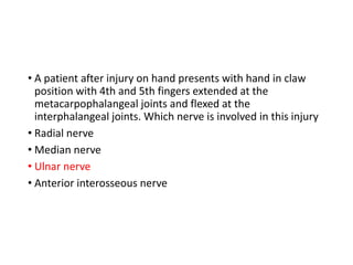
The ulnar nerve is involved. Damage to the ulnar nerve results in a characteristic claw hand deformity with the 4th and 5th fingers hyperextended at the MCP joints and flexed at the IP joints due to paralysis of the interossei and lumbrical muscles.
Learning Objectives
1. Ulnarnerve formation , root value and important
relations
2. Motor and sensory supply
3. Important sites of injuries/entrapment of nerve
4. Effects of injury of ulnar nerve
5. How to clinically test ulnar nerve injury
3.
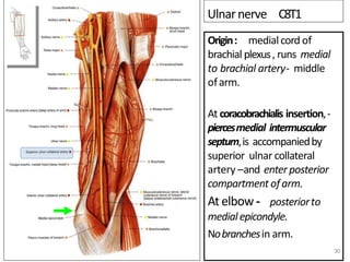
Ulnarnerve C8T1
Origin: medialcordof
brachial plexus, runs medial
to brachial artery- middle
ofarm.
At coracobrachialis insertion,-
piercesmedial intermuscular
septum,is accompaniedby
superior ulnar collateral
artery –and enter posterior
compartment ofarm.
At elbow - posteriorto
medialepicondyle.
Nobranchesin arm.
30
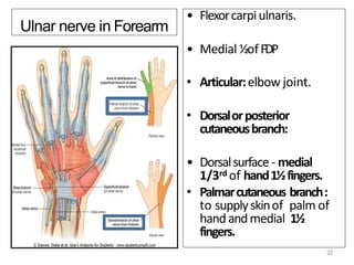
4.
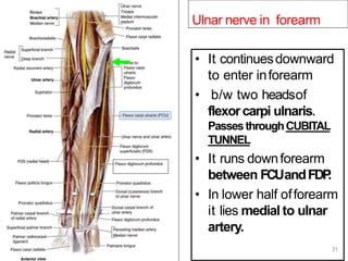
Ulnar nerve inforearm
• It continuesdownward
to enter inforearm
• b/w two headsof
flexorcarpi ulnaris.
PassesthroughCUBITAL
TUNNEL
• It runs downforearm
between FCUandFDP
.
• In lower half offorearm
it lies medial to ulnar
artery.
31
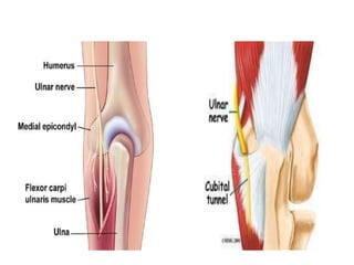
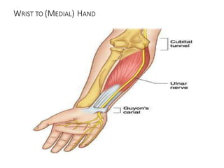
5.
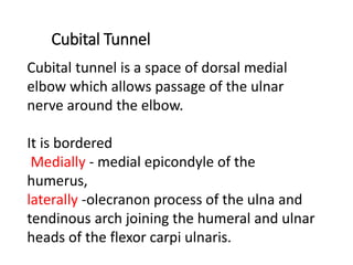
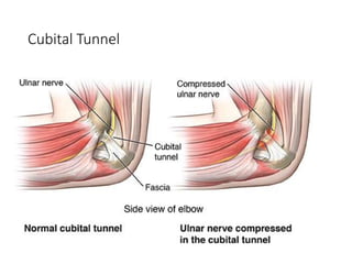
Cubital Tunnel
Cubital tunnelis a space of dorsal medial
elbow which allows passage of the ulnar
nerve around the elbow.
It is bordered
Medially - medial epicondyle of the
humerus,
laterally -olecranon process of the ulna and
tendinous arch joining the humeral and ulnar
heads of the flexor carpi ulnaris.
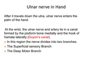
After it travelsdown the ulna, ulnar nerve enters the
palm of the hand.
At the wrist, the ulnar nerve and artery lie in a canal
formed by the pisiform bone medially and the hook of
hamate laterally (Guyon’s canal).
In this region the nerve divides into two branches.
The Superficial sensory Branch
The Deep Motor Branch
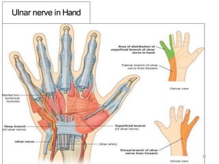
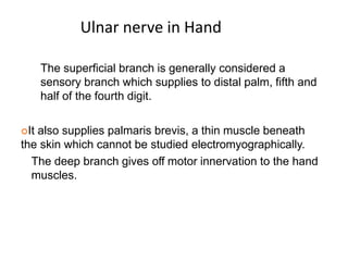
Ulnar nerve in Hand
11.
The superficial branchis generally considered a
sensory branch which supplies to distal palm, fifth and
half of the fourth digit.
It also supplies palmaris brevis, a thin muscle beneath
the skin which cannot be studied electromyographically.
The deep branch gives off motor innervation to the hand
muscles.
Ulnar nerve in Hand
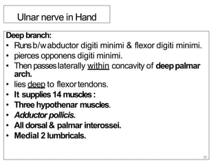
Ulnar nerve inHand
Deep branch:
• Runsb/wabductor digiti minimi & flexor digiti minimi.
• pierces opponens digiti minimi.
• Then passeslaterally within concavity of deeppalmar
arch.
• lies deep to flexortendons.
• It supplies14 muscles:
• Threehypothenar muscles.
• Adductor pollicis.
• All dorsal& palmar interossei.
• Medial 2 lumbricals.
35
15.
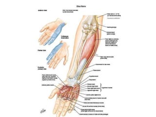
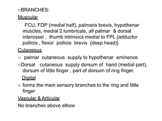
BRANCHES:
Muscular
FCU, FDP (medialhalf), palmaris brevis, hypothenar
muscles, medial 2 lumbricals, all palmar & dorsal
interossei , thumb intrinsics medial to FPL {adductor
pollicis , flexor pollicis brevis (deep head)}
Cutaneous
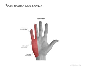
palmar cutaneous supply to hypothenar eminence
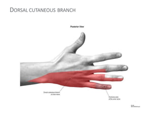
Dorsal cutaneous supply dorsum of hand (medial part),
dorsum of little finger , part of dorsum of ring finger.
Digital
forms the main sensory branches to the ring and little
finger
Vascular & Articular
No branches above elbow
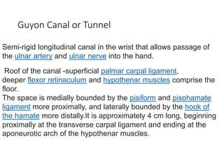
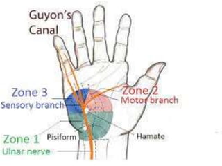
Guyon Canal orTunnel
Semi-rigid longitudinal canal in the wrist that allows passage of
the ulnar artery and ulnar nerve into the hand.
Roof of the canal -superficial palmar carpal ligament,
deeper flexor retinaculum and hypothenar muscles comprise the
floor.
The space is medially bounded by the pisiform and pisohamate
ligament more proximally, and laterally bounded by the hook of
the hamate more distally.It is approximately 4 cm long, beginning
proximally at the transverse carpal ligament and ending at the
aponeurotic arch of the hypothenar muscles.
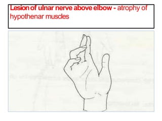
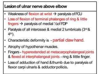
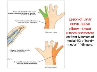
Lesionof ulnar nerveaboveelbow
• Weaknessof flexion at wrist paralysis ofFCU
• Lossof flexion of terminal phalanges of ring & little
fingers paralysis of medial ½ofFDP
• Paralysisof all interossei & medial 2 lumbricals (3rd&
4th).
• Characteristic deformity is - partial clawhand.
• Atrophy of hypothenar muscles.
• Fingers- hyperextended at metacarpophalangeal joints
& flexed at interphalangeal joints - ring & little finger.
• Lossof adduction of hand &thumb due to paralysis of
flexor carpi ulnaris & adductorpollicis.
23.
Lesion of ulnar
nerveabove
elbow - Lossof
cutaneous sensations
on front &dorsum of
medial 1/3 of hand+
medial 1 ½fingers.
24.
• It leadstoparalysisof
intrinsic musclesof handas
describedabove.
• deformity ‘clawhand’
• Lossof cutaneoussensations
of medial 1 ½ fingers.
Lesionof ulnar
nerve above
wrist
Test for Palmar interossei
for adduction of fingers.
Test for adductor &
opponens pollicis.
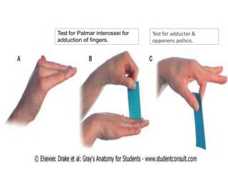
25.
Test for Palmarinterossei for
adduction of fingers.
Test for adductor &
opponens pollicis.
27.
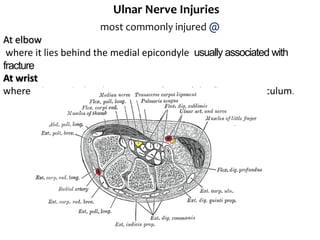
Ulnar Nerve Injuries
mostcommonly injured @
At elbow
where it lies behind the medial epicondyle usually associated with
fracture
At wrist
where it lies with the ulnar artery in front of the flexor retinaculum.
28.
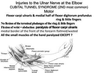
Injuries to theUlnar Nerve at the Elbow
CUBITAL TUNNEL SYNDROME (2ND most common)
Motor
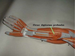
Flexor carpi ulnaris & medial half of flexor digitorum profundus
ring & little fingers
Noflexionoftheterminalphalangesofthering&littlefingers
Flexionofwrist=abduction paralysis of flexor carpi ulnaris
medial border of the front of the forearm flattned/wasted
All the small muscles of the hand paralyzed EXCEPT ?
29.
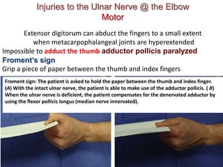
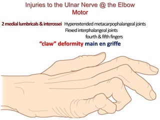
Injuries to theUlnar Nerve @ the Elbow
Motor
Extensor digitorum can abduct the fingers to a small extent
when metacarpophalangeal joints are hyperextended
Impossible to adduct the thumb adductor pollicis paralyzed
Froment’s sign
Grip a piece of paper between the thumb and index fingers
Froment sign: The patient is asked to hold the paper between the thumb and index finger.
(A) With the intact ulnar nerve, the patient is able to make use of the adductor pollicis. ( B)
When the ulnar nerve is deficient, the patient compensates for the denervated adductor by
using the flexor pollicis longus (median nerve innervated).
30.
Injuries to theUlnar Nerve @ the Elbow
Motor
2mediallumbricals&interossei Hyperextendedmetacarpophalangealjoints
Flexedinterphalangealjoints
fourth&fifthfingers
“claw” deformity main en griffe
31.
Injuries to theUlnar Nerve @ the Elbow
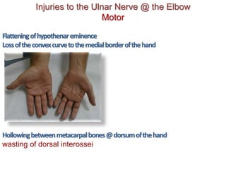
Motor
Flatteningofhypothenareminence
Lossoftheconvexcurvetothemedialborderofthehand
Hollowingbetweenmetacarpalbones@dorsumofthehand
wasting of dorsal interossei
32.
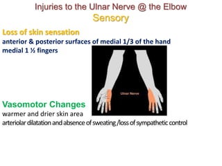
Loss of skinsensation
anterior & posterior surfaces of medial 1/3 of the hand
medial 1 ½ fingers
Vasomotor Changes
warmer and drier skin area
arteriolardilatationandabsenceofsweating/lossofsympatheticcontrol
Injuries to the Ulnar Nerve @ the Elbow
Sensory
33.
Injuries to theUlnar Nerve @ the Wrist
Motor
Smallhandmusclesparalyzed,wasted–EXCEPT3thenar@first2lumbricals
Claw hand
Moreobvious
Flexordigitorumprofundusintact
Markedflexionoftheterminalphalanges
Ulnar paradox
Higher lesion
Less obvious claw deformity
More proximal injury
Less claw
34.
PROXIMAL/ @ ELBOW
CUBITALTUNNEL SYNDROME
BETWEEN MEDIAL EPICONDYLE & FLEXOR CARPI ULNARIS
DISTAL/ @ WRIST
GUYON’S CANAL Roof: Palmaris brevis, hamate,pisiforme bones &
FCU
Q: Medial half of Flexor digitorum profundus
affected in which one most?
35.
Froment’s sign
To performthe test, a patient is
asked to hold an object, usually a flat
object such as a piece of paper,
between their thumb and index finger
(pinch grip). The examiner then
attempts to pull the object out of the
subject's hands.[2]
Froment’s sign : hyperflexion of IP jt of thumb
while attempting a lateral pinch(indicates
paralysis of adductor pollicis, 1st DI , with
replacement of pinch function by FPL)
36.
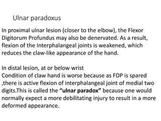
Ulnar paradoxus
In proximalulnar lesion (closer to the elbow), the Flexor
Digitorum Profundus may also be denervated. As a result,
flexion of the Interphalangeal joints is weakened, which
reduces the claw-like appearance of the hand.
In distal lesion, at or below wrist
Condition of claw hand is worse because as FDP is spared
,there is active flexion of interphalangeal joint of medial two
digits.This is called the “ulnar paradox” because one would
normally expect a more debilitating injury to result in a more
deformed appearance.
37.
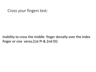
Cross your fingerstest:
Inability to cross the middle finger dorsally over the index
finger or vise versa.(1st PI & 2nd DI)
38.
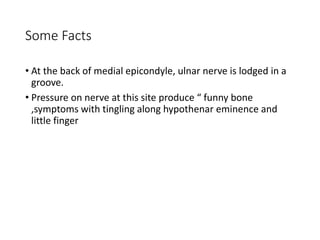
Some Facts
• Atthe back of medial epicondyle, ulnar nerve is lodged in a
groove.
• Pressure on nerve at this site produce “ funny bone
,symptoms with tingling along hypothenar eminence and
little finger
39.
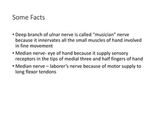
Some Facts
• Deepbranch of ulnar nerve is called “musician” nerve
because it innervates all the small muscles of hand involved
in fine movement
• Median nerve- eye of hand because it supply sensory
receptors in the tips of medial three and half fingers of hand
• Median nerve – laborer’s nerve because of motor supply to
long flexor tendons
40.
which of thesesymptoms is not caused by damage to the
median nerve at the wrist?
A
ape/simian hand
B
loss of pronation
C
loss of sensation in most of thumb and digits 2 and 3.
D
thenar muscle paralysis
41.
which of thesesymptoms is not caused by damage to the
median nerve at the wrist?
A
ape/simian hand
B
loss of pronation
C
loss of sensation in most of thumb and digits 2 and 3.
D
thenar muscle paralysis
42.
• Injury tothe ulnar nerve at the ____ causes ______, and at
the ____ causes ____
• A
• elbow, radial deviation, wrist, wrist drop
• B
• elbow, radial wrist deviation, wrist, severe clawing of hand
• C
• elbow, severe clawing of hand, wrist, radial deviation
• D
• elbow, wrist drop, wrist, radial deviation
43.
• Injury tothe ulnar nerve at the ____ causes ______, and at
the ____ causes ____
• A
• elbow, radial deviation, wrist, wrist drop
• B
• elbow, radial wrist deviation, wrist, severe clawing of hand
• C
• elbow, severe clawing of hand, wrist, radial deviation
• D
• elbow, wrist drop, wrist, radial deviation
44.
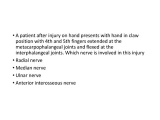
• A patientafter injury on hand presents with hand in claw
position with 4th and 5th fingers extended at the
metacarpophalangeal joints and flexed at the
interphalangeal joints. Which nerve is involved in this injury
• Radial nerve
• Median nerve
• Ulnar nerve
• Anterior interosseous nerve
45.
• A patientafter injury on hand presents with hand in claw
position with 4th and 5th fingers extended at the
metacarpophalangeal joints and flexed at the
interphalangeal joints. Which nerve is involved in this injury
• Radial nerve
• Median nerve
• Ulnar nerve
• Anterior interosseous nerve
46.
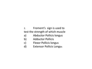
2. Froment’s signis used to
test the strength of which muscle
a) Abductor Pollicis longus
b) Adductor Pollicis
c) Flexor Pollicis longus
d) Extensor Pollicis Longus
47.
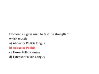
Froment’s sign isused to test the strength of
which muscle
a) Abductor Pollicis longus
b) Adductor Pollicis
c) Flexor Pollicis longus
d) Extensor Pollicis Longus
48.
• A patientis asked by his physician to hold their wrist in
complete and forced flexion (pushing the dorsal surfaces of
both hands together) for 30–60 seconds. This maneuver
compress the nerve within the carpal tunnel and
characteristic symptoms (such as burning, tingling or numb
sensation over the thumb, index, middle and ring fingers)
conveys a positive test result and suggests carpal tunnel
syndrome. What is the name of manoeuvre physician is
performing
• Turning Circle maneure
• Phalen’s Manoeuvre
• Collision Avoidance Manoeuvre
• Zig-zag Test Manoeuvre.
49.
1. A patientis asked by his physician to hold their wrist in
complete and forced flexion (pushing the dorsal surfaces of
both hands together) for 30–60 seconds. This maneuver
compress the nerve within the carpal tunnel and
characteristic symptoms (such as burning, tingling or numb
sensation over the thumb, index, middle and ring fingers)
conveys a positive test result and suggests carpal tunnel
syndrome. What is the name of manoeuvre physician is
performing
a) Turning Circle maneure
b) Phalen’s Manoeuvre
c) Collision Avoidance Manoeuvre
d) Zig-zag Test Manoeuvre.
50.
• A clinicalcondition in which patient shows inability to
abduct the thumb due to median nerve lesion is called
• a. Pollock ‘s Sign
• b. Pointing Index
• c. Ape thumb deformity
• d. Andre- Thomas Sign
51.
A clinical conditionin which patient shows inability to abduct
the thumb due to median nerve lesion is called
a. Pollock ‘s Sign
b. Pointing Index
c. Ape thumb deformity
d. Andre- Thomas Sign
52.
The index fingeris not flexed at the proximal
interphalangeal (PIP) and distal interphalangeal (DIP)
joints. This clinical condition is called pointing index. This
condition is due to lesion of
a) Ulnar nerve
b) Median nerve
c) Radial nerve
d) Posterior interosseous nerve
53.
The index fingeris not flexed at the proximal interphalangeal
(PIP) and distal interphalangeal (DIP) joints. This clinical
condition is called pointing index. This condition is due to
lesion of
a) Ulnar nerve
b) Median nerve
c) Radial nerve
d) Posterior interosseous nerve
54.
Pen test inhand is performed to assess the
neuromuscular status of:
a) Opponens pollicis
b) Flexor pollicis brevis
c) Abductor pollicis brevis
d) First palmar interossei
55.
Pen test inhand is performed to assess the neuromuscular
status of:
a) Opponens pollicis
b) Flexor pollicis brevis
c) Abductor pollicis brevis
d) First palmar interossei
56.
In the phalen’stest, the suspected compression of
median nerve is elicited. Which of the following are the other
structures passing through the carpal tunnel?
• a. Ulnar nerve
• b. Superficial cutaneous branch
• c. Flexor digitorum profundud tendons
• d. Palmaris longus tendon
57.
In the phalen’stest, the suspected compression of median
nerve is elicited. Which of the following are the other
structures passing through the carpal tunnel?
a. Ulnar nerve
b. Superficial cutaneous branch
c. Flexor digitorum profundud tendons
d. Palmaris longus tendon

![Froment’s sign
To perform the test, a patient is
asked to hold an object, usually a flat
object such as a piece of paper,
between their thumb and index finger
(pinch grip). The examiner then
attempts to pull the object out of the
subject's hands.[2]
Froment’s sign : hyperflexion of IP jt of thumb
while attempting a lateral pinch(indicates
paralysis of adductor pollicis, 1st DI , with
replacement of pinch function by FPL)](https://image.slidesharecdn.com/807ulnar-nerve-and-its-lesions-230320003204-fc0a0581/85/807_Ulnar-nerve-and-its-lesions-pptx-35-320.jpg)