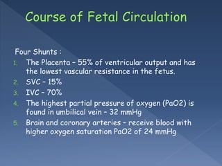
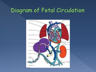
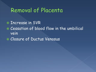
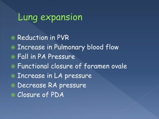
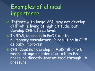
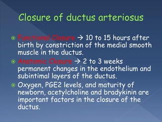
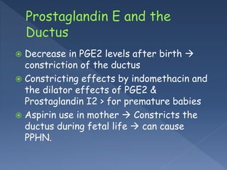
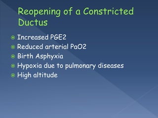
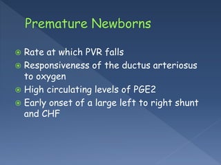
 The placenta provides the exchange of
gases and nutrients in the fetus.
Four Shunts :
1. The Placenta – 55% of ventricular output and has
the lowest vascular resistance in the fetus.
2. SVC – 15%
3. IVC – 70%
4. The highest partial pressure of oxygen (PaO2) is
found in umbilical vein – 32 mmHg
5. Brain and coronary arteries – receive blood with
higher oxygen saturation PaO2 of 24 mmHg
 Lungs receive 15% of combined ventricular
output.
 RV > LV
 RV- 55% LV – 45%
 Fetal C.O directly proportional to HR
 Low compliance of Fetal Heart .
 Primary change – Shift of blood flow from
placenta to lungs for oxygenation.
 Establishment of Pulmonary circulation
 Increase in SVR
 Cessation of blood flow in the umbilical
vein
 Closure of Ductus Venosus
 Reduction in PVR
 Increase in Pulmonary blood flow
 Fall in PA Pressure
 Functional closure of foramen ovale
 Increase in LA pressure
 Decrease RA pressure
 Closure of PDA
1.Hypoxia and /or altitude
2. RDS or Congenital Pneumonia
3. Metabolic Acidosis
4. Increased pulmonary artery pressure
secondary to VSD or PDA
5. Increased pressure in the left atrium or
pulmonary vein.
 Infants with large VSD may not develop
CHF while living at high altitude, but
develop CHF at sea level.
 In RDS, increase in PaO2 dilates
pulmonary vasculature  resulting in CHF
as baby improves
 CHF does not develop in VSD till 6 to 8
weeks of age or older due to high PA
pressure directly transmitted through LV
pressure.
 Functional Closure  10 to 15 hours after
birth by constriction of the medial smooth
muscle in the ductus.
 Anatomic Closure  2 to 3 weeks
permanent changes in the endothelium and
subintimal layers of the ductus.
 Oxygen, PGE2 levels, and maturity of
newborn, acetylcholine and bradykinin are
important factors in the closure of the
ductus.
 Strongest stimulus for constriction of the
ductal smooth muscle  postnatal
increase in PaO2 from 25 mmHg to 50
mmHg
 Ductal tissue is less responsive to the
oxygenation changes in premature infant
due to immature ductus and smooth
muscles in the media of ductus.
 High levels of PGE2 in preterm infants.
 Decrease in PGE2 levels after birth 
constriction of the ductus
 Constricting effects by indomethacin and
the dilator effects of PGE2 &
Prostaglandin I2 > for premature babies
 Aspirin use in mother  Constricts the
ductus during fetal life  can cause
PPHN.
 Increased PGE2
 Reduced arterial PaO2
 Birth Asphyxia
 Hypoxia due to pulmonary diseases
 High altitude
PA constricted by
 O2 and Metabolic acidosis
 Epinephrine
 Nor-epinephrine
PA dilated by
 Vagal stimulation
 Isoproterenol
 Bradykinin
 Rate at which PVR falls
 Responsiveness of the ductus arteriosus
to oxygen
 High circulating levels of PGE2
 Early onset of a large left to right shunt
and CHF
Fetal and perinatal circulation

Fetal and perinatal circulation

  • 2.
     The placentaprovides the exchange of gases and nutrients in the fetus.
  • 3.
    Four Shunts : 1.The Placenta – 55% of ventricular output and has the lowest vascular resistance in the fetus. 2. SVC – 15% 3. IVC – 70% 4. The highest partial pressure of oxygen (PaO2) is found in umbilical vein – 32 mmHg 5. Brain and coronary arteries – receive blood with higher oxygen saturation PaO2 of 24 mmHg
  • 5.
     Lungs receive15% of combined ventricular output.  RV > LV  RV- 55% LV – 45%
  • 6.
     Fetal C.Odirectly proportional to HR  Low compliance of Fetal Heart .
  • 7.
     Primary change– Shift of blood flow from placenta to lungs for oxygenation.  Establishment of Pulmonary circulation
  • 8.
     Increase inSVR  Cessation of blood flow in the umbilical vein  Closure of Ductus Venosus
  • 9.
     Reduction inPVR  Increase in Pulmonary blood flow  Fall in PA Pressure  Functional closure of foramen ovale  Increase in LA pressure  Decrease RA pressure  Closure of PDA
  • 11.
    1.Hypoxia and /oraltitude 2. RDS or Congenital Pneumonia 3. Metabolic Acidosis 4. Increased pulmonary artery pressure secondary to VSD or PDA 5. Increased pressure in the left atrium or pulmonary vein.
  • 12.
     Infants withlarge VSD may not develop CHF while living at high altitude, but develop CHF at sea level.  In RDS, increase in PaO2 dilates pulmonary vasculature  resulting in CHF as baby improves  CHF does not develop in VSD till 6 to 8 weeks of age or older due to high PA pressure directly transmitted through LV pressure.
  • 13.
     Functional Closure 10 to 15 hours after birth by constriction of the medial smooth muscle in the ductus.  Anatomic Closure  2 to 3 weeks permanent changes in the endothelium and subintimal layers of the ductus.  Oxygen, PGE2 levels, and maturity of newborn, acetylcholine and bradykinin are important factors in the closure of the ductus.
  • 14.
     Strongest stimulusfor constriction of the ductal smooth muscle  postnatal increase in PaO2 from 25 mmHg to 50 mmHg  Ductal tissue is less responsive to the oxygenation changes in premature infant due to immature ductus and smooth muscles in the media of ductus.  High levels of PGE2 in preterm infants.
  • 15.
     Decrease inPGE2 levels after birth  constriction of the ductus  Constricting effects by indomethacin and the dilator effects of PGE2 & Prostaglandin I2 > for premature babies  Aspirin use in mother  Constricts the ductus during fetal life  can cause PPHN.
  • 16.
     Increased PGE2 Reduced arterial PaO2  Birth Asphyxia  Hypoxia due to pulmonary diseases  High altitude
  • 17.
    PA constricted by O2 and Metabolic acidosis  Epinephrine  Nor-epinephrine PA dilated by  Vagal stimulation  Isoproterenol  Bradykinin
  • 18.
     Rate atwhich PVR falls  Responsiveness of the ductus arteriosus to oxygen  High circulating levels of PGE2  Early onset of a large left to right shunt and CHF