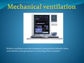
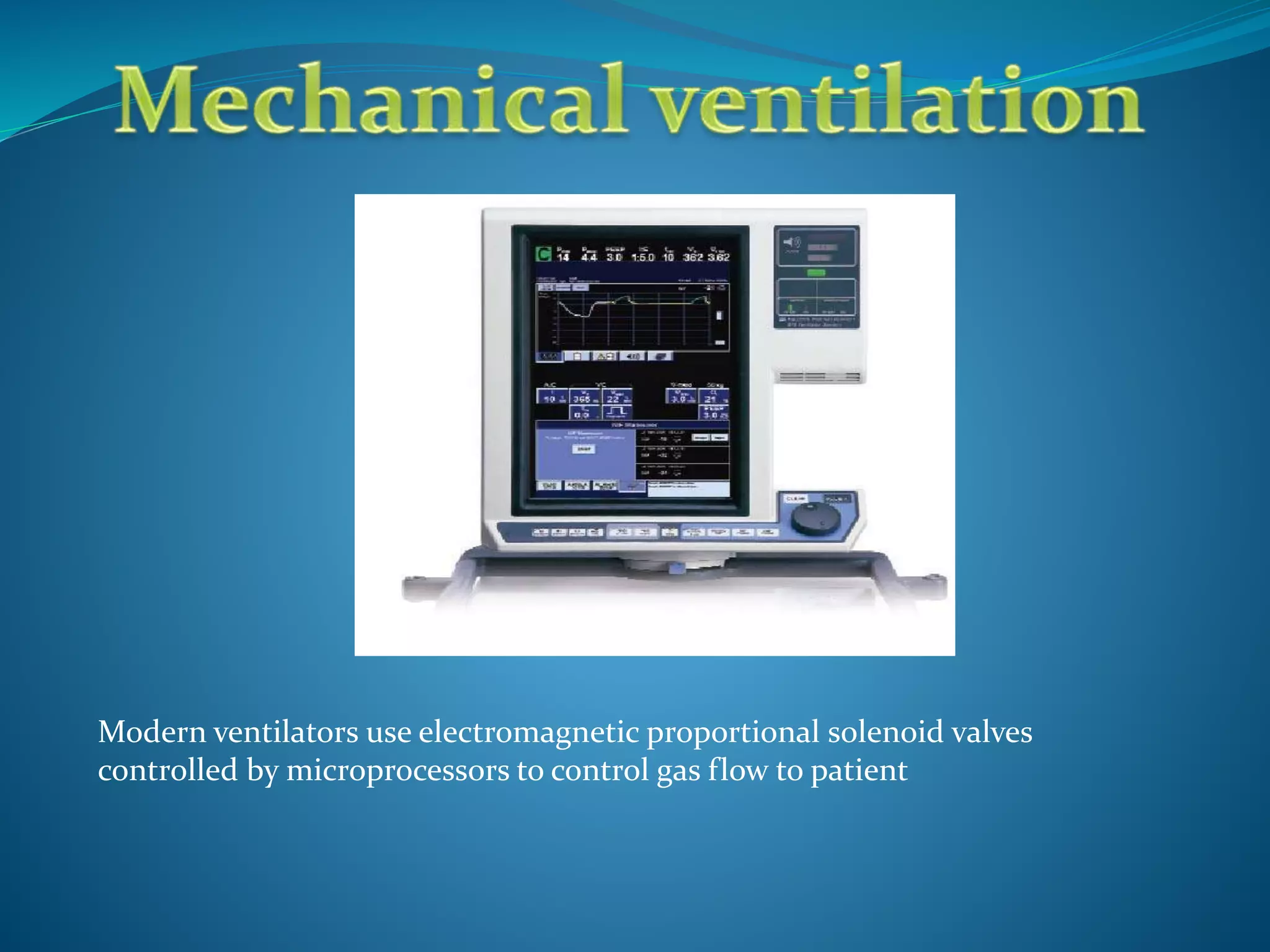
TataKelola dan KamSiber Kecerdasan Buatan v022.pdf
Mechanical ventilation
1. Modern ventilators use electromagnetic proportional solenoid valves
controlled by microprocessors to control gas flow to patient
2. FIO2
FiO2 is the fraction of inspired oxygen. It is the
percentatge of oxygen delivered on inspiration.
Ranges from 21% to 100%.
The oxygen analysers In the inspiratory limb is used to
ensure the accuracy of
FiO2 delivered
3. Tidal volume (TV or VT)
Measured volume with each breath
Normal tidal volume should be kept at
6 – 8 ml/kg of patients ideal body weight
4. Minute volume (MV)
MV is the total volume inspired or expires over a minute
It is the product of tidal volume (TV) and respiratory rate (RR) or
frequency
MV = TV x RR
6. Ratio of the duration of inspiration to expiration.
Inspiratory time (Ti) is commonly set at a third of the
total inspiratory expiratory cycle (1:3).
The mechanism by which the operator adjusts the
inspiratory time in AC mode is by controlling the
inspiratory flow rate.
Increase in inspiratory flow rate will decrease
inspiratory time
Inspiratory expiratory ratio
7. Peep
Positive end expiratory pressure is the positive pressure applied at the end of
expiration during mechanical ventilation.
The use of PEEP improves gas exchange by recruiting collapsed alveoli and
thereby improving functional residual capacity.
PEEP also helps in redistributing lung water from the alveolar space into the
pulmonary circulation and improving ventilation perfusion mismatch.
All these contributes to reduction in dead space ventilation and thereby
improving PaO2.
PEEP increases intra-thoracic pressure resulting in decreased venous return to
the right heart and increasing the right ventricular after load (the force needed
to eject blood from right ventricle) .
8. AUTO PEEP
Auto PEEP is the actual pressure difference between the
higher pressure and the baseline pressure (set PEEP level or
0) at the end of full expiration
It is measured by pressing the expiratory hold button on
the ventilator.
9. Peak airway pressure
Pressure needed initially to assure the flow of gas
down the airways and open up the alveoli.
Depends upon resistance and compliance
10. PLATEAU PRESSURE
Pressure required to hold the lungs and chest wall open when there is no air flow during
the inspiratory cycle
Measured at the end of a full inspiration during a breath hold (no air flow)
How to measure Plateau pressure
1. Switch to volume controlled mode ventilation
2. Press the end-inspiratory hold button
3. The machine will display plateau pressure. It is automatically done by the ventilator by
sensing the pressure which is required to keep the alveoli open at the end of inspiration
when there is no airflow which will be generally 10-15cm H2O less than Peak airway
pressure.
With decreased compliance, the plateau pressure will increase.
11. RESISTANCE
Degree of easiness to move gases down the airways
High resistance --- difficult to move gases in and out
Low resistance --- easy to move gases in and out.
Measured using the difference between peak airway
pressure and plateau pressure.
12. COMPLIANCE
Degree of easiness to distend the alveoli, lungs and
chest wall.
High compliance --- easy to distend the alveoli
Low compliance ---- hard to distend the alveoli
Compliance is measured with plateau pressure
13. RECRUITMENT
Ventilation strategy whereby the non ventilated alveoli are opened by
progressively introducing higher pressures over a short period. The aim is
reinflate atelectatic alveoli without distending the normal lung units.
Techniques
1. PEEP is increased in increments of 5cm H2O from a baseline PEEP to 35cm
H2O reducing tidal volume to limit peak inspiratory pressure to 35cm H2O.
CPAP is maintained for 30s.
2. Intermittent higher tidal volume in pressure controlled ventilation applied
with escalating PEEP and constant driving pressure: peak pressure of 45cm
H2O, I:E ratio of 1:2 and PEEP level of 16cm H2O for 2min.
Sustained inflation by application of CPAP at 30-40cm H20 for 40s.
14. Weaning
Weaning is the process when the support of the ventilator is gradually
decreased to re-establish spontaneous breathing. Readiness for
weaning is evaluated by assessing level of consciousness, adequacy of
oxygen and ventilation, spontaneous breathing strength etc.
15. Anatomic dead space
Anatomic Dead space is the portion of the respiratory
system not involved in gas exchange.
It is be divided into alveolar and airway dead space.
Airway dead space is also called tracheal dead space as it is the
part of the respiratory system (mainly trachea) where tidal
volume remains in the conducting passages at the end of
inspiration and therefore does not participate in gas exchange.
Alveolar dead space is a measurement of the ventilated alveoli
without adequate blood flow for gas exchange. It is negligible in
healthy individuals.