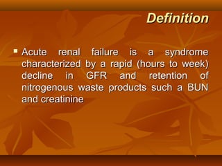
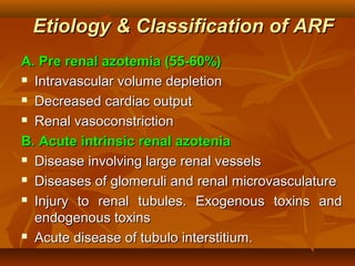
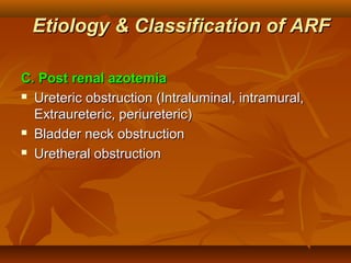
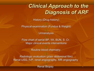
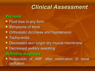
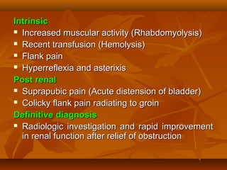
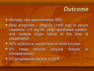
Management of
        Acute Renal Failure
                  Dr. Sachin Verma MD, FICM, FCCS, ICFC
                    Fellowship in Intensive Care Medicine
                      Infection Control Fellows Course
                Consultant Internal Medicine and Critical Care
              Web:- http://www.medicinedoctorinchandigarh.com
                           Mob:- +91-7508677495


References
 Brenner & Rector’s The Kidney, 7 th ed.

 Harrison’s Principles of Internal Medicine, 16th ed.

                                                                 29/9/05
Definition

   Acute renal failure is a syndrome
    characterized by a rapid (hours to week)
    decline in GFR and retention of
    nitrogenous waste products such a BUN
    and creatinine
Etiology & Classification of ARF
A. Pre renal azotemia (55-60%)
 Intravascular volume depletion

 Decreased cardiac output

 Renal vasoconstriction

B. Acute intrinsic renal azotenia
 Disease involving large renal vessels

 Diseases of glomeruli and renal microvasculature

 Injury to renal tubules. Exogenous toxins and

  endogenous toxins
 Acute disease of tubulo interstitium.
Etiology & Classification of ARF

C. Post renal azotemia
 Ureteric obstruction (Intraluminal, intramural,

  Extraureteric, periureteric)
 Bladder neck obstruction

 Uretheral obstruction
Clinical Approach to the
                  Diagnosis of ARF
                History (Drug history)
                           ↓
      Physical examination (Fundus & Weight)
                           ↓
                     Urinanalysis
                           ↓
      Flow chart of serial BP, Wt, BUN, S. Cr.
         Major clinical events interventions
                           ↓
             Routine blood chemistry
                           ↓
    Radiologic evaluation (plain abdominal film)
Renal USG, IVP, renal angiography, MR angiography
                           ↓
                   Renal Biopsy
Clinical Assessment
Pre renal
 Fluid loss in any form

 Symptoms of thirst

 Orthostatic dizziness and hypotension

 Tachycardia

 Decreased skin turgor dry mucus membrane

 Decreased axillary sweating

Definitive diagnosis
 Resolution of ARF after restoration of renal

  perfusion
Intrinsic
 Increased muscular activity (Rhabdomyolysis)

 Recent transfusion (Hemolysis)

 Flank pain

 Hyperreflexia and asterixis

Post renal
 Suprapubic pain (Acute distension of bladder)

 Colicky flank pain radiating to groin

Definitive diagnosis
 Radiologic investigation and rapid improvement

  in renal function after relief of obstruction
Urinanalysis
1. Urine volume
2. Urine sediment
     Acellular / Transparent hyaline cast (pre renal)

     Pigmented “muddy brown” granular cast, tubule

      epithelial cell cast (renal)
     Benign sediment, hematuria, pyuria (post renal)

     Broad granular cast characteristics of chronic renal

      disease and reflect interstitial fibrosis and dilatation of
      tubules
 Granular cast

     ATN, GN / vasculitis, Interstitial nephritis

 RBC cast

     GN / Vasculitis, Malignant hypertension
  WBC cast
     Acute interstitial nephritis, Severe pyelonephritis,

       Marked leukemic or lymphomatous infiltration
3. Eosinophiluria (>5%)
     Drug induced allergic interstitial nephritis

4. Crystalluria
     Uric acid crystals (pleomorphic), oxalate (envelop

       shaped), Hippurate (needle shaped)
5. Tubule proteinuria (<1g/d) : proximal tubule cell injury,
   glomerular proteinuria (>1g/d) injury to glomerular
   ultrafiltration barrier
6. Haemoglobinuria
7. Myoglobinuria
Confirmatory test
   Plain abdominal film
   USG
   CT Scan
   Radio nuclide scan
   MRA
    Doppler USG and Spiral CT
   Contrast angiography (Gold standard)
   Renal biopsy
FENa (Fractional Excretion
                           of Na+(%)

   Most sensitive index to differentiate pre renal
    azotemia from ATN
    UNa X Pcr          <1 prerenal
               X100
    PNa X Ucr          >1 ATN
Treatment
Pre renal azotemia
 Correction of Hypovolemia by packed red cells,

  isotonic saline, Hypotonic saline (0.45%)
 Loop blocking diuretic, (Frusemide high dose 20

  – 160 mg orally or IV twice daily) to effect
  adequate diuresis and convert oliguric to non-
  oliguric RF.
 ARF with cirrhosis (fluid challenge) paracentesis

  with albumin administration
 Renal dose dopamine (1-3 mg/kg/min)
Treatment
Intrinsic ATN
 Optimization   of CV function & intravascular
  volume
 Prophylactic oral acetylcysteine (600 mg BD 24

  hour before and after procedure)
 Use     of less nephrotoxic contrast agent
  (Gadolinium and CO2)
 Cautious use of diuretics, NSAIDs, ACE inhibitors

 Lipid encapsulated formulation of amphotericin B

 Allopurinol (Acute urate nephropathy)

 Amifostine an organic thiophosphate (Cisplatin)
   Forced diuresis and alkanization of urine
    (Rhabdomyolysis)
   N      Acetylcysteine      within   24     hour
    (Acetaminophen)
   Dimercaprol (Chelating agent) (heavy metal)
   Ethanol (ethylene glycol toxicity)
   Plasma pharesis (Myeloma cast nephropathy)
   Systemic arterial pressure control (malignant
    htpertensive nephrosclerosis)
   Acute GN (pulse glucocorticoid therapy)
ANP
 28 amino acid polypeptide. Synthesized in

  cardiac atrial muscle. Increased GFR by
  triggering afferent arteriolar vasodilatation and
  increasing ultrafiltration. Inhibits Na transport
  and lower oxygen requirement.
Post renal ARF
 Transuretheral    or suprapubic placement of
  bladder catheter (obstruction of urethra or
  bladder neck)
 Percutaneous catheterization of dilated renal

  pelvis or ureter (ureteric obstruction)
 Removal of obstructing lesion percutaneously or

  bypassed by insertion of ureteric stent
Management of complication
Intravascular volume overload
 Salt (1-2 gm/day) and water (<1 lt/day) restriction

 Diuretics, usually loop + thiazide

 Ultrafiltration or dialysis

Hyponatremia
 Restriction of enteral free water intake (<1lt/day)

 Avoid hypotonic intravenous solution (including

  dextrose)
Hyperkalemia
 Restriction of dietary K+ intake (<40 mmol/day)

 Eliminate K+ supplement and K+ sparing diuretic,

 Potassium     binding ion-exchange resin (Na
  polystyrene sulphonate)
 Glucose (50 ml of 50% Dextrose) and insulin (10

  U regular)
 NaCO (50-100 mmol)
        3
   Calcium gluconate (10 ml of 10% solution) over 5
    minute
   Dialysis (with low K+ dialysate)
Metabolic acidosis
 Restriction of dietary protein (0.6 g/Kg/day of high

  biologic value)
 Na bicarbonate (maintain serum bicarbonate >15

  mmol/L or arterial pH >7.2)
 Dialysis

Hyperphosphatemia
 Restriction of dietary phosphate intake (<800

  mg/day)
 Phosphate     binding agents (Ca carbonate,
  Aluminium hydroxide)
Hypocalcemia
 Calcium Carbonate

 Calcium gluconate (10 – 20 ml of 10% solution)

Hypermagnesemia
 Avoid Mg2+ containing antacids

Hyperuricemia
 Treatment usually not necessary (<15 mg/dl)

Nutrition
 Restriction of dietary protein (0.6 g/kg/day)

 Carbohydrate (100 g/day)

 Enteral / Parenteral nutrition
Indication for Dialysis

   Clinical evidence (signs & symptoms) of uremia
   Intractable intravascular volume over load
   Hyperkalemia
   Severe acidosis (resistant to conservative
    measures)
   Prophylactic dialysis when urea >100-150 mg/dl
    or creatinine >8-10 mg/dl
Outcome
   Mortality rate approximately 50%
   Poor prognosis – Oliguria (<400 mg) or serum
    creatinine (>3 mg/dl), older debilitated patient
    and multiple organ failure at the time of
    presentation
   50% subclinical impairment of renal function
   5% never recover (require dialysis or
    transplantation)
   5% progressive decline in GFR
Management of arf

Management of arf

  • 1.
    Management of Acute Renal Failure Dr. Sachin Verma MD, FICM, FCCS, ICFC Fellowship in Intensive Care Medicine Infection Control Fellows Course Consultant Internal Medicine and Critical Care Web:- http://www.medicinedoctorinchandigarh.com Mob:- +91-7508677495 References  Brenner & Rector’s The Kidney, 7 th ed.  Harrison’s Principles of Internal Medicine, 16th ed. 29/9/05
  • 2.
    Definition  Acute renal failure is a syndrome characterized by a rapid (hours to week) decline in GFR and retention of nitrogenous waste products such a BUN and creatinine
  • 3.
    Etiology & Classificationof ARF A. Pre renal azotemia (55-60%)  Intravascular volume depletion  Decreased cardiac output  Renal vasoconstriction B. Acute intrinsic renal azotenia  Disease involving large renal vessels  Diseases of glomeruli and renal microvasculature  Injury to renal tubules. Exogenous toxins and endogenous toxins  Acute disease of tubulo interstitium.
  • 4.
    Etiology & Classificationof ARF C. Post renal azotemia  Ureteric obstruction (Intraluminal, intramural, Extraureteric, periureteric)  Bladder neck obstruction  Uretheral obstruction
  • 5.
    Clinical Approach tothe Diagnosis of ARF History (Drug history) ↓ Physical examination (Fundus & Weight) ↓ Urinanalysis ↓ Flow chart of serial BP, Wt, BUN, S. Cr. Major clinical events interventions ↓ Routine blood chemistry ↓ Radiologic evaluation (plain abdominal film) Renal USG, IVP, renal angiography, MR angiography ↓ Renal Biopsy
  • 6.
    Clinical Assessment Pre renal Fluid loss in any form  Symptoms of thirst  Orthostatic dizziness and hypotension  Tachycardia  Decreased skin turgor dry mucus membrane  Decreased axillary sweating Definitive diagnosis  Resolution of ARF after restoration of renal perfusion
  • 7.
    Intrinsic  Increased muscularactivity (Rhabdomyolysis)  Recent transfusion (Hemolysis)  Flank pain  Hyperreflexia and asterixis Post renal  Suprapubic pain (Acute distension of bladder)  Colicky flank pain radiating to groin Definitive diagnosis  Radiologic investigation and rapid improvement in renal function after relief of obstruction
  • 8.
    Urinanalysis 1. Urine volume 2.Urine sediment  Acellular / Transparent hyaline cast (pre renal)  Pigmented “muddy brown” granular cast, tubule epithelial cell cast (renal)  Benign sediment, hematuria, pyuria (post renal)  Broad granular cast characteristics of chronic renal disease and reflect interstitial fibrosis and dilatation of tubules  Granular cast  ATN, GN / vasculitis, Interstitial nephritis  RBC cast  GN / Vasculitis, Malignant hypertension
  • 9.
     WBCcast  Acute interstitial nephritis, Severe pyelonephritis, Marked leukemic or lymphomatous infiltration 3. Eosinophiluria (>5%)  Drug induced allergic interstitial nephritis 4. Crystalluria  Uric acid crystals (pleomorphic), oxalate (envelop shaped), Hippurate (needle shaped) 5. Tubule proteinuria (<1g/d) : proximal tubule cell injury, glomerular proteinuria (>1g/d) injury to glomerular ultrafiltration barrier 6. Haemoglobinuria 7. Myoglobinuria
  • 10.
    Confirmatory test  Plain abdominal film  USG  CT Scan  Radio nuclide scan  MRA Doppler USG and Spiral CT  Contrast angiography (Gold standard)  Renal biopsy
  • 11.
    FENa (Fractional Excretion of Na+(%)  Most sensitive index to differentiate pre renal azotemia from ATN UNa X Pcr <1 prerenal X100 PNa X Ucr >1 ATN
  • 12.
    Treatment Pre renal azotemia Correction of Hypovolemia by packed red cells, isotonic saline, Hypotonic saline (0.45%)  Loop blocking diuretic, (Frusemide high dose 20 – 160 mg orally or IV twice daily) to effect adequate diuresis and convert oliguric to non- oliguric RF.  ARF with cirrhosis (fluid challenge) paracentesis with albumin administration  Renal dose dopamine (1-3 mg/kg/min)
  • 13.
    Treatment Intrinsic ATN  Optimization of CV function & intravascular volume  Prophylactic oral acetylcysteine (600 mg BD 24 hour before and after procedure)  Use of less nephrotoxic contrast agent (Gadolinium and CO2)  Cautious use of diuretics, NSAIDs, ACE inhibitors  Lipid encapsulated formulation of amphotericin B  Allopurinol (Acute urate nephropathy)  Amifostine an organic thiophosphate (Cisplatin)
  • 14.
    Forced diuresis and alkanization of urine (Rhabdomyolysis)  N Acetylcysteine within 24 hour (Acetaminophen)  Dimercaprol (Chelating agent) (heavy metal)  Ethanol (ethylene glycol toxicity)  Plasma pharesis (Myeloma cast nephropathy)  Systemic arterial pressure control (malignant htpertensive nephrosclerosis)  Acute GN (pulse glucocorticoid therapy)
  • 15.
    ANP  28 aminoacid polypeptide. Synthesized in cardiac atrial muscle. Increased GFR by triggering afferent arteriolar vasodilatation and increasing ultrafiltration. Inhibits Na transport and lower oxygen requirement. Post renal ARF  Transuretheral or suprapubic placement of bladder catheter (obstruction of urethra or bladder neck)  Percutaneous catheterization of dilated renal pelvis or ureter (ureteric obstruction)  Removal of obstructing lesion percutaneously or bypassed by insertion of ureteric stent
  • 16.
    Management of complication Intravascularvolume overload  Salt (1-2 gm/day) and water (<1 lt/day) restriction  Diuretics, usually loop + thiazide  Ultrafiltration or dialysis Hyponatremia  Restriction of enteral free water intake (<1lt/day)  Avoid hypotonic intravenous solution (including dextrose)
  • 17.
    Hyperkalemia  Restriction ofdietary K+ intake (<40 mmol/day)  Eliminate K+ supplement and K+ sparing diuretic,  Potassium binding ion-exchange resin (Na polystyrene sulphonate)  Glucose (50 ml of 50% Dextrose) and insulin (10 U regular)  NaCO (50-100 mmol) 3  Calcium gluconate (10 ml of 10% solution) over 5 minute  Dialysis (with low K+ dialysate)
  • 18.
    Metabolic acidosis  Restrictionof dietary protein (0.6 g/Kg/day of high biologic value)  Na bicarbonate (maintain serum bicarbonate >15 mmol/L or arterial pH >7.2)  Dialysis Hyperphosphatemia  Restriction of dietary phosphate intake (<800 mg/day)  Phosphate binding agents (Ca carbonate, Aluminium hydroxide)
  • 19.
    Hypocalcemia  Calcium Carbonate Calcium gluconate (10 – 20 ml of 10% solution) Hypermagnesemia  Avoid Mg2+ containing antacids Hyperuricemia  Treatment usually not necessary (<15 mg/dl) Nutrition  Restriction of dietary protein (0.6 g/kg/day)  Carbohydrate (100 g/day)  Enteral / Parenteral nutrition
  • 20.
    Indication for Dialysis  Clinical evidence (signs & symptoms) of uremia  Intractable intravascular volume over load  Hyperkalemia  Severe acidosis (resistant to conservative measures)  Prophylactic dialysis when urea >100-150 mg/dl or creatinine >8-10 mg/dl
  • 21.
    Outcome  Mortality rate approximately 50%  Poor prognosis – Oliguria (<400 mg) or serum creatinine (>3 mg/dl), older debilitated patient and multiple organ failure at the time of presentation  50% subclinical impairment of renal function  5% never recover (require dialysis or transplantation)  5% progressive decline in GFR