SlideShare a Scribd company logo
Mammography
BY HEENA PARVEEN
RADIOLOGY FACULTY
CONTENTS
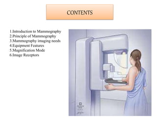
1.Introduction to Mammography
2.Principle of Mammography
3.Mammography imaging needs
4.Equipment Features
5.Magnification Mode
6.Image Receptors
What is Mammography?
Mammography is the process to examine the human breast for
diagnosis and screening by using low-energy X-rays .
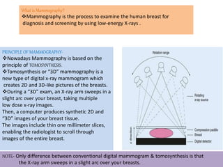
PRINCIPLE OF MAMMOGRAPHY-
Nowadays Mammography is based on the
principle of TOMOSYNTHESIS.
Tomosynthesis or “3D” mammography is a
new type of digital x-ray mammogram which
creates 2D and 3D-like pictures of the breasts.
During a “3D” exam, an X-ray arm sweeps in a
slight arc over your breast, taking multiple
low dose x-ray images.
Then, a computer produces synthetic 2D and
“3D” images of your breast tissue.
The images include thin one millimeter slices,
enabling the radiologist to scroll through
images of the entire breast.
NOTE- Only difference between conventional digital mammogram & tomosynthesis is that
the X-ray arm sweeps in a slight arc over your breasts.
Excellent Spatial Resolution-
•Shape and spatial configuration of calcification (0.15mm)
•Achieved with-
-Dedicated Digital detector
-X-ray tube with small focal spot
Excellent Contrast Resolution
•Required to see minimal attenuation difference
between normal glandular tissue & breast tissue
•Achieved with-Low Kvp technique
•Compression
•Grid
Mammography imaging needs
Noise should be low
•Required for accurate detector of subtle contrast difference & tiny structure
•Achieved with- Appropriate mAs (balance between noise & dose)
-Compression (Reduces breast thickness)
Dose should be low
•As breast is radiosensitive organ
•Must balance with resolution,contrast & noise
•Achieved with- Appropriate exposure factors
-Appropriate target & filter material
-AEC
-Collimation
-Shielding
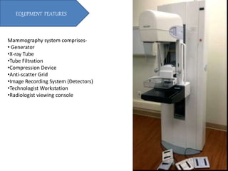
Mammography system comprises-
• Generator
•X-ray Tube
•Tube Filtration
•Compression Device
•Anti-scatter Grid
•Image Recording System (Detectors)
•Technologist Workstation
•Radiologist viewing console
EQUIPMENT FEATURES
•Stationery
•Anode Angle-6 degree
•Tube angle- 23 degree
•Focal Spot size- small (0.1mm – 0.3mm)
•Target Material- Tungsten(W)
-Molybdenum (Mo)
-Rhodium (Rh)
When tungsten target is used- Only Bremstrahlung xrays
predominate.
L-shell xrays are of no value (too low to penetrate)
Contribute only to patient radiation dose.
•Exposure factors- Low Kvp & High mAs
•X-ray most useful for enhancing differential absorption in
breast tissue & maximising
radio-contrast are in the range of 25-28 Kvp.
Source to Image Receptor Distance (SID)- 60-70 cm
X-ray Tube
The conic shape of the breast requires that the radiation intensity near the chest wall
must be higher than that to the nipple side to ensure near uniform exposure of the image
Receptor.
Accomplished by positioning the cathode to the chest wall.
However its not absolutely necessary because compression ensures imaging of a uniform
thickness of tissue
HEEL EFFECT APPLICATION
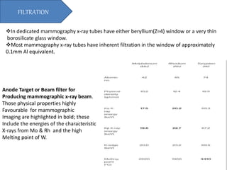
In dedicated mammography x-ray tubes have either beryllium(Z=4) window or a very thin
borosilicate glass window.
Most mammography x-ray tubes have inherent filtration in the window of approximately
0.1mm Al equivalent.
FILTRATION
Anode Target or Beam filter for
Producing mammographic x-ray beam.
Those physical properties highly
Favourable for mammographic
Imaging are highlighted in bold; these
Include the energies of the characteristic
X-rays from Mo & Rh and the high
Melting point of W.
COMPRESSION DEVICE
Routinely used in Mammography.
Most system now have a moving grid with a ratio of 4:1 to 5:1.
Grid frequency- 40lines /cm
Use of such grid increases patient dose compared with nongrid contact mammography.
Dose is still acceptably low, & improvement in contrast is signifant.
Unique grid that has been developed especially for mammography is HTC
(High Transmission Cellular Grid)
•Cross-Hatched grid
•Cleanup characteristics of crossed grid
•Grid strip material- Copper
•Air- Interspace material
Anti-Scatter Grid
Air gap between breast and detector
not only increases magnification but also
reduces amount of scatter radiation.
NO grid used.
MAGNIFICATION MODE
Phototimers- measure not only image intensity but also x-ray quality.
Position of AEC- after Image Receptor to minimize OID & improve spatial resolution.
AUTOMATIC EXPOSURE CONTROL
Indirect Conversion
•Xrays are converted in two step process,
•Xray energy is collected by CsI & converted into light.
•Light photons are then converted into electric signals by photodiode array.
•Some earlier models, such as GE 2000D or Fisher SenoScan use this type of detector s with CsI.
•This process is less efficient & results in some light scatter during conversion.
Direct Conversion
•Xrays are converted in a one step process.
•The layer of a-Se in the detector absorbs the xray energy & converts it into electronic
signal directly.
•Based on efficiency of direct conversion & its elimination of light scatter, a-Se offer
high resolution.
•Later system like Hologic Dimensions uses direct flat panel detectors with a-Selenium.
Image Receptors/Detectors
Mammography.pptx

More Related Content

What's hot

A Brief Overview of Mammography
A Brief Overview of MammographyA Brief Overview of Mammography
A Brief Overview of Mammography
Sam Shaikh
 
Mammography
Mammography Mammography
Mammography technique
Mammography techniqueMammography technique
Mammography technique
vishwanath0908
 
Imaging breast mammogram
Imaging breast mammogramImaging breast mammogram
Imaging breast mammogram
REKHAKHARE
 
Mammographic equipment
Mammographic equipmentMammographic equipment
Mammographic equipment
SharmaRajan4
 
Luc Rotenberg : Digital Breast Tomosynthesis
Luc Rotenberg : Digital Breast TomosynthesisLuc Rotenberg : Digital Breast Tomosynthesis
Luc Rotenberg : Digital Breast Tomosynthesis
breastcancerupdatecongress
 
Mammography
MammographyMammography
Mammography
Clinic
 
Mammography
Mammography Mammography
Mammography
عبدالله فهد
 
Digital Breast Tomosynthesis
Digital Breast TomosynthesisDigital Breast Tomosynthesis
Digital Breast Tomosynthesis
Allina Health
 
Mammography Positioning Technique for Additional Views
Mammography Positioning Technique for Additional Views Mammography Positioning Technique for Additional Views
Mammography Positioning Technique for Additional Views
Selin Prasad
 
Digital breast tomosynthesis
Digital breast tomosynthesisDigital breast tomosynthesis
Digital breast tomosynthesis
Yashawant Yadav
 
Mammography
MammographyMammography
Mammography
DORIKYADAV2
 
Mri artifacts
Mri artifactsMri artifacts
Mri artifacts
shajitha khan
 
Mammography positioning technique for Cranio Caudal (CC)
Mammography positioning technique for Cranio Caudal (CC) Mammography positioning technique for Cranio Caudal (CC)
Mammography positioning technique for Cranio Caudal (CC)
Selin Prasad
 
Chapter 6 image quality in ct
Chapter 6 image quality in ct Chapter 6 image quality in ct
Chapter 6 image quality in ct
Muntaser S.Ahmad
 
Flat Panel Digital Fluoroscopy by Akash Das
Flat Panel Digital Fluoroscopy by Akash Das Flat Panel Digital Fluoroscopy by Akash Das
Flat Panel Digital Fluoroscopy by Akash Das
AkashDas169
 
Mammogram
MammogramMammogram
Mammogram
NadhirahSayuti1
 
TOMOSYNTHESIS.pptx
TOMOSYNTHESIS.pptxTOMOSYNTHESIS.pptx
TOMOSYNTHESIS.pptx
sahebjana5
 
Mammography : quality control (quality assurance)
Mammography : quality control (quality assurance)Mammography : quality control (quality assurance)
Mammography : quality control (quality assurance)
Kajal Jha
 
Mammography
MammographyMammography
Mammography
Akari Kyaw
 

What's hot (20)

A Brief Overview of Mammography
A Brief Overview of MammographyA Brief Overview of Mammography
A Brief Overview of Mammography
 
Mammography
Mammography Mammography
Mammography
 
Mammography technique
Mammography techniqueMammography technique
Mammography technique
 
Imaging breast mammogram
Imaging breast mammogramImaging breast mammogram
Imaging breast mammogram
 
Mammographic equipment
Mammographic equipmentMammographic equipment
Mammographic equipment
 
Luc Rotenberg : Digital Breast Tomosynthesis
Luc Rotenberg : Digital Breast TomosynthesisLuc Rotenberg : Digital Breast Tomosynthesis
Luc Rotenberg : Digital Breast Tomosynthesis
 
Mammography
MammographyMammography
Mammography
 
Mammography
Mammography Mammography
Mammography
 
Digital Breast Tomosynthesis
Digital Breast TomosynthesisDigital Breast Tomosynthesis
Digital Breast Tomosynthesis
 
Mammography Positioning Technique for Additional Views
Mammography Positioning Technique for Additional Views Mammography Positioning Technique for Additional Views
Mammography Positioning Technique for Additional Views
 
Digital breast tomosynthesis
Digital breast tomosynthesisDigital breast tomosynthesis
Digital breast tomosynthesis
 
Mammography
MammographyMammography
Mammography
 
Mri artifacts
Mri artifactsMri artifacts
Mri artifacts
 
Mammography positioning technique for Cranio Caudal (CC)
Mammography positioning technique for Cranio Caudal (CC) Mammography positioning technique for Cranio Caudal (CC)
Mammography positioning technique for Cranio Caudal (CC)
 
Chapter 6 image quality in ct
Chapter 6 image quality in ct Chapter 6 image quality in ct
Chapter 6 image quality in ct
 
Flat Panel Digital Fluoroscopy by Akash Das
Flat Panel Digital Fluoroscopy by Akash Das Flat Panel Digital Fluoroscopy by Akash Das
Flat Panel Digital Fluoroscopy by Akash Das
 
Mammogram
MammogramMammogram
Mammogram
 
TOMOSYNTHESIS.pptx
TOMOSYNTHESIS.pptxTOMOSYNTHESIS.pptx
TOMOSYNTHESIS.pptx
 
Mammography : quality control (quality assurance)
Mammography : quality control (quality assurance)Mammography : quality control (quality assurance)
Mammography : quality control (quality assurance)
 
Mammography
MammographyMammography
Mammography
 

Similar to Mammography.pptx

Mammographic equipment and its advancement
Mammographic equipment and its advancementMammographic equipment and its advancement
Mammographic equipment and its advancement
Yashawant Yadav
 
MAMMOGRAPHIC EQUIPMENT.pptx//////////////
MAMMOGRAPHIC EQUIPMENT.pptx//////////////MAMMOGRAPHIC EQUIPMENT.pptx//////////////
MAMMOGRAPHIC EQUIPMENT.pptx//////////////
justinfan550
 
25632789 01-basics1-advanced-mammo-system
25632789 01-basics1-advanced-mammo-system25632789 01-basics1-advanced-mammo-system
25632789 01-basics1-advanced-mammo-system
Klaus19
 
Mammography-Physics-and-Technique-PDF.pdf
Mammography-Physics-and-Technique-PDF.pdfMammography-Physics-and-Technique-PDF.pdf
Mammography-Physics-and-Technique-PDF.pdf
UmaMaheswariC4
 
Mammography physics and technique
Mammography  physics and techniqueMammography  physics and technique
Mammography physics and technique
Archana Koshy
 
MAMMOGRAPHY.pptx
MAMMOGRAPHY.pptxMAMMOGRAPHY.pptx
MAMMOGRAPHY.pptx
rohanjohnjacob
 
Mammography.pdf
Mammography.pdfMammography.pdf
Mammography.pdf
AfaqAhmed849251
 
Breast mammography and tomosynthesis
Breast mammography and tomosynthesisBreast mammography and tomosynthesis
Breast mammography and tomosynthesis
shamimhyder161
 
mammophysics.ppt
mammophysics.pptmammophysics.ppt
mammophysics.ppt
dido_pegio
 
allisonmammophysics2018.ppt
allisonmammophysics2018.pptallisonmammophysics2018.ppt
allisonmammophysics2018.ppt
PHCSADRAWADI
 
X-ray Machine
X-ray Machine X-ray Machine
X-ray Machine
SamyaQadi
 
Cbct
CbctCbct
FINAL MAMMO.pptx
FINAL MAMMO.pptxFINAL MAMMO.pptx
FINAL MAMMO.pptx
JayaAditya6
 
Mammography -A ppt bt J K PATIL, Prof,dept of radiology
Mammography -A ppt bt J K PATIL, Prof,dept of radiologyMammography -A ppt bt J K PATIL, Prof,dept of radiology
Mammography -A ppt bt J K PATIL, Prof,dept of radiology
dypradio
 
SRT AND SRS - Arun.pdf
SRT AND SRS - Arun.pdfSRT AND SRS - Arun.pdf
SRT AND SRS - Arun.pdf
BBAdhikari
 
rpad seminar
rpad seminarrpad seminar
rpad seminar
K.K.D.RAMESH -
 
Biomedical applications of lasers (1)
Biomedical applications of lasers (1)Biomedical applications of lasers (1)
Biomedical applications of lasers (1)
Srihari KN
 
Dose reduction in Conventional Radiography and Fluoroscopy
Dose reduction in Conventional Radiography and FluoroscopyDose reduction in Conventional Radiography and Fluoroscopy
Dose reduction in Conventional Radiography and Fluoroscopy
Tarun Goyal
 
The innovation of breast scanning
The innovation of breast scanningThe innovation of breast scanning
The innovation of breast scanning
Ktn Aye
 
QUALITY-ASSURANCE---MAMMOGRAPHY.pdf
QUALITY-ASSURANCE---MAMMOGRAPHY.pdfQUALITY-ASSURANCE---MAMMOGRAPHY.pdf
QUALITY-ASSURANCE---MAMMOGRAPHY.pdf
Angelina Protik
 

Similar to Mammography.pptx (20)

Mammographic equipment and its advancement
Mammographic equipment and its advancementMammographic equipment and its advancement
Mammographic equipment and its advancement
 
MAMMOGRAPHIC EQUIPMENT.pptx//////////////
MAMMOGRAPHIC EQUIPMENT.pptx//////////////MAMMOGRAPHIC EQUIPMENT.pptx//////////////
MAMMOGRAPHIC EQUIPMENT.pptx//////////////
 
25632789 01-basics1-advanced-mammo-system
25632789 01-basics1-advanced-mammo-system25632789 01-basics1-advanced-mammo-system
25632789 01-basics1-advanced-mammo-system
 
Mammography-Physics-and-Technique-PDF.pdf
Mammography-Physics-and-Technique-PDF.pdfMammography-Physics-and-Technique-PDF.pdf
Mammography-Physics-and-Technique-PDF.pdf
 
Mammography physics and technique
Mammography  physics and techniqueMammography  physics and technique
Mammography physics and technique
 
MAMMOGRAPHY.pptx
MAMMOGRAPHY.pptxMAMMOGRAPHY.pptx
MAMMOGRAPHY.pptx
 
Mammography.pdf
Mammography.pdfMammography.pdf
Mammography.pdf
 
Breast mammography and tomosynthesis
Breast mammography and tomosynthesisBreast mammography and tomosynthesis
Breast mammography and tomosynthesis
 
mammophysics.ppt
mammophysics.pptmammophysics.ppt
mammophysics.ppt
 
allisonmammophysics2018.ppt
allisonmammophysics2018.pptallisonmammophysics2018.ppt
allisonmammophysics2018.ppt
 
X-ray Machine
X-ray Machine X-ray Machine
X-ray Machine
 
Cbct
CbctCbct
Cbct
 
FINAL MAMMO.pptx
FINAL MAMMO.pptxFINAL MAMMO.pptx
FINAL MAMMO.pptx
 
Mammography -A ppt bt J K PATIL, Prof,dept of radiology
Mammography -A ppt bt J K PATIL, Prof,dept of radiologyMammography -A ppt bt J K PATIL, Prof,dept of radiology
Mammography -A ppt bt J K PATIL, Prof,dept of radiology
 
SRT AND SRS - Arun.pdf
SRT AND SRS - Arun.pdfSRT AND SRS - Arun.pdf
SRT AND SRS - Arun.pdf
 
rpad seminar
rpad seminarrpad seminar
rpad seminar
 
Biomedical applications of lasers (1)
Biomedical applications of lasers (1)Biomedical applications of lasers (1)
Biomedical applications of lasers (1)
 
Dose reduction in Conventional Radiography and Fluoroscopy
Dose reduction in Conventional Radiography and FluoroscopyDose reduction in Conventional Radiography and Fluoroscopy
Dose reduction in Conventional Radiography and Fluoroscopy
 
The innovation of breast scanning
The innovation of breast scanningThe innovation of breast scanning
The innovation of breast scanning
 
QUALITY-ASSURANCE---MAMMOGRAPHY.pdf
QUALITY-ASSURANCE---MAMMOGRAPHY.pdfQUALITY-ASSURANCE---MAMMOGRAPHY.pdf
QUALITY-ASSURANCE---MAMMOGRAPHY.pdf
 

Recently uploaded

Natural birth techniques - Mrs.Akanksha Trivedi Rama University
Natural birth techniques - Mrs.Akanksha Trivedi Rama UniversityNatural birth techniques - Mrs.Akanksha Trivedi Rama University
Natural birth techniques - Mrs.Akanksha Trivedi Rama University
Akanksha trivedi rama nursing college kanpur.
 
The basics of sentences session 5pptx.pptx
The basics of sentences session 5pptx.pptxThe basics of sentences session 5pptx.pptx
The basics of sentences session 5pptx.pptx
heathfieldcps1
 
South African Journal of Science: Writing with integrity workshop (2024)
South African Journal of Science: Writing with integrity workshop (2024)South African Journal of Science: Writing with integrity workshop (2024)
South African Journal of Science: Writing with integrity workshop (2024)
Academy of Science of South Africa
 
PCOS corelations and management through Ayurveda.
PCOS corelations and management through Ayurveda.PCOS corelations and management through Ayurveda.
PCOS corelations and management through Ayurveda.
Dr. Shivangi Singh Parihar
 
Introduction to AI for Nonprofits with Tapp Network
Introduction to AI for Nonprofits with Tapp NetworkIntroduction to AI for Nonprofits with Tapp Network
Introduction to AI for Nonprofits with Tapp Network
TechSoup
 
Life upper-Intermediate B2 Workbook for student
Life upper-Intermediate B2 Workbook for studentLife upper-Intermediate B2 Workbook for student
Life upper-Intermediate B2 Workbook for student
NgcHiNguyn25
 
CACJapan - GROUP Presentation 1- Wk 4.pdf
CACJapan - GROUP Presentation 1- Wk 4.pdfCACJapan - GROUP Presentation 1- Wk 4.pdf
CACJapan - GROUP Presentation 1- Wk 4.pdf
camakaiclarkmusic
 
ANATOMY AND BIOMECHANICS OF HIP JOINT.pdf
ANATOMY AND BIOMECHANICS OF HIP JOINT.pdfANATOMY AND BIOMECHANICS OF HIP JOINT.pdf
ANATOMY AND BIOMECHANICS OF HIP JOINT.pdf
Priyankaranawat4
 
MARY JANE WILSON, A “BOA MÃE” .
MARY JANE WILSON, A “BOA MÃE”           .MARY JANE WILSON, A “BOA MÃE”           .
MARY JANE WILSON, A “BOA MÃE” .
Colégio Santa Teresinha
 
DRUGS AND ITS classification slide share
DRUGS AND ITS classification slide shareDRUGS AND ITS classification slide share
DRUGS AND ITS classification slide share
taiba qazi
 
Digital Artefact 1 - Tiny Home Environmental Design
Digital Artefact 1 - Tiny Home Environmental DesignDigital Artefact 1 - Tiny Home Environmental Design
Digital Artefact 1 - Tiny Home Environmental Design
amberjdewit93
 
The basics of sentences session 6pptx.pptx
The basics of sentences session 6pptx.pptxThe basics of sentences session 6pptx.pptx
The basics of sentences session 6pptx.pptx
heathfieldcps1
 
Advanced Java[Extra Concepts, Not Difficult].docx
Advanced Java[Extra Concepts, Not Difficult].docxAdvanced Java[Extra Concepts, Not Difficult].docx
Advanced Java[Extra Concepts, Not Difficult].docx
adhitya5119
 
Hindi varnamala | hindi alphabet PPT.pdf
Hindi varnamala | hindi alphabet PPT.pdfHindi varnamala | hindi alphabet PPT.pdf
Hindi varnamala | hindi alphabet PPT.pdf
Dr. Mulla Adam Ali
 
clinical examination of hip joint (1).pdf
clinical examination of hip joint (1).pdfclinical examination of hip joint (1).pdf
clinical examination of hip joint (1).pdf
Priyankaranawat4
 
BÀI TẬP BỔ TRỢ TIẾNG ANH 8 CẢ NĂM - GLOBAL SUCCESS - NĂM HỌC 2023-2024 (CÓ FI...
BÀI TẬP BỔ TRỢ TIẾNG ANH 8 CẢ NĂM - GLOBAL SUCCESS - NĂM HỌC 2023-2024 (CÓ FI...BÀI TẬP BỔ TRỢ TIẾNG ANH 8 CẢ NĂM - GLOBAL SUCCESS - NĂM HỌC 2023-2024 (CÓ FI...
BÀI TẬP BỔ TRỢ TIẾNG ANH 8 CẢ NĂM - GLOBAL SUCCESS - NĂM HỌC 2023-2024 (CÓ FI...
Nguyen Thanh Tu Collection
 
Pollock and Snow "DEIA in the Scholarly Landscape, Session One: Setting Expec...
Pollock and Snow "DEIA in the Scholarly Landscape, Session One: Setting Expec...Pollock and Snow "DEIA in the Scholarly Landscape, Session One: Setting Expec...
Pollock and Snow "DEIA in the Scholarly Landscape, Session One: Setting Expec...
National Information Standards Organization (NISO)
 
Assessment and Planning in Educational technology.pptx
Assessment and Planning in Educational technology.pptxAssessment and Planning in Educational technology.pptx
Assessment and Planning in Educational technology.pptx
Kavitha Krishnan
 
Chapter 4 - Islamic Financial Institutions in Malaysia.pptx
Chapter 4 - Islamic Financial Institutions in Malaysia.pptxChapter 4 - Islamic Financial Institutions in Malaysia.pptx
Chapter 4 - Islamic Financial Institutions in Malaysia.pptx
Mohd Adib Abd Muin, Senior Lecturer at Universiti Utara Malaysia
 
Digital Artifact 1 - 10VCD Environments Unit
Digital Artifact 1 - 10VCD Environments UnitDigital Artifact 1 - 10VCD Environments Unit
Digital Artifact 1 - 10VCD Environments Unit
chanes7
 

Recently uploaded (20)

Natural birth techniques - Mrs.Akanksha Trivedi Rama University
Natural birth techniques - Mrs.Akanksha Trivedi Rama UniversityNatural birth techniques - Mrs.Akanksha Trivedi Rama University
Natural birth techniques - Mrs.Akanksha Trivedi Rama University
 
The basics of sentences session 5pptx.pptx
The basics of sentences session 5pptx.pptxThe basics of sentences session 5pptx.pptx
The basics of sentences session 5pptx.pptx
 
South African Journal of Science: Writing with integrity workshop (2024)
South African Journal of Science: Writing with integrity workshop (2024)South African Journal of Science: Writing with integrity workshop (2024)
South African Journal of Science: Writing with integrity workshop (2024)
 
PCOS corelations and management through Ayurveda.
PCOS corelations and management through Ayurveda.PCOS corelations and management through Ayurveda.
PCOS corelations and management through Ayurveda.
 
Introduction to AI for Nonprofits with Tapp Network
Introduction to AI for Nonprofits with Tapp NetworkIntroduction to AI for Nonprofits with Tapp Network
Introduction to AI for Nonprofits with Tapp Network
 
Life upper-Intermediate B2 Workbook for student
Life upper-Intermediate B2 Workbook for studentLife upper-Intermediate B2 Workbook for student
Life upper-Intermediate B2 Workbook for student
 
CACJapan - GROUP Presentation 1- Wk 4.pdf
CACJapan - GROUP Presentation 1- Wk 4.pdfCACJapan - GROUP Presentation 1- Wk 4.pdf
CACJapan - GROUP Presentation 1- Wk 4.pdf
 
ANATOMY AND BIOMECHANICS OF HIP JOINT.pdf
ANATOMY AND BIOMECHANICS OF HIP JOINT.pdfANATOMY AND BIOMECHANICS OF HIP JOINT.pdf
ANATOMY AND BIOMECHANICS OF HIP JOINT.pdf
 
MARY JANE WILSON, A “BOA MÃE” .
MARY JANE WILSON, A “BOA MÃE”           .MARY JANE WILSON, A “BOA MÃE”           .
MARY JANE WILSON, A “BOA MÃE” .
 
DRUGS AND ITS classification slide share
DRUGS AND ITS classification slide shareDRUGS AND ITS classification slide share
DRUGS AND ITS classification slide share
 
Digital Artefact 1 - Tiny Home Environmental Design
Digital Artefact 1 - Tiny Home Environmental DesignDigital Artefact 1 - Tiny Home Environmental Design
Digital Artefact 1 - Tiny Home Environmental Design
 
The basics of sentences session 6pptx.pptx
The basics of sentences session 6pptx.pptxThe basics of sentences session 6pptx.pptx
The basics of sentences session 6pptx.pptx
 
Advanced Java[Extra Concepts, Not Difficult].docx
Advanced Java[Extra Concepts, Not Difficult].docxAdvanced Java[Extra Concepts, Not Difficult].docx
Advanced Java[Extra Concepts, Not Difficult].docx
 
Hindi varnamala | hindi alphabet PPT.pdf
Hindi varnamala | hindi alphabet PPT.pdfHindi varnamala | hindi alphabet PPT.pdf
Hindi varnamala | hindi alphabet PPT.pdf
 
clinical examination of hip joint (1).pdf
clinical examination of hip joint (1).pdfclinical examination of hip joint (1).pdf
clinical examination of hip joint (1).pdf
 
BÀI TẬP BỔ TRỢ TIẾNG ANH 8 CẢ NĂM - GLOBAL SUCCESS - NĂM HỌC 2023-2024 (CÓ FI...
BÀI TẬP BỔ TRỢ TIẾNG ANH 8 CẢ NĂM - GLOBAL SUCCESS - NĂM HỌC 2023-2024 (CÓ FI...BÀI TẬP BỔ TRỢ TIẾNG ANH 8 CẢ NĂM - GLOBAL SUCCESS - NĂM HỌC 2023-2024 (CÓ FI...
BÀI TẬP BỔ TRỢ TIẾNG ANH 8 CẢ NĂM - GLOBAL SUCCESS - NĂM HỌC 2023-2024 (CÓ FI...
 
Pollock and Snow "DEIA in the Scholarly Landscape, Session One: Setting Expec...
Pollock and Snow "DEIA in the Scholarly Landscape, Session One: Setting Expec...Pollock and Snow "DEIA in the Scholarly Landscape, Session One: Setting Expec...
Pollock and Snow "DEIA in the Scholarly Landscape, Session One: Setting Expec...
 
Assessment and Planning in Educational technology.pptx
Assessment and Planning in Educational technology.pptxAssessment and Planning in Educational technology.pptx
Assessment and Planning in Educational technology.pptx
 
Chapter 4 - Islamic Financial Institutions in Malaysia.pptx
Chapter 4 - Islamic Financial Institutions in Malaysia.pptxChapter 4 - Islamic Financial Institutions in Malaysia.pptx
Chapter 4 - Islamic Financial Institutions in Malaysia.pptx
 
Digital Artifact 1 - 10VCD Environments Unit
Digital Artifact 1 - 10VCD Environments UnitDigital Artifact 1 - 10VCD Environments Unit
Digital Artifact 1 - 10VCD Environments Unit
 

Mammography.pptx

  • 2. CONTENTS 1.Introduction to Mammography 2.Principle of Mammography 3.Mammography imaging needs 4.Equipment Features 5.Magnification Mode 6.Image Receptors
  • 3. What is Mammography? Mammography is the process to examine the human breast for diagnosis and screening by using low-energy X-rays . PRINCIPLE OF MAMMOGRAPHY- Nowadays Mammography is based on the principle of TOMOSYNTHESIS. Tomosynthesis or “3D” mammography is a new type of digital x-ray mammogram which creates 2D and 3D-like pictures of the breasts. During a “3D” exam, an X-ray arm sweeps in a slight arc over your breast, taking multiple low dose x-ray images. Then, a computer produces synthetic 2D and “3D” images of your breast tissue. The images include thin one millimeter slices, enabling the radiologist to scroll through images of the entire breast. NOTE- Only difference between conventional digital mammogram & tomosynthesis is that the X-ray arm sweeps in a slight arc over your breasts.
  • 4.
  • 5.
  • 6. Excellent Spatial Resolution- •Shape and spatial configuration of calcification (0.15mm) •Achieved with- -Dedicated Digital detector -X-ray tube with small focal spot Excellent Contrast Resolution •Required to see minimal attenuation difference between normal glandular tissue & breast tissue •Achieved with-Low Kvp technique •Compression •Grid Mammography imaging needs
  • 7. Noise should be low •Required for accurate detector of subtle contrast difference & tiny structure •Achieved with- Appropriate mAs (balance between noise & dose) -Compression (Reduces breast thickness) Dose should be low •As breast is radiosensitive organ •Must balance with resolution,contrast & noise •Achieved with- Appropriate exposure factors -Appropriate target & filter material -AEC -Collimation -Shielding
  • 8. Mammography system comprises- • Generator •X-ray Tube •Tube Filtration •Compression Device •Anti-scatter Grid •Image Recording System (Detectors) •Technologist Workstation •Radiologist viewing console EQUIPMENT FEATURES
  • 9.
  • 10. •Stationery •Anode Angle-6 degree •Tube angle- 23 degree •Focal Spot size- small (0.1mm – 0.3mm) •Target Material- Tungsten(W) -Molybdenum (Mo) -Rhodium (Rh) When tungsten target is used- Only Bremstrahlung xrays predominate. L-shell xrays are of no value (too low to penetrate) Contribute only to patient radiation dose. •Exposure factors- Low Kvp & High mAs •X-ray most useful for enhancing differential absorption in breast tissue & maximising radio-contrast are in the range of 25-28 Kvp. Source to Image Receptor Distance (SID)- 60-70 cm X-ray Tube
  • 11. The conic shape of the breast requires that the radiation intensity near the chest wall must be higher than that to the nipple side to ensure near uniform exposure of the image Receptor. Accomplished by positioning the cathode to the chest wall. However its not absolutely necessary because compression ensures imaging of a uniform thickness of tissue HEEL EFFECT APPLICATION
  • 12. In dedicated mammography x-ray tubes have either beryllium(Z=4) window or a very thin borosilicate glass window. Most mammography x-ray tubes have inherent filtration in the window of approximately 0.1mm Al equivalent. FILTRATION Anode Target or Beam filter for Producing mammographic x-ray beam. Those physical properties highly Favourable for mammographic Imaging are highlighted in bold; these Include the energies of the characteristic X-rays from Mo & Rh and the high Melting point of W.
  • 13.
  • 15.
  • 16. Routinely used in Mammography. Most system now have a moving grid with a ratio of 4:1 to 5:1. Grid frequency- 40lines /cm Use of such grid increases patient dose compared with nongrid contact mammography. Dose is still acceptably low, & improvement in contrast is signifant. Unique grid that has been developed especially for mammography is HTC (High Transmission Cellular Grid) •Cross-Hatched grid •Cleanup characteristics of crossed grid •Grid strip material- Copper •Air- Interspace material Anti-Scatter Grid
  • 17. Air gap between breast and detector not only increases magnification but also reduces amount of scatter radiation. NO grid used. MAGNIFICATION MODE
  • 18. Phototimers- measure not only image intensity but also x-ray quality. Position of AEC- after Image Receptor to minimize OID & improve spatial resolution. AUTOMATIC EXPOSURE CONTROL
  • 19. Indirect Conversion •Xrays are converted in two step process, •Xray energy is collected by CsI & converted into light. •Light photons are then converted into electric signals by photodiode array. •Some earlier models, such as GE 2000D or Fisher SenoScan use this type of detector s with CsI. •This process is less efficient & results in some light scatter during conversion. Direct Conversion •Xrays are converted in a one step process. •The layer of a-Se in the detector absorbs the xray energy & converts it into electronic signal directly. •Based on efficiency of direct conversion & its elimination of light scatter, a-Se offer high resolution. •Later system like Hologic Dimensions uses direct flat panel detectors with a-Selenium. Image Receptors/Detectors