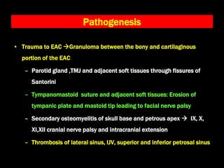
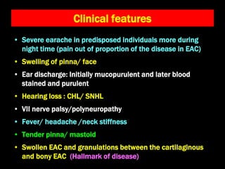
• Rapidly progressive infection of external auditory meatus
spreading to surrounding soft tissues and bones of skull
base
• Malignant is a misnomer (Chandler 1968)
– Aggressive clinical behavior, poor treatment outcome
and high mortality in elderly uncontrolled diabetics
• Synonyms :
– Invasive/ granulomatous/ necrotizing otitis externa
– Skull base osteomyelitis
Malignant Otitis externa
• Predisposing factors
− Elderly , poorly controlled diabetics, prolonged steroid
use, atherosclerosis, immunosuppressed, AIDS
• Causative organisms
– Pseudomonas aeruginosa (95%)
– Staph. epidermidis
– Aspergillus fumigatus
• Hallmark of disease
– Granulation tissue at the junction of cartilaginous and
bony EAC
Pathogenesis
• Trauma to EAC Granuloma between the bony and cartilaginous
portion of the EAC
– Parotid gland ,TMJ and adjacent soft tissues through fissures of
Santorini
– Tympanomastoid suture and adjacent soft tissues: Erosion of
tympanic plate and mastoid tip leading to facial nerve palsy
– Secondary osteomyelitis of skull base and petrous apex  IX, X,
XI,XII cranial nerve palsy and intracranial extension
– Thrombosis of lateral sinus, IJV, superior and inferior petrosal sinus
Trauma to EAC Granuloma between the bony and
cartilaginous portion of the EAC
Secondary osteomyelitis of
petrous Apex
Thrombosis of lateral sinus, IJV,
superior and inferior petrosal sinus
IX, X, XI,XII cranial nerve
palsy
Erosion of Tympanic plate,
Mastoid tip
Tympanomastoid suture, Adjacent soft tissues
Parotid gland ,TMJ and adjacent soft
tissues through fissures of Santorini
Facial Palsy
Pathogenesis
Floor of MCF, Basisphenoid.
Intracranial extension
Clinical features
• Severe earache in predisposed individuals more during
night time (pain out of proportion of the disease in EAC)
• Swelling of pinna/ face
• Ear discharge: Initially mucopurulent and later blood
stained and purulent
• Hearing loss : CHL/ SNHL
• VII nerve palsy/polyneuropathy
• Fever/ headache /neck stiffness
• Tender pinna/ mastoid
• Swollen EAC and granulations between the cartilaginous
and bony EAC (Hallmark of disease)
Investigations
• CBC : raised total counts
• ESR : raised ESR signifies active disease
• FBS/ PPBS : Indicator of diabetic control
• Ear swab culture for Pseudomonas
• CT Scan / MRI: Extent of disease, bone and soft tissue
involvement
• Gallium and Technetium bone scan:
– Better than CT/MRI
– Radiotracer concentrates in areas with increased osteoblastic
activity
Treatment
• Control of diabetes and other predisposing factors
• Debridement of necrotic tissues
• Administration of antibiotics against pseudomonas (high
dose , broad spectrum)
– Piperacillin-tazobactam: 4 to 6 g IV every 4 to 6 hours
– Ciprofloxacin 750 mg twice daily for 6-12 weeks
– Ceftazidime 2 gm iv TDS
• Hyperbaric oxygen therapy ???
– Improves hypoxia and leads to greater oxidative killing
of bacteria

Malignant otitis externa

  • 1.
    • Rapidly progressiveinfection of external auditory meatus spreading to surrounding soft tissues and bones of skull base • Malignant is a misnomer (Chandler 1968) – Aggressive clinical behavior, poor treatment outcome and high mortality in elderly uncontrolled diabetics • Synonyms : – Invasive/ granulomatous/ necrotizing otitis externa – Skull base osteomyelitis Malignant Otitis externa
  • 2.
    • Predisposing factors −Elderly , poorly controlled diabetics, prolonged steroid use, atherosclerosis, immunosuppressed, AIDS • Causative organisms – Pseudomonas aeruginosa (95%) – Staph. epidermidis – Aspergillus fumigatus • Hallmark of disease – Granulation tissue at the junction of cartilaginous and bony EAC
  • 3.
    Pathogenesis • Trauma toEAC Granuloma between the bony and cartilaginous portion of the EAC – Parotid gland ,TMJ and adjacent soft tissues through fissures of Santorini – Tympanomastoid suture and adjacent soft tissues: Erosion of tympanic plate and mastoid tip leading to facial nerve palsy – Secondary osteomyelitis of skull base and petrous apex  IX, X, XI,XII cranial nerve palsy and intracranial extension – Thrombosis of lateral sinus, IJV, superior and inferior petrosal sinus
  • 4.
    Trauma to EACGranuloma between the bony and cartilaginous portion of the EAC Secondary osteomyelitis of petrous Apex Thrombosis of lateral sinus, IJV, superior and inferior petrosal sinus IX, X, XI,XII cranial nerve palsy Erosion of Tympanic plate, Mastoid tip Tympanomastoid suture, Adjacent soft tissues Parotid gland ,TMJ and adjacent soft tissues through fissures of Santorini Facial Palsy Pathogenesis Floor of MCF, Basisphenoid. Intracranial extension
  • 5.
    Clinical features • Severeearache in predisposed individuals more during night time (pain out of proportion of the disease in EAC) • Swelling of pinna/ face • Ear discharge: Initially mucopurulent and later blood stained and purulent • Hearing loss : CHL/ SNHL • VII nerve palsy/polyneuropathy • Fever/ headache /neck stiffness • Tender pinna/ mastoid • Swollen EAC and granulations between the cartilaginous and bony EAC (Hallmark of disease)
  • 6.
    Investigations • CBC :raised total counts • ESR : raised ESR signifies active disease • FBS/ PPBS : Indicator of diabetic control • Ear swab culture for Pseudomonas • CT Scan / MRI: Extent of disease, bone and soft tissue involvement • Gallium and Technetium bone scan: – Better than CT/MRI – Radiotracer concentrates in areas with increased osteoblastic activity
  • 7.
    Treatment • Control ofdiabetes and other predisposing factors • Debridement of necrotic tissues • Administration of antibiotics against pseudomonas (high dose , broad spectrum) – Piperacillin-tazobactam: 4 to 6 g IV every 4 to 6 hours – Ciprofloxacin 750 mg twice daily for 6-12 weeks – Ceftazidime 2 gm iv TDS • Hyperbaric oxygen therapy ??? – Improves hypoxia and leads to greater oxidative killing of bacteria