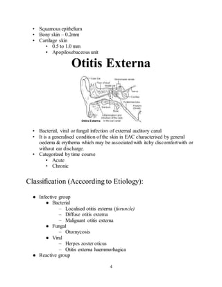
1. The document provides information on the anatomy and physiology of the external ear canal and discusses various types of otitis externa including acute otitis externa, chronic otitis externa, necrotizing external otitis, fungal otitis externa, and herpes zoster oticus.
2. It describes the symptoms, signs, causative agents, diagnosis, and treatment for each type of otitis externa. For acute otitis externa, examples of treatment mentioned include ear toilet, medicated wicks, antibiotic-steroid preparations, and analgesics.
3. Necrotizing external otitis is described
1. 1
Ministry of Healthof the Republic of Belarus
Vitebsk State Medical University
Department of Otorhinolaryngology
Topic:- Otitis Externa (OE)
Head of Department:
Teacher:
Student: Dinoosh De Livera
4th
Course, Group 49
Vitebsk, VSMU
2015
3. 3
Anatomy & Physiology
• Consists of the auricle and EAM
• Skin-lined apparatus
• Approximately 2.5 cm in length
• Ends at tympanic membrane
• Auricle is mostly skin-lined cartilage
• External auditory meatus
• Cartilage: ~40%, Bony: ~60%
• S-shaped, Narrowest portion at bony-cartilage junction
● Outer part – upwards, backwards & medially
● Inner part – downwards,forwards & medially
• EAC is related to various contiguous structures
• Tympanic membrane
• Mastoid
• Glenoid fossa
• Cranial fossa
• Infratemporal fossa
• Innervation: cranial nerves V, VII, IX, X, and greater auricular nerve
• Arterial supply: superficial temporal, posterior and deep auricular branches
• Venous drainage: superficial temporal and posterior auricular veins
• Lymphatics
4. 4
• Squamous epithelium
• Bony skin – 0.2mm
• Cartilage skin
• 0.5 to 1.0 mm
• Apopilosebaceous unit
Otitis Externa
• Bacterial, viral or fungal infection of external auditory canal
• It is a generalised condition of the skin in EAC characterised by general
oedema & erythema which may be associated with itchy discomfortwith or
without ear discharge.
• Categorized by time course
• Acute
• Chronic
Classification (Acccording to Etiology):
● Infective group
● Bacterial
– Localised otitis externa (furuncle)
– Diffuse otitis externa
– Malignant otitis externa
● Fungal
– Otomycosis
● Viral
– Herpes zoster oticus
– Otitis externa haemmorhagica
● Reactive group
5. 5
● Eczematous otitis externa
● Seborrhoeic otitis externa
● Neurodermatitis
SpeculumFindings:
• the canal may be so swollen that a view into the ear is impossible
• In swimmers, divers and surfers, chronic water exposure can lead to the
growth of bony swellings in the canal known as exostoses. These can
interfere with the drainage of wax and predisposeto infection.
Differential Diagnosis:
•
• Otitis media
• Ramsay Hunt syndrome
• Furuncle
• Skull base osteomyelitis
• Preauricular cyst and fistula
• Lacerations
• Atopic dermatitis
• Cerumen impaction
• Exostosis and osteoma
• Foreign body
• Acute (bullous) and chronic (granular) myringitis
Causative Agents:
1. Pseudomonas species
2. Staphylococci
3. Streptococci/Gram negative rods
4. Fungi (Aspergillus/Candida species)
LaboratoryDiagnosis:
•
• bacterial and fungal culture
6. 6
• Adults with otitis externa: screening blood glucose and/or a urine dipstick
test to rule out occult diabetes.
• Additional tests (if available):
• Gram stain of d/c
• KOH prep smear (within 10 min)
Complications:
● Cellulitis/ Perichondritis/ Chondritis
● Medial canal fibrosis
● Tympanic membrane perforation
● Malignant otitis externa
7. 7
Acute Otitis Externa
• “swimmer’s ear”
• Preinflammatory stage
• Acute inflammatory stage
• Mild
• Moderate
• Severe
Etiology:
• High humidity
• Water exposure
• Maceration of canal skin
• High environmental temperature
• Local trauma
• Perespiration
• Allergy
• Stress
• Removal of normal skin lipids
• Absence of cerumen
• Alkaline pH of canal
Clinical Features:
• Ostalgia
• Tenderness on palpation or manipulation (tragus sign)
• Ear fullness
8. 8
• Conductive hearing loss.
• Erythaema of meatus and canal
• Swelling and obstructionof canal
• Crusting and discharge
• Odor!
Pathogenesis:
● Infection of a hair follicle (furuncle)
● begin as folliculitis-->small abscess/furuncle
● Staphylococcus aureus
● Lateral cartilaginous (outer 1/3rd) portion of EAC
1)PreInflammatory Stage:
• Oedema of stratum corneum and plugging of apopilosebaceous unit
• Symptoms: pruritus and sense of fullness
• Signs: mild edema
• Starts the itch/scratch cycle
2)Mild to ModerateStage:
• Progressive infection
• Symptoms
• Pain
• Increased pruritus
• Signs
• Erythema
• Increasing edema
• Canal debris, discharge
3)Severe Stage:
• Severe pain, worse with ear movement
• Signs
9. 9
• Lumen obliteration
• Purulent otorrhoea
• Involvement of periauricular soft tissue
Treatment:
• Most common pathogens: P. aeruginosa and S. aureus, E.coli and proteus.!
Treatment:
● Ear toilet
● Medicated wicks -
● Antibiotic-steroid prepration
● Acidifying/antiseptic agents – gentian violet
● Mild astingent – 8% aluminium acetate/3% silver nitrate
● Antibiotics -
● Topical antibiotics – (neomycin/ciprofloxacin/ofloxacin) with/without
corticosteroids
● Broad spectrum systemic antibiotics
● Analgesics
● Avoid water entry/avoid usind cotton buds/avoid digital manipulation of ear
canal
Herpes zoster oticus:
● HSV – most frequent virus to affect EAC
● HSV stay dominant in sensory ganglia – reactivates in decreased
immunocompetence
● Blisters/vesicles on auricle, EAC, TM
● Blisters – short lived, rupture, dry & heal spontaneously
● May develop CN VII, VIII palsy
● 'Ramsay Hunt Syndrome' – clinical syndrome with facial N palsy with or
without hearing loss and dizziness owing to herpes zoster
10. 10
Treatment:
● Self limiting, primarily supportive
● Antivirals (acyclovir) & steroids can be used
Chronic Otitis Externa:
• Acute otitis externa occurs in 4 of every 1000 people per year
• Otitis externa is defined as chronic when the duration of the infection
exceeds 4 weeks or when more than 4 episodes occurin 1 year
• Bacterial, fungal, dermatological aetiologies
Clinical Features:
• Unrelenting pruritus
• Mild discomfort
• Dryness, Crusting, and flaking of canal skin
• Asteatosis
• Dry, flaky skin
• Hypertrophied skin
• Mucopurulent otorrhoea (occasional)
Treatment:
Conservative treatment: Similar to that of AOE
prevent stenosis & restore normal skin in EAC
● Frequent inspection & debridment of EAC
● Antibiotic-corticosteroid topical applications
● EAC can be painted with gentian violet/ triamcinolone/ nystatin
● Treat underlying causes – seborrhoea, psoriasis, neurodermatitis
Surgical treatment: When Conservative treatment fails:
In case of medical treatment failure with canal stenosis
11. 11
● Canalplasty with skin grafting – restore canal patency and hearing
Procedure:
➔ Abnormal skin is removed entirely
➔ Denuded canal is enlarged using diamond bur
➔ Split thickness graft is harvested from medial surface of upper arm with a
dermatome
➔ Graft placed on exposed suface
➔ 'rosebud' typeof packing is done over skin graft and left for 2 weeks
➔ Crusting may occurfor several weeks, requires removal till complete healing
Necrotizing (malignant)
External Otitis(NEO)
• Potentially lethal infection of EAC and surrounding structures
• Pseudomonasaeruginosa is the usual culprit
• Risk Factors:
- Elderly
- Human Immunodeficiency Virus (HIV)
• Typically seen in diabetics and immunocompromised patients
Pathogenesis:
●
● Infection begins in EAC --> cellulitis, chondritis, osteitis, osteomyelitis
● May spread to osseus auditory canal & skull base through fissures of
Santorini --> replacement of compactbone with granulation tissue
● Facial N paalysis – stylomastoid foramen involvement
● CN IX, X, XI palsies - jugular foramen involvement
● Jugular V thrombosis-->lateral sinus thrombosis
Clinical Features:
• Similar to Otitis Externa except
• Severe, unrelenting Ear Pain and Headache
• Persistent discharge
12. 12
• Does not respond to topical medications
• Commonly associated with Diabetes Mellitus
• Granulation tissue in posterior and inferior canal
• Pathognomonic for necrotizing otitis
• Occurs at bone-cartilage junction
• Extra-auricular findings
• Cervical Lymphadenopathy
• Trismus (TMJ involvement)
• Facial Nerve Palsy or paralysis (Bell's Palsy)
• Associated with poorprognosis
Diagnosis & Prevention:
• Prognosis; Reportedly mortality 20-53%
• Dx: Hx, PE, Labs and Imaging:
- Labs; FBC, Culture of discharge, ESR, Serum glucose, Serum
creatinine.
- Radiology; CT, or MRI (ear),Tc 99m medronate methylene bone
scanning, Ga 67 scintography.
• Prevention:
- Avoid use of cottonswabs in ear and other canal trauma.
- Use caution when irrigating ear of high risk patients.
- Treat eczema of ear canal and other pruritic dermatitis
- Cohen and Friedman – criteria from review: They were divided into
two categories: obligatory and occasional. The obligatory criteria are:
pain, edema, exudate, granulations, microabscess (when operated),
positive bone scan or failure of local treatment often more than 1
week, and possibly pseudomonas in culture. The occasionalcriteria
are diabetes, cranial nerve involvement, positive radiograph,
debilitating condition and old age. All of the obligatory criteria must
be present in order to establish the diagnosis. The presence of
occasionalcriteria alone does not establish it. The importance of Tc99
scan in detecting osteomyelitis is stressed. When bone scan is not
available, a trial of 1-3 weeks of local treatment is suggested. Failure
to respond to such treatment may assist in making the diagnosis of
MEO
Treatment:
13. 13
• Intravenous antibiotics for at least 4 weeks – with serial gallium scans
monthly
• Local canal debridement until healed
• Pain control
• Use of topical agents controversial
• Hyperbaric oxygen experimental
• Surgical debridement for refractory cases
Fungal Otitis Externa
(Otomycosis)
● Fungal infection of EAC
● Aspergillus niger – black headed filamented growth
● Aspergillus fumigatus – brown
● Candidaalbicans – white/creamy deposits
● Secondaryfungal infection may be seen in pts using topical antibiotics for
otitis externa/ middle ear suppuration
Clinical Features:
● pruritis/ pain / discomfort in ear/ watery discharge with odour/ ear block
● erythematous canal with black/grey fungal mass – 'wet piece of filter paper'
Treatment:
● Ear toilet
● Antifungal agents – nystatin/clotrimazole
● 2% salicylic acid
● Ear must be kept dry
● Secondarybacterial infections – antibiotic + steroid prepration
● Oral antifungals – refractory to topical agents