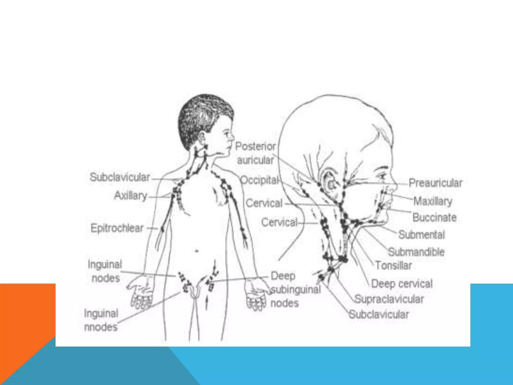
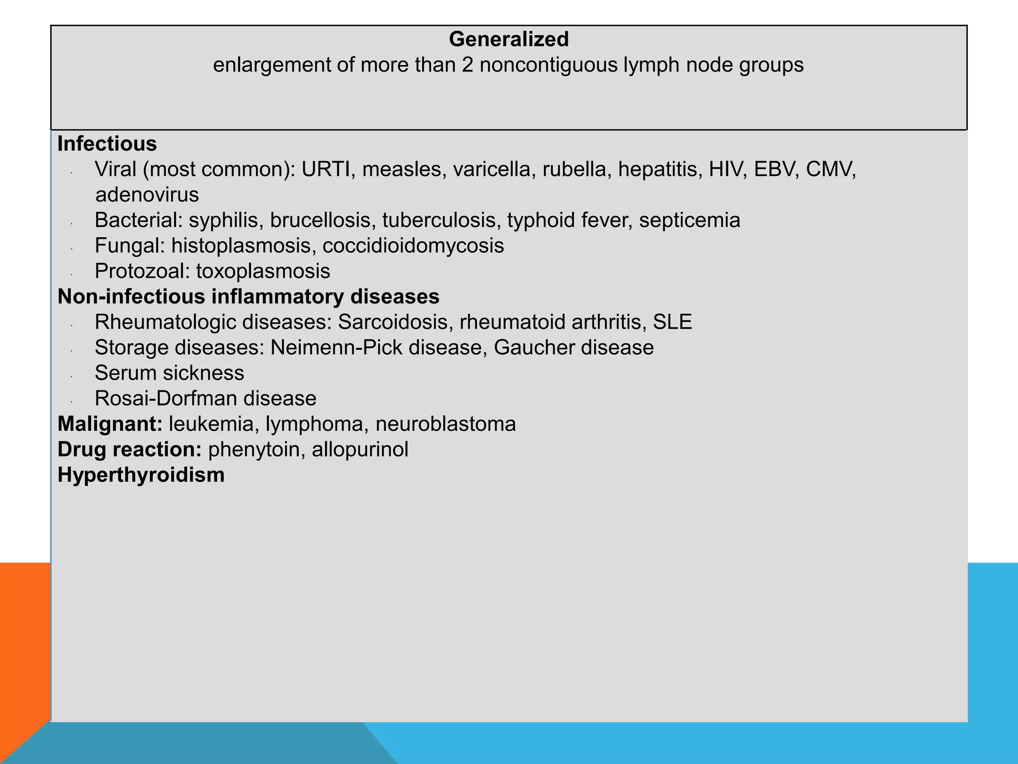
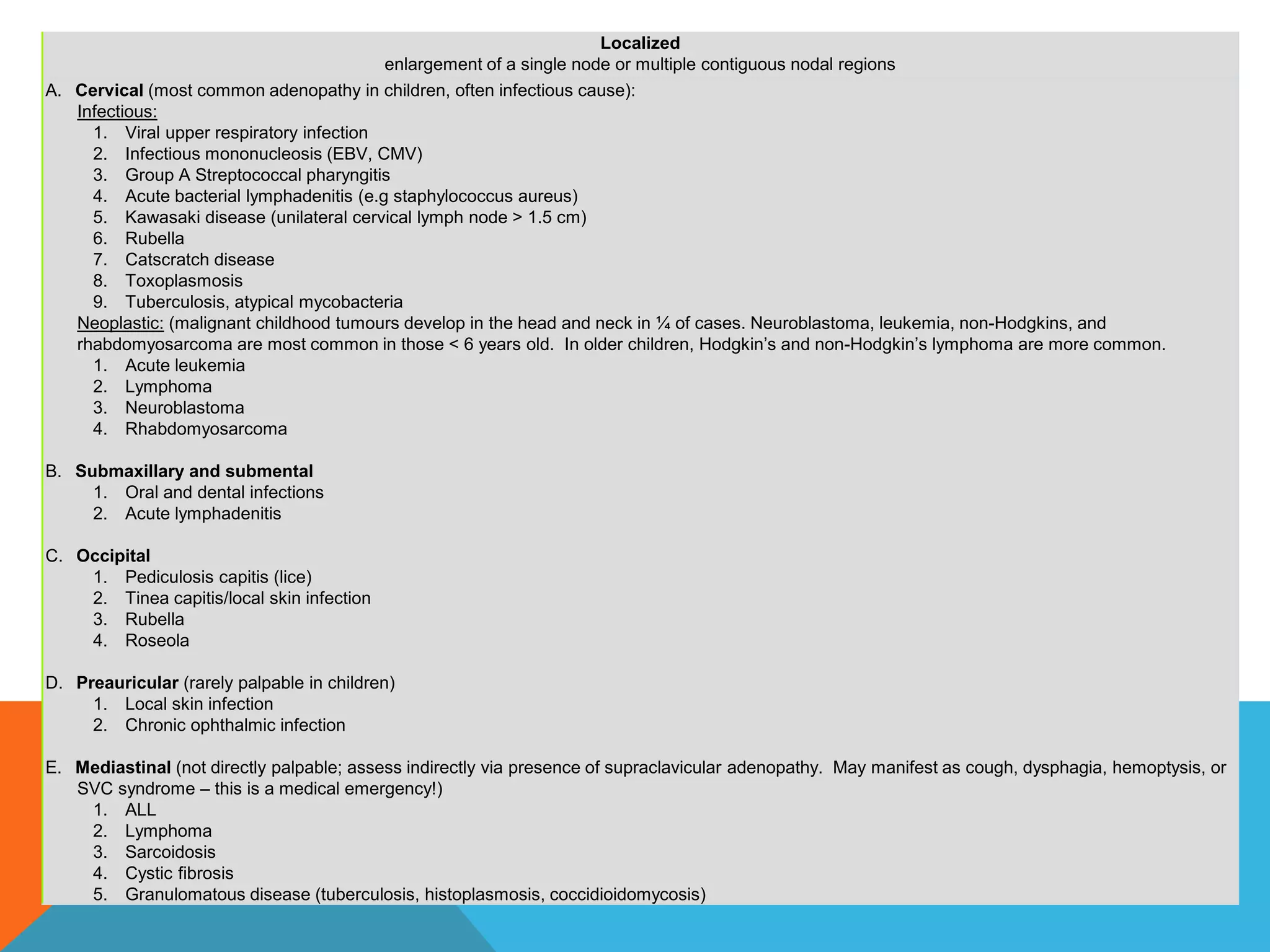
DEFINITION
Lymphadenopathy
 enlargement of lymph nodes.
 Viral or bacterial infections lead to localized responses from lymphocytes and
macrophages, leading to enlargement of nodes
 Palpable lymph nodes are normal in anterior cervical, axillary and inguinal
regions in healthy children whose naïve immune systems respond more
frequently to newly encountered infections.
Lymphadenitis
 localized infiltration by inflammatory cells in response to an infection of the
nodes themselves
HISTORY
Onset, size, duration
Is it painful or erythematous?
Generalized or local?
Associated symptoms?
Hx of Recent infections
Constitutional symptoms? Fever, night sweats, weight loss?
Skin lesions or trauma? Eg: Cat scratch? Animal/insect bites? Other open wounds? Dental
abscesses?
Recent Travel & Exposures
Immunization status. MMR?
Medications.Carbemazepine or phenytoin?
Allergies.
PHYSICAL EXAMINATION
Antropometry
Head and Neck – Examine closely for:
– Scalp infection (e.g. seborrheic dermatitis, tinea capitius)
– Conjunctivitis injection
– Oropharynx for pharyngitis, dental problems, HSV
ginivostomatitis
– Ears for acute otitis media
Abdomen – Examine closely for:
– Hepatosplenomegaly (this is actually considered part of your lymph
node exam!)
– Abdominal masses (e.g. neuroblastoma)
Skin – Examine closely for:
– Any rashes
– Petechiae, purpura, eccyhmoses (e.g. thrombocytopenia)
Lymph Node Exam:
When palpating a lymph node it is important to consider the following:
– Size (measure them)
– Location
– Fixation
– Consistency
– Tenderness
Less concerning findings Worrisome findings which increase risk
of malignancy
 Localized
 <1-2cm (depending on location)
 Cervical, inguinal, and axillary
regions
 Erythema
 Tender
 Warm
 Fluctuant
 Generalized adenopathy
 >2cm
 Occipital, auricular, supraclavicular,
mediastinal, epitrochlear or posterior
cervical nodes
 Firm
 Matted
 Nontender
 Systemic symptoms
Generalized
enlargement of more than 2 noncontiguous lymph node groups
Infectious
· Viral (most common): URTI, measles, varicella, rubella, hepatitis, HIV, EBV, CMV,
adenovirus
· Bacterial: syphilis, brucellosis, tuberculosis, typhoid fever, septicemia
· Fungal: histoplasmosis, coccidioidomycosis
· Protozoal: toxoplasmosis
Non-infectious inflammatory diseases
· Rheumatologic diseases: Sarcoidosis, rheumatoid arthritis, SLE
· Storage diseases: Neimenn-Pick disease, Gaucher disease
· Serum sickness
· Rosai-Dorfman disease
Malignant: leukemia, lymphoma, neuroblastoma
Drug reaction: phenytoin, allopurinol
Hyperthyroidism
Localized
enlargement of a single node or multiple contiguous nodal regions
A. Cervical (most common adenopathy in children, often infectious cause):
Infectious:
1. Viral upper respiratory infection
2. Infectious mononucleosis (EBV, CMV)
3. Group A Streptococcal pharyngitis
4. Acute bacterial lymphadenitis (e.g staphylococcus aureus)
5. Kawasaki disease (unilateral cervical lymph node > 1.5 cm)
6. Rubella
7. Catscratch disease
8. Toxoplasmosis
9. Tuberculosis, atypical mycobacteria
Neoplastic: (malignant childhood tumours develop in the head and neck in ¼ of cases. Neuroblastoma, leukemia, non-Hodgkins, and
rhabdomyosarcoma are most common in those < 6 years old. In older children, Hodgkin’s and non-Hodgkin’s lymphoma are more common.
1. Acute leukemia
2. Lymphoma
3. Neuroblastoma
4. Rhabdomyosarcoma
B. Submaxillary and submental
1. Oral and dental infections
2. Acute lymphadenitis
C. Occipital
1. Pediculosis capitis (lice)
2. Tinea capitis/local skin infection
3. Rubella
4. Roseola
D. Preauricular (rarely palpable in children)
1. Local skin infection
2. Chronic ophthalmic infection
E. Mediastinal (not directly palpable; assess indirectly via presence of supraclavicular adenopathy. May manifest as cough, dysphagia, hemoptysis, or
SVC syndrome – this is a medical emergency!)
1. ALL
2. Lymphoma
3. Sarcoidosis
4. Cystic fibrosis
5. Granulomatous disease (tuberculosis, histoplasmosis, coccidioidomycosis)
F. Supraclavicular (associated with serious underlying disease)
1. Lymphoma
2. Tuberculosis
3. Histoplasmosis
4. Coccidioidomycosis
G. Axillary
1. Local infection
2. Cat scratch disease
3. Brucellosis
4. Reactions to immunizations
5. Non Hodgkin lymphoma
6. Juvenile rheumatoid arthritis
7. Hidradenitis suppurativa
H. Abdominal (may manifest as abdominal pain, backache, urinary frequency, constipation, or intestinal
obstruction due to intussuception)
1. Acute mesenteric adenitis
2. Lymphoma
I. Inguinal
1. Local infection
2. Diaper dermatitis
3. Syphilis
INVESTIGATIONS
Complete blood count, peripheral blood smear
ESR (non-specific)
BLOOD C&S
Rule out infectious causes: Monospot, CMV, EBV, & toxoplasma, bartonella
titres, TB skin test, Anti-HIV test, CRP, ESR
Hepatic and renal function + urinalysis (systemic disorders that can cause
lymphadenopathy)
Lactate dehydrogenase, uric acid, calcium, phosphate, magnesium if
malignancy suspected
Bone marrow, liver biopsies, CT or US guided lymph node biopsy
Chest X-ray.
MANAGEMENT
Infectious cause: Treatment with antibiotics
 Start on antibiotics that cover the bacterial pathogens frequently implicated in
lymphadenitis, including staphylococcus aureus and streptococcus pyogenes.
 Reevaluate in 2-4 weeks. Biopsy if unchanged or larger
If malignancy is a strong possibility excisional biopsy should be
considered immediately.
If lymphadenitis is present, aspirate may be needed for culture.
REFERENCES
McClain, K and Fletcher R. Approach to the child with peripheral
lymphadenopathy. UpToDate 2009. www.uptodate.com.
McClain, K and Fletcher R. Causes of Peripheral lymphadenopathy in
children. UpToDate 2009. www.uptodate.com.
Kleigman RM, Marcdante KJ, Jensen HB, Behrman RE. Nelson Essentials
of Pediatrics 5th Edition. Elselvier Saunders, 2006.

Approach in lymphadenopathy in children

  • 3.
    DEFINITION Lymphadenopathy  enlargement oflymph nodes.  Viral or bacterial infections lead to localized responses from lymphocytes and macrophages, leading to enlargement of nodes  Palpable lymph nodes are normal in anterior cervical, axillary and inguinal regions in healthy children whose naïve immune systems respond more frequently to newly encountered infections. Lymphadenitis  localized infiltration by inflammatory cells in response to an infection of the nodes themselves
  • 4.
    HISTORY Onset, size, duration Isit painful or erythematous? Generalized or local? Associated symptoms? Hx of Recent infections Constitutional symptoms? Fever, night sweats, weight loss? Skin lesions or trauma? Eg: Cat scratch? Animal/insect bites? Other open wounds? Dental abscesses? Recent Travel & Exposures Immunization status. MMR? Medications.Carbemazepine or phenytoin? Allergies.
  • 5.
    PHYSICAL EXAMINATION Antropometry Head andNeck – Examine closely for: – Scalp infection (e.g. seborrheic dermatitis, tinea capitius) – Conjunctivitis injection – Oropharynx for pharyngitis, dental problems, HSV ginivostomatitis – Ears for acute otitis media
  • 6.
    Abdomen – Examineclosely for: – Hepatosplenomegaly (this is actually considered part of your lymph node exam!) – Abdominal masses (e.g. neuroblastoma) Skin – Examine closely for: – Any rashes – Petechiae, purpura, eccyhmoses (e.g. thrombocytopenia)
  • 7.
    Lymph Node Exam: Whenpalpating a lymph node it is important to consider the following: – Size (measure them) – Location – Fixation – Consistency – Tenderness
  • 8.
    Less concerning findingsWorrisome findings which increase risk of malignancy  Localized  <1-2cm (depending on location)  Cervical, inguinal, and axillary regions  Erythema  Tender  Warm  Fluctuant  Generalized adenopathy  >2cm  Occipital, auricular, supraclavicular, mediastinal, epitrochlear or posterior cervical nodes  Firm  Matted  Nontender  Systemic symptoms
  • 9.
    Generalized enlargement of morethan 2 noncontiguous lymph node groups Infectious · Viral (most common): URTI, measles, varicella, rubella, hepatitis, HIV, EBV, CMV, adenovirus · Bacterial: syphilis, brucellosis, tuberculosis, typhoid fever, septicemia · Fungal: histoplasmosis, coccidioidomycosis · Protozoal: toxoplasmosis Non-infectious inflammatory diseases · Rheumatologic diseases: Sarcoidosis, rheumatoid arthritis, SLE · Storage diseases: Neimenn-Pick disease, Gaucher disease · Serum sickness · Rosai-Dorfman disease Malignant: leukemia, lymphoma, neuroblastoma Drug reaction: phenytoin, allopurinol Hyperthyroidism
  • 10.
    Localized enlargement of asingle node or multiple contiguous nodal regions A. Cervical (most common adenopathy in children, often infectious cause): Infectious: 1. Viral upper respiratory infection 2. Infectious mononucleosis (EBV, CMV) 3. Group A Streptococcal pharyngitis 4. Acute bacterial lymphadenitis (e.g staphylococcus aureus) 5. Kawasaki disease (unilateral cervical lymph node > 1.5 cm) 6. Rubella 7. Catscratch disease 8. Toxoplasmosis 9. Tuberculosis, atypical mycobacteria Neoplastic: (malignant childhood tumours develop in the head and neck in ¼ of cases. Neuroblastoma, leukemia, non-Hodgkins, and rhabdomyosarcoma are most common in those < 6 years old. In older children, Hodgkin’s and non-Hodgkin’s lymphoma are more common. 1. Acute leukemia 2. Lymphoma 3. Neuroblastoma 4. Rhabdomyosarcoma B. Submaxillary and submental 1. Oral and dental infections 2. Acute lymphadenitis C. Occipital 1. Pediculosis capitis (lice) 2. Tinea capitis/local skin infection 3. Rubella 4. Roseola D. Preauricular (rarely palpable in children) 1. Local skin infection 2. Chronic ophthalmic infection E. Mediastinal (not directly palpable; assess indirectly via presence of supraclavicular adenopathy. May manifest as cough, dysphagia, hemoptysis, or SVC syndrome – this is a medical emergency!) 1. ALL 2. Lymphoma 3. Sarcoidosis 4. Cystic fibrosis 5. Granulomatous disease (tuberculosis, histoplasmosis, coccidioidomycosis)
  • 11.
    F. Supraclavicular (associatedwith serious underlying disease) 1. Lymphoma 2. Tuberculosis 3. Histoplasmosis 4. Coccidioidomycosis G. Axillary 1. Local infection 2. Cat scratch disease 3. Brucellosis 4. Reactions to immunizations 5. Non Hodgkin lymphoma 6. Juvenile rheumatoid arthritis 7. Hidradenitis suppurativa H. Abdominal (may manifest as abdominal pain, backache, urinary frequency, constipation, or intestinal obstruction due to intussuception) 1. Acute mesenteric adenitis 2. Lymphoma I. Inguinal 1. Local infection 2. Diaper dermatitis 3. Syphilis
  • 12.
    INVESTIGATIONS Complete blood count,peripheral blood smear ESR (non-specific) BLOOD C&S Rule out infectious causes: Monospot, CMV, EBV, & toxoplasma, bartonella titres, TB skin test, Anti-HIV test, CRP, ESR Hepatic and renal function + urinalysis (systemic disorders that can cause lymphadenopathy) Lactate dehydrogenase, uric acid, calcium, phosphate, magnesium if malignancy suspected Bone marrow, liver biopsies, CT or US guided lymph node biopsy Chest X-ray.
  • 13.
    MANAGEMENT Infectious cause: Treatmentwith antibiotics  Start on antibiotics that cover the bacterial pathogens frequently implicated in lymphadenitis, including staphylococcus aureus and streptococcus pyogenes.  Reevaluate in 2-4 weeks. Biopsy if unchanged or larger If malignancy is a strong possibility excisional biopsy should be considered immediately. If lymphadenitis is present, aspirate may be needed for culture.
  • 14.
    REFERENCES McClain, K andFletcher R. Approach to the child with peripheral lymphadenopathy. UpToDate 2009. www.uptodate.com. McClain, K and Fletcher R. Causes of Peripheral lymphadenopathy in children. UpToDate 2009. www.uptodate.com. Kleigman RM, Marcdante KJ, Jensen HB, Behrman RE. Nelson Essentials of Pediatrics 5th Edition. Elselvier Saunders, 2006.