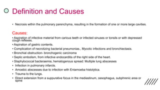
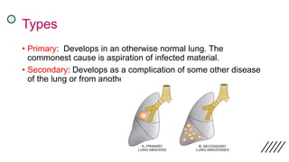
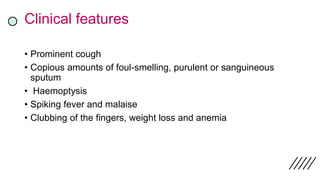
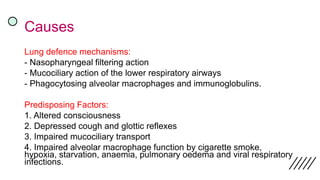
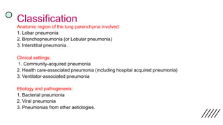
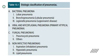
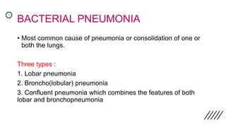
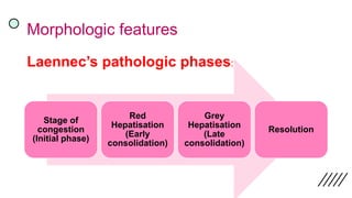
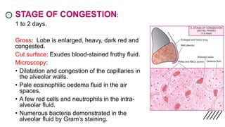
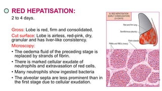
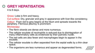
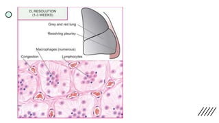
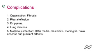
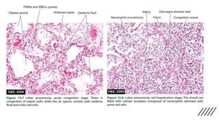
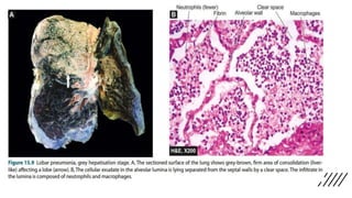
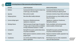
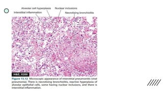
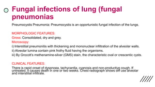
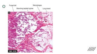
L U N G A B S C E S S
D R R E E N A Z S H A I K
M D PAT H O L O G Y
Definition and Causes
• Necrosis within the pulmonary parenchyma, resulting in the formation of one or more large cavities.
Causes:
• Aspiration of infective material from carious teeth or infected sinuses or tonsils or with depressed
cough reflexes.
• Aspiration of gastric contents.
• Complication of necrotizing bacterial pneumonias., Mycotic infections and bronchiectasis.
• Bronchial obstruction- bronchogenic carcinoma
• Septic embolism, from infective endocarditis of the right side of the heart.
• Staphylococcal bacteraemia, hematogenous spread: Multiple lung abscesses
• Infection in pulmonary infarcts.
• Amoebic abscesses due to infection with Entamoeba histolytica.
• Trauma to the lungs.
• Direct extension from a suppurative focus in the mediastinum, oesophagus, subphrenic area or
spine
Types
• Primary: Develops in an otherwise normal lung. The
commonest cause is aspiration of infected material.
• Secondary: Develops as a complication of some other disease
of the lung or from another site.
Clinical features
• Prominent cough
• Copious amounts of foul-smelling, purulent or sanguineous
sputum
• Haemoptysis
• Spiking fever and malaise
• Clubbing of the fingers, weight loss and anemia
Morphology
• Size: Few millimetres to large cavities 5 to 6 cm across.
• Localization and number: Depends on their mode of development.
• Aspiration: more common on the right side and occurs in the posterior
segment of the upper lobe and in the apical segments of the lower lobe,
• Septic emboli, hematogenous seeding: multiple and affect any region of
the lungs.
• Gross:
-Abscesses may be of variable size.
-The cavity contains exudate.
- Poorly defined ragged wall.
- With passage of time, the abscess becomes chronic and develops fibrous
wall
• Microscopy:
- Destruction of lung parenchyma with suppurative exudate in the lung
cavity.
- The cavity is initially surrounded by acute inflammation in the wall but later
there is replacement by chronic inflammatory cell infiltrate composed of
lymphocytes, plasma cells and macrophages.
- In more chronic cases, there is considerable fibroblastic proliferation
forming a fibrocollagenic wall.
Complications
• Ruptures into airways, pleural cavity and produce
bronchopleural fistulas, the consequence of which is
pneumothorax or empyema.
• Embolization of septic material to the brain, giving rise to
meningitis or brain abscess.
Histologic examination:
1. Fibrous scarring
2. Mononuclear infiltration (lymphocytes, plasma cells,
macrophages)
Treatment
• Intravenous antibiotics have been used successfully for the
treatment of lung abscesses, particularly in children.
• For small unilocular abscesses, aspiration and drainage or
partial resection of the lobe may be curative.
• For larger lesions, lobectomy.
P N E U M O N I A
Definition
• An acute inflammation of the lung parenchyma distal to the
terminal bronchioles
• The terms ‘pneumonia’ and ‘pneumonitis’ are often used
synonymously for inflammation of the lungs,
• ‘Consolidation’ (meaning solidification) is the term used for
gross and radiologic appearance of the lungs in pneumonia
Pathogenesis
Entry of organism:
1. Inhalation of the microbes present in the air.
2. Aspiration of organisms from the nasopharynx or oropharynx.
3. Haematogenous spread from a distant focus of infection.
4. Direct spread from an adjoining site of infection.
Causes
Lung defence mechanisms:
- Nasopharyngeal filtering action
- Mucociliary action of the lower respiratory airways
- Phagocytosing alveolar macrophages and immunoglobulins.
Predisposing Factors:
1. Altered consciousness
2. Depressed cough and glottic reflexes
3. Impaired mucociliary transport
4. Impaired alveolar macrophage function by cigarette smoke,
hypoxia, starvation, anaemia, pulmonary oedema and viral respiratory
infections.
Classification
Anatomic region of the lung parenchyma involved:
1. Lobar pneumonia
2. Bronchopneumonia (or Lobular pneumonia)
3. Interstitial pneumonia.
Clinical settings:
1. Community-acquired pneumonia
2. Health care-associated pneumonia (including hospital acquired pneumonia)
3. Ventilator-associated pneumonia
Etiology and pathogenesis:
1. Bacterial pneumonia
2. Viral pneumonia
3. Pneumonias from other aetiologies.
BACTERIAL PNEUMONIA
• Most common cause of pneumonia or consolidation of one or
both the lungs.
Three types :
1. Lobar pneumonia
2. Broncho(lobular) pneumonia
3. Confluent pneumonia which combines the features of both
lobar and bronchopneumonia
Lobar Pneumonia
• Acute bacterial infection of a part of a lobe, the entire lobe, or
even two lobes of one or both the lungs.
ETIOLOGY:
• 1. Pneumococcal pneumonia: More than 90%
• 2. Staphylococcal pneumonia
• 3. Streptococcal pneumonia
• 4. Pneumonia by gram-negative aerobic bacteria
Clinical features
• Shaking chills, fever, malaise with pleuritic chest pain, dyspnoea
and cough with expectoration which may be mucoid, purulent or
even bloody.
• CBP: Marked neutrophilic leucocytosis.
• Blood cultures: Positive in about 30% of cases.
• Chest Xray: Consolidation.
Morphologic features
Laennec’s pathologic phases:
Stage of
congestion
(Initial phase)
Red
Hepatisation
(Early
consolidation)
Grey
Hepatisation
(Late
consolidation)
Resolution
STAGE OF CONGESTION:
1 to 2 days.
Gross: Lobe is enlarged, heavy, dark red and
congested.
Cut surface: Exudes blood-stained frothy fluid.
Microscopy:
• Dilatation and congestion of the capillaries in
the alveolar walls.
• Pale eosinophilic oedema fluid in the air
spaces.
• A few red cells and neutrophils in the intra-
alveolar fluid.
• Numerous bacteria demonstrated in the
alveolar fluid by Gram’s staining.
RED HEPATISATION:
2 to 4 days.
Gross: Lobe is red, firm and consolidated.
Cut surface: Lobe is airless, red-pink, dry,
granular and has liver-like consistency.
Microscopy:
• The oedema fluid of the preceding stage is
replaced by strands of fibrin.
• There is marked cellular exudate of
neutrophils and extravasation of red cells.
• Many neutrophils show ingested bacteria
• The alveolar septa are less prominent than in
the first stage due to cellular exudation.
GREY HEPATISATION:
4 to 8 days.
Gross: Lobe is firm and heavy.
Cut surface: Dry, granular and grey in appearance with liver-like consistency.
Colour: From red to grey begins at the hilum and spreads towards the
periphery. Fibrinous pleurisy is prominent.
Microscopy:
• The fibrin strands are dense and more numerous.
• The cellular exudate of neutrophils is reduced due to disintegration of
many inflammatory cells as evidenced by their pyknotic nuclei.
• The red cells are also fewer and macrophages begin to appear in the
exudate.
• The cellular exudate is often separated from the septal walls by a thin clear
space.
• The organisms are less numerous and appear as degenerated forms.
RESOLUTION
8 - 9 days if no chemotherapy is administered and is completed in 1 to
3 weeks.
With antibiotic therapy induces resolution on about 3rd day.
Gross: Solid fibrinous constituent is liquefied by enzymatic action.
Cut surface: Grey-red or dirty brown and frothy, yellow, creamy fluid
can be expressed on pressing.
Microscopy:
• Macrophages are the predominant cells in the alveolar spaces, while
neutrophils diminish in number. They contain engulfed neutrophils
and debris.
• Granular and fragmented strands of fibrin in the alveolar spaces are
seen due to progressive enzymatic digestion.
• Alveolar capillaries are engorged.
Complications
1. Organisation: Fibrosis
2. Pleural effusion
3. Empyema
4. Lung abscess
5. Metastatic infection: Otitis media, mastoiditis, meningitis, brain
abscess and purulent arthritis
Bronchopneumonia or lobular
pneumonia
• Infection of the terminal bronchioles that extends into the
surrounding alveoli resulting in patchy consolidation of the lung.
ETIOLOGY:
Staphylococci, Streptococci, Pneumococci, Klebsiella
pneumoniae, Haemophilus influenzae, and Gram negative bacilli
like Pseudomonas and coliform bacteria.
Morphologic features
• Gross: Patchy areas of red or grey consolidation affecting one or
more lobes.
• Cut surface: Dry, granular, firm, red or grey in colour, 3 to 4 cm in
diameter, slightly elevated over the sur face and are often centred
around a bronchiole.
• Microscopy:
• i) Acute bronchiolitis.
• ii) Suppurative exudate, consisting chiefly of neutrophils in the
peribronchiolar alveoli.
• iii) Thickening of the alveolar septa by congested capillaries and
leucocytic infiltration.
• iv) Less involved alveoli contain oedema fluid
Complications:
Destruction of the bronchioles resulting in foci of bronchiolar
fibrosis that may eventually cause bronchiectasis.
Clinical features:
History of preceding bed-ridden illness, chronic debility,
aspiration of gastric contents or upper respiratory infection.
For initial 2 to 3 days, there are features of acute bronchitis but
subsequently signs and symptoms similar to those of lobar
pneumonia appear.
CBP: Neutrophilic leucocytosis.
Chest Xray: Mottled, focal opacities in both the lungs, chiefly in
the lower zones.
Legionella Pneumonia
Legionella pneumonia or Legionnaire’s disease is an epidemic illness
caused by gram-negative bacilli.
Gross: Bronchopneumonia involving many lobes and there may be
consolidation of the entire lung. Pleural effusion +.
Microscopy:
• Intra-alveolar exudate
• Alveolar septa show foci of hyperplasia of the lining epithelium and
thrombosis of vessels in the septa.
Clinical features: Malaise, headache and muscle-aches followed by high
fever, chills, cough and tachypnoea.
Viral and Mycoplasma pneumonia
• Patchy inflammatory changes in interstitial tissue of the lungs,
without any alveolar exudate.
Etiology:
Interstitial pneumonitis is caused by most common: Respiratory
syncytial virus (RSV).
Others: Influenza and para influenza viruses, Adenoviruses,
Rhinoviruses, Coxsackieviruses and Cytomegaloviruses (CMV).
Psittacosis (Chlamydia) and Q fever (Coxiella) are also
associated with interstitial pneumonitis.
Morphologic features:
Gross: Patchy to massive and widespread consolidation of one
or both the lungs
Cut Section: Sectioned surface of the lung exudes small amount
of frothy or bloody fluid.
Microscopy:
i) Interstitial inflammation.
ii) Necrotising bronchiolitis
iii) Reactive changes
iv) Alveolar changes: Oedema fluid, fibrin, scanty inflammatory
exudate and coating of alveolar walls by pink, hyaline membrane
similar to the one seen in respiratory distress syndrome.
Fungal infections of lung (fungal
pneumonias
Pneumocystis Pneumonia: Pneumocystis is an opportunistic fungal infection of the lungs.
MORPHOLOGIC FEATURES:
Gross: Consolidated, dry and grey.
Microscopy:
i) Interstitial pneumonitis with thickening and mononuclear infiltration of the alveolar walls.
ii) Alveolar lumina contain pink frothy fluid having the organisms.
iii) By Grocott’s methenamine-silver (GMS) stain, the characteristic oval or crescentic cysts.
CLINICAL FEATURES:
There is rapid onset of dyspnoea, tachycardia, cyanosis and non-productive cough. If
untreated, it causes death in one or two weeks. Chest radiograph shows diff use alveolar
and interstitial infiltrate.
Non infective pneumonias
Aspiration (Inhalation) Pneumonia:
Factors: Unconsciousness, drunkenness, neurological disorders affecting
swallowing, drowning, necrotic oropharyngeal tumours, in premature infants and
congenital tracheo-oesophageal fistula.
Morphologic features :
• Chemical pneumonitis: Haemorrhagic pulmonary oedema with presence of
particles in the bronchioles.
• Patients rapidly develop cyanosis, dyspnoea, shock and bloody sputum and are
often likely to die of cardiac failure.
• Non-sterile aspirate causes widespread bronchopneumonia with multiple areas of
necrosis and suppuration.
• A granulomatous reaction with foreign body giant cells may surround the
aspirated vegetable matter.
Hypostatic Pneumonia
Collection of oedema fluid and secretions in the dependent parts of
the lungs in severely debilitated, bed-ridden patients.
Lipid Pneumonia
1. Exogenous lipid pneumonia: Aspiration of a variety of oily
materials. Th ese are: inhalation of oily nasal drops, regurgitation
of oily medicines from stomach (e.g. liquid paraffin), administration
of oily vitamin preparation to reluctant children or to debilitated old
patients.
2. Endogenous lipid pneumonia: Tissue breakdown following
obstruction to airways e.g. obstruction by bronchogenic cancer,
tuberculosis and bronchiectasis
THANK
YOU

Lung Abscess and Pneumonia (Pathology)

  • 1.
    L U NG A B S C E S S D R R E E N A Z S H A I K M D PAT H O L O G Y
  • 2.
    Definition and Causes •Necrosis within the pulmonary parenchyma, resulting in the formation of one or more large cavities. Causes: • Aspiration of infective material from carious teeth or infected sinuses or tonsils or with depressed cough reflexes. • Aspiration of gastric contents. • Complication of necrotizing bacterial pneumonias., Mycotic infections and bronchiectasis. • Bronchial obstruction- bronchogenic carcinoma • Septic embolism, from infective endocarditis of the right side of the heart. • Staphylococcal bacteraemia, hematogenous spread: Multiple lung abscesses • Infection in pulmonary infarcts. • Amoebic abscesses due to infection with Entamoeba histolytica. • Trauma to the lungs. • Direct extension from a suppurative focus in the mediastinum, oesophagus, subphrenic area or spine
  • 3.
    Types • Primary: Developsin an otherwise normal lung. The commonest cause is aspiration of infected material. • Secondary: Develops as a complication of some other disease of the lung or from another site.
  • 4.
    Clinical features • Prominentcough • Copious amounts of foul-smelling, purulent or sanguineous sputum • Haemoptysis • Spiking fever and malaise • Clubbing of the fingers, weight loss and anemia
  • 5.
    Morphology • Size: Fewmillimetres to large cavities 5 to 6 cm across. • Localization and number: Depends on their mode of development. • Aspiration: more common on the right side and occurs in the posterior segment of the upper lobe and in the apical segments of the lower lobe, • Septic emboli, hematogenous seeding: multiple and affect any region of the lungs.
  • 6.
    • Gross: -Abscesses maybe of variable size. -The cavity contains exudate. - Poorly defined ragged wall. - With passage of time, the abscess becomes chronic and develops fibrous wall • Microscopy: - Destruction of lung parenchyma with suppurative exudate in the lung cavity. - The cavity is initially surrounded by acute inflammation in the wall but later there is replacement by chronic inflammatory cell infiltrate composed of lymphocytes, plasma cells and macrophages. - In more chronic cases, there is considerable fibroblastic proliferation forming a fibrocollagenic wall.
  • 7.
    Complications • Ruptures intoairways, pleural cavity and produce bronchopleural fistulas, the consequence of which is pneumothorax or empyema. • Embolization of septic material to the brain, giving rise to meningitis or brain abscess. Histologic examination: 1. Fibrous scarring 2. Mononuclear infiltration (lymphocytes, plasma cells, macrophages)
  • 9.
    Treatment • Intravenous antibioticshave been used successfully for the treatment of lung abscesses, particularly in children. • For small unilocular abscesses, aspiration and drainage or partial resection of the lobe may be curative. • For larger lesions, lobectomy.
  • 10.
    P N EU M O N I A
  • 11.
    Definition • An acuteinflammation of the lung parenchyma distal to the terminal bronchioles • The terms ‘pneumonia’ and ‘pneumonitis’ are often used synonymously for inflammation of the lungs, • ‘Consolidation’ (meaning solidification) is the term used for gross and radiologic appearance of the lungs in pneumonia
  • 12.
    Pathogenesis Entry of organism: 1.Inhalation of the microbes present in the air. 2. Aspiration of organisms from the nasopharynx or oropharynx. 3. Haematogenous spread from a distant focus of infection. 4. Direct spread from an adjoining site of infection.
  • 13.
    Causes Lung defence mechanisms: -Nasopharyngeal filtering action - Mucociliary action of the lower respiratory airways - Phagocytosing alveolar macrophages and immunoglobulins. Predisposing Factors: 1. Altered consciousness 2. Depressed cough and glottic reflexes 3. Impaired mucociliary transport 4. Impaired alveolar macrophage function by cigarette smoke, hypoxia, starvation, anaemia, pulmonary oedema and viral respiratory infections.
  • 14.
    Classification Anatomic region ofthe lung parenchyma involved: 1. Lobar pneumonia 2. Bronchopneumonia (or Lobular pneumonia) 3. Interstitial pneumonia. Clinical settings: 1. Community-acquired pneumonia 2. Health care-associated pneumonia (including hospital acquired pneumonia) 3. Ventilator-associated pneumonia Etiology and pathogenesis: 1. Bacterial pneumonia 2. Viral pneumonia 3. Pneumonias from other aetiologies.
  • 16.
    BACTERIAL PNEUMONIA • Mostcommon cause of pneumonia or consolidation of one or both the lungs. Three types : 1. Lobar pneumonia 2. Broncho(lobular) pneumonia 3. Confluent pneumonia which combines the features of both lobar and bronchopneumonia
  • 17.
    Lobar Pneumonia • Acutebacterial infection of a part of a lobe, the entire lobe, or even two lobes of one or both the lungs. ETIOLOGY: • 1. Pneumococcal pneumonia: More than 90% • 2. Staphylococcal pneumonia • 3. Streptococcal pneumonia • 4. Pneumonia by gram-negative aerobic bacteria
  • 18.
    Clinical features • Shakingchills, fever, malaise with pleuritic chest pain, dyspnoea and cough with expectoration which may be mucoid, purulent or even bloody. • CBP: Marked neutrophilic leucocytosis. • Blood cultures: Positive in about 30% of cases. • Chest Xray: Consolidation.
  • 19.
    Morphologic features Laennec’s pathologicphases: Stage of congestion (Initial phase) Red Hepatisation (Early consolidation) Grey Hepatisation (Late consolidation) Resolution
  • 20.
    STAGE OF CONGESTION: 1to 2 days. Gross: Lobe is enlarged, heavy, dark red and congested. Cut surface: Exudes blood-stained frothy fluid. Microscopy: • Dilatation and congestion of the capillaries in the alveolar walls. • Pale eosinophilic oedema fluid in the air spaces. • A few red cells and neutrophils in the intra- alveolar fluid. • Numerous bacteria demonstrated in the alveolar fluid by Gram’s staining.
  • 21.
    RED HEPATISATION: 2 to4 days. Gross: Lobe is red, firm and consolidated. Cut surface: Lobe is airless, red-pink, dry, granular and has liver-like consistency. Microscopy: • The oedema fluid of the preceding stage is replaced by strands of fibrin. • There is marked cellular exudate of neutrophils and extravasation of red cells. • Many neutrophils show ingested bacteria • The alveolar septa are less prominent than in the first stage due to cellular exudation.
  • 22.
    GREY HEPATISATION: 4 to8 days. Gross: Lobe is firm and heavy. Cut surface: Dry, granular and grey in appearance with liver-like consistency. Colour: From red to grey begins at the hilum and spreads towards the periphery. Fibrinous pleurisy is prominent. Microscopy: • The fibrin strands are dense and more numerous. • The cellular exudate of neutrophils is reduced due to disintegration of many inflammatory cells as evidenced by their pyknotic nuclei. • The red cells are also fewer and macrophages begin to appear in the exudate. • The cellular exudate is often separated from the septal walls by a thin clear space. • The organisms are less numerous and appear as degenerated forms.
  • 24.
    RESOLUTION 8 - 9days if no chemotherapy is administered and is completed in 1 to 3 weeks. With antibiotic therapy induces resolution on about 3rd day. Gross: Solid fibrinous constituent is liquefied by enzymatic action. Cut surface: Grey-red or dirty brown and frothy, yellow, creamy fluid can be expressed on pressing. Microscopy: • Macrophages are the predominant cells in the alveolar spaces, while neutrophils diminish in number. They contain engulfed neutrophils and debris. • Granular and fragmented strands of fibrin in the alveolar spaces are seen due to progressive enzymatic digestion. • Alveolar capillaries are engorged.
  • 26.
    Complications 1. Organisation: Fibrosis 2.Pleural effusion 3. Empyema 4. Lung abscess 5. Metastatic infection: Otitis media, mastoiditis, meningitis, brain abscess and purulent arthritis
  • 29.
    Bronchopneumonia or lobular pneumonia •Infection of the terminal bronchioles that extends into the surrounding alveoli resulting in patchy consolidation of the lung. ETIOLOGY: Staphylococci, Streptococci, Pneumococci, Klebsiella pneumoniae, Haemophilus influenzae, and Gram negative bacilli like Pseudomonas and coliform bacteria.
  • 30.
    Morphologic features • Gross:Patchy areas of red or grey consolidation affecting one or more lobes. • Cut surface: Dry, granular, firm, red or grey in colour, 3 to 4 cm in diameter, slightly elevated over the sur face and are often centred around a bronchiole. • Microscopy: • i) Acute bronchiolitis. • ii) Suppurative exudate, consisting chiefly of neutrophils in the peribronchiolar alveoli. • iii) Thickening of the alveolar septa by congested capillaries and leucocytic infiltration. • iv) Less involved alveoli contain oedema fluid
  • 31.
    Complications: Destruction of thebronchioles resulting in foci of bronchiolar fibrosis that may eventually cause bronchiectasis. Clinical features: History of preceding bed-ridden illness, chronic debility, aspiration of gastric contents or upper respiratory infection. For initial 2 to 3 days, there are features of acute bronchitis but subsequently signs and symptoms similar to those of lobar pneumonia appear. CBP: Neutrophilic leucocytosis. Chest Xray: Mottled, focal opacities in both the lungs, chiefly in the lower zones.
  • 34.
    Legionella Pneumonia Legionella pneumoniaor Legionnaire’s disease is an epidemic illness caused by gram-negative bacilli. Gross: Bronchopneumonia involving many lobes and there may be consolidation of the entire lung. Pleural effusion +. Microscopy: • Intra-alveolar exudate • Alveolar septa show foci of hyperplasia of the lining epithelium and thrombosis of vessels in the septa. Clinical features: Malaise, headache and muscle-aches followed by high fever, chills, cough and tachypnoea.
  • 35.
    Viral and Mycoplasmapneumonia • Patchy inflammatory changes in interstitial tissue of the lungs, without any alveolar exudate. Etiology: Interstitial pneumonitis is caused by most common: Respiratory syncytial virus (RSV). Others: Influenza and para influenza viruses, Adenoviruses, Rhinoviruses, Coxsackieviruses and Cytomegaloviruses (CMV). Psittacosis (Chlamydia) and Q fever (Coxiella) are also associated with interstitial pneumonitis.
  • 36.
    Morphologic features: Gross: Patchyto massive and widespread consolidation of one or both the lungs Cut Section: Sectioned surface of the lung exudes small amount of frothy or bloody fluid. Microscopy: i) Interstitial inflammation. ii) Necrotising bronchiolitis iii) Reactive changes iv) Alveolar changes: Oedema fluid, fibrin, scanty inflammatory exudate and coating of alveolar walls by pink, hyaline membrane similar to the one seen in respiratory distress syndrome.
  • 38.
    Fungal infections oflung (fungal pneumonias Pneumocystis Pneumonia: Pneumocystis is an opportunistic fungal infection of the lungs. MORPHOLOGIC FEATURES: Gross: Consolidated, dry and grey. Microscopy: i) Interstitial pneumonitis with thickening and mononuclear infiltration of the alveolar walls. ii) Alveolar lumina contain pink frothy fluid having the organisms. iii) By Grocott’s methenamine-silver (GMS) stain, the characteristic oval or crescentic cysts. CLINICAL FEATURES: There is rapid onset of dyspnoea, tachycardia, cyanosis and non-productive cough. If untreated, it causes death in one or two weeks. Chest radiograph shows diff use alveolar and interstitial infiltrate.
  • 40.
    Non infective pneumonias Aspiration(Inhalation) Pneumonia: Factors: Unconsciousness, drunkenness, neurological disorders affecting swallowing, drowning, necrotic oropharyngeal tumours, in premature infants and congenital tracheo-oesophageal fistula. Morphologic features : • Chemical pneumonitis: Haemorrhagic pulmonary oedema with presence of particles in the bronchioles. • Patients rapidly develop cyanosis, dyspnoea, shock and bloody sputum and are often likely to die of cardiac failure. • Non-sterile aspirate causes widespread bronchopneumonia with multiple areas of necrosis and suppuration. • A granulomatous reaction with foreign body giant cells may surround the aspirated vegetable matter.
  • 41.
    Hypostatic Pneumonia Collection ofoedema fluid and secretions in the dependent parts of the lungs in severely debilitated, bed-ridden patients. Lipid Pneumonia 1. Exogenous lipid pneumonia: Aspiration of a variety of oily materials. Th ese are: inhalation of oily nasal drops, regurgitation of oily medicines from stomach (e.g. liquid paraffin), administration of oily vitamin preparation to reluctant children or to debilitated old patients. 2. Endogenous lipid pneumonia: Tissue breakdown following obstruction to airways e.g. obstruction by bronchogenic cancer, tuberculosis and bronchiectasis
  • 42.