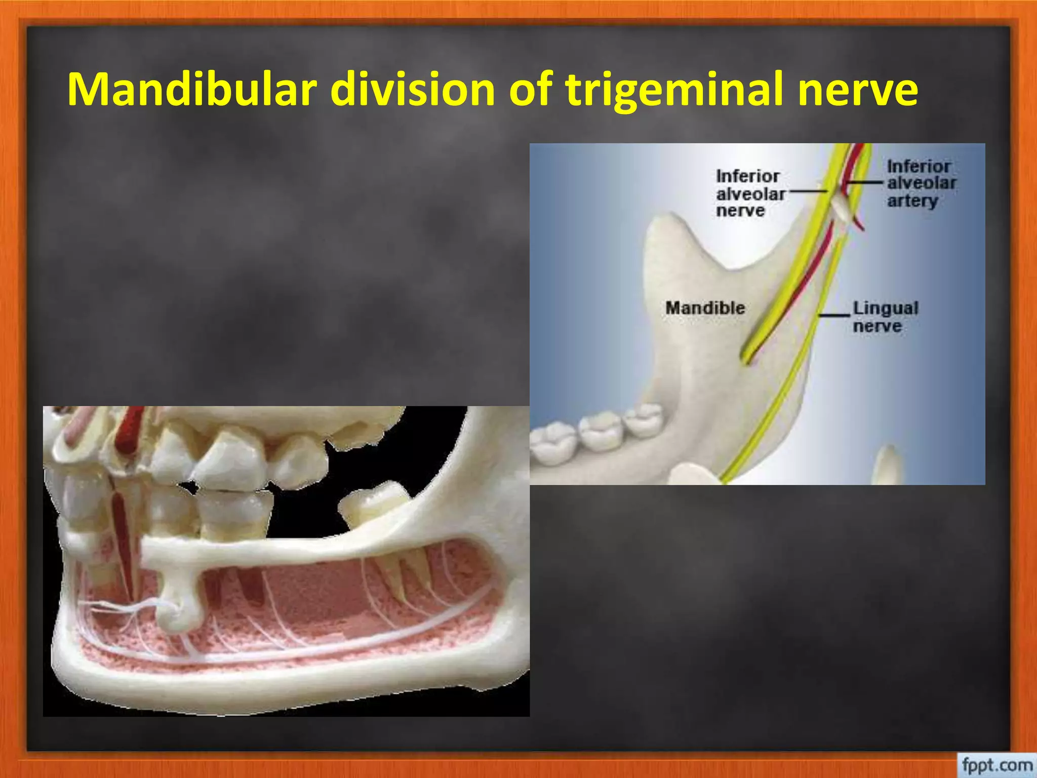
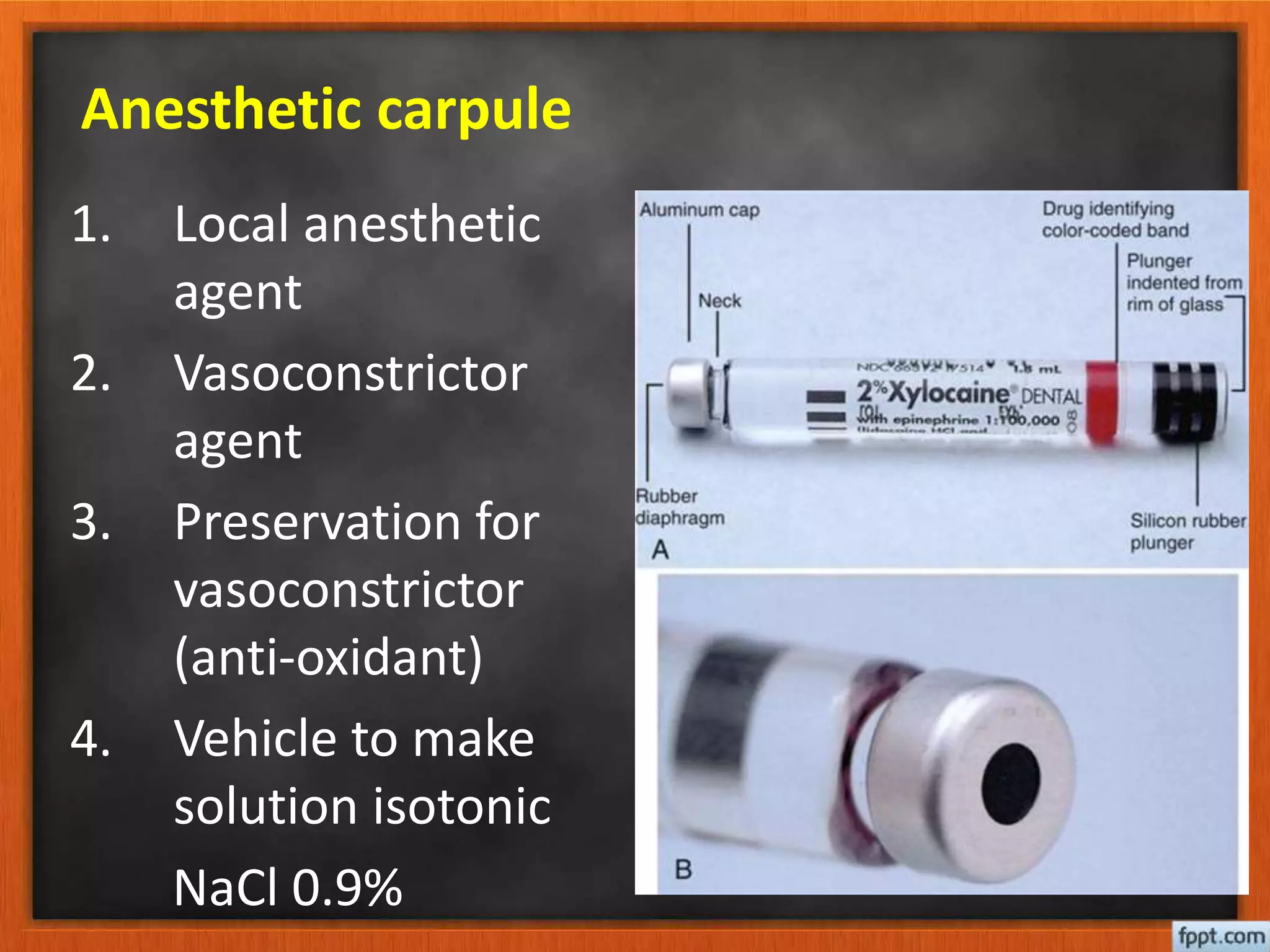
Local anesthesia
anatomy review & injection technique
Dr. Haydar Muneer Salih
B.D.S., F.I.B.M.S
Nothing that is done by a dentist for
patient is of greater importance than
the administration of a drug for
prevention of pain during dental
treatment yet the act of administration
of local anesthesia frequently
associated with great anxiety and pain
Local Anesthetics
• Role:
• Decrease intraoperative and
postoperative pain
• Increase patients cooperation
• Diagnostic testing/examination
Anatomical considerations:
• Trigeminal nerve:
– Sensory divisions:
• Ophthalmic division V1
• Maxillary division V2
• Mandibular division V3
– Motor division:
• Muscles of mastication- masseter,
temporalis, medial and lateral pterygoids
• Mylohyoid, Anterior belly of the digastric ,
Tensor tympani, Tensor veli palatini
Maxillary division of trigeminal nerve
Mandibular division of trigeminal nerve
Local anesthetic instruments:
• Anesthetic carpule
• Syringe
• Needle
Anesthetic carpule
1. Local anesthetic
agent
2. Vasoconstrictor
agent
3. Preservation for
vasoconstrictor
(anti-oxidant)
4. Vehicle to make
solution isotonic
NaCl 0.9%
Anesthetic syringe
Anesthetic needle
Injection technique
1. Use sterile sharp needle
2. Check the follow of the solution
3. Warm the anesthetic carpule and syringe to
the room temperature
4. Position the patient
5. Dry the tissue
6. Apply topical anesthesia
7. Communicate with the patient
8. Establish a firm hand rest
9. Make the tissue taut
10. Keep the syringe out of patient line of
sight
11. Insert the needle to the mucosa
12. Watch and communicate with patient
13. Slowly advance and withdraw
14. Observe the patient after injection
Maxillary anesthesia:
• Infiltration:
Able to be performed in the maxilla due to the
thin cortical nature of the bone
• Target area: apical region of the tooth to be
anesthetize
• Land marks:
a. Mucosal fold
b. Crown of the tooth
c. Root contour of the tooth
• Orientation of the needle bevel so it faces
the bone
Maxillary anesthesia:
• Anesthesia of the hard palate is
necessary for dental extraction of
maxillary teeth
• For many patient palatal injection
proved to be very traumatic and for
many dentist is the one of the most
traumatic procedures
1. Inform the patient that this procedure
is little uncomfortable so he will be
psychological prepared
2. Provide adequate topical anesthesia
3. Maintain control over the needle
4. Deposit the anesthesia slowly
5. Trust yourself
Anesthesia for greater palatine
Naso palatine nerve block:
Mandibular anesthesia:
• Infiltration techniques do not work in the
adult mandible due to the dense cortical bone
• Nerve blocks are utilized to anesthetize the
inferior alveolar, lingual, and buccal nerves
• Provides anesthesia to the pulpal, alveolar,
lingual and buccal gingival tissue, and skin of
lower lip and medial aspect of chin on side
injected
Inferior alveolar nerve block (IAN)
Technique involves blocking the inferior alveolar
nerve prior to entry into the mandibular lingula
on the medial aspect of the mandibular ramus.
Technique:
• Area of insertion is the mucous membrane on
the medial border of the mandibular ramus
• Height of injection injection- 6-10 mm above
the occlusal table of the mandibular teeth
• Mouth must be open for this technique
Inferior alveolar nerve block (IAN)
• Depth of injection: 25mm
• Approach area of injection from
contralateral premolar region
• Use the non-dominant hand to retract the
buccal soft tissue (thumb in coronoid notch
of mandible; index finger on posterior
border of extraoral mandible
• Inject ~0.5 0.5-1.0cc of local anesthetic
• Continue to inject ~0.5cc on removal from
injection site to anesthetize the lingual
branch
Mental nerve block
• Mental and incisive nerves are the
terminal branches for the inferior
alveolar nerve
• Provides sensory input for the lower lip
skin, mucous membrane, pulpal/alveolar
tissue for the premolars, canine, and
incisors on side blocked
Mental nerve block
Technique:
• Area of injection mucobuccal fold at or
anterior to the mental foramen. This lies
between the mandibular premolars
• Depth of injection ~5 5-6mm
• Inject 0.5 0.5-1.0cc of local anesthesia
• Message local anesthesia into tissue to
manipulate into mental foramen to anesthetize
the incisive branch
Local anesthesia
Local anesthesia

Local anesthesia

  • 1.
    Local anesthesia anatomy review& injection technique Dr. Haydar Muneer Salih B.D.S., F.I.B.M.S
  • 2.
    Nothing that isdone by a dentist for patient is of greater importance than the administration of a drug for prevention of pain during dental treatment yet the act of administration of local anesthesia frequently associated with great anxiety and pain
  • 3.
    Local Anesthetics • Role: •Decrease intraoperative and postoperative pain • Increase patients cooperation • Diagnostic testing/examination
  • 4.
    Anatomical considerations: • Trigeminalnerve: – Sensory divisions: • Ophthalmic division V1 • Maxillary division V2 • Mandibular division V3 – Motor division: • Muscles of mastication- masseter, temporalis, medial and lateral pterygoids • Mylohyoid, Anterior belly of the digastric , Tensor tympani, Tensor veli palatini
  • 5.
    Maxillary division oftrigeminal nerve
  • 6.
    Mandibular division oftrigeminal nerve
  • 7.
    Local anesthetic instruments: •Anesthetic carpule • Syringe • Needle
  • 8.
    Anesthetic carpule 1. Localanesthetic agent 2. Vasoconstrictor agent 3. Preservation for vasoconstrictor (anti-oxidant) 4. Vehicle to make solution isotonic NaCl 0.9%
  • 9.
  • 10.
  • 12.
    Injection technique 1. Usesterile sharp needle 2. Check the follow of the solution 3. Warm the anesthetic carpule and syringe to the room temperature 4. Position the patient 5. Dry the tissue 6. Apply topical anesthesia 7. Communicate with the patient
  • 13.
    8. Establish afirm hand rest 9. Make the tissue taut 10. Keep the syringe out of patient line of sight 11. Insert the needle to the mucosa 12. Watch and communicate with patient 13. Slowly advance and withdraw 14. Observe the patient after injection
  • 15.
    Maxillary anesthesia: • Infiltration: Ableto be performed in the maxilla due to the thin cortical nature of the bone • Target area: apical region of the tooth to be anesthetize • Land marks: a. Mucosal fold b. Crown of the tooth c. Root contour of the tooth • Orientation of the needle bevel so it faces the bone
  • 16.
  • 17.
    • Anesthesia ofthe hard palate is necessary for dental extraction of maxillary teeth • For many patient palatal injection proved to be very traumatic and for many dentist is the one of the most traumatic procedures
  • 18.
    1. Inform thepatient that this procedure is little uncomfortable so he will be psychological prepared 2. Provide adequate topical anesthesia 3. Maintain control over the needle 4. Deposit the anesthesia slowly 5. Trust yourself
  • 19.
  • 20.
  • 21.
    Mandibular anesthesia: • Infiltrationtechniques do not work in the adult mandible due to the dense cortical bone • Nerve blocks are utilized to anesthetize the inferior alveolar, lingual, and buccal nerves • Provides anesthesia to the pulpal, alveolar, lingual and buccal gingival tissue, and skin of lower lip and medial aspect of chin on side injected
  • 23.
    Inferior alveolar nerveblock (IAN) Technique involves blocking the inferior alveolar nerve prior to entry into the mandibular lingula on the medial aspect of the mandibular ramus. Technique: • Area of insertion is the mucous membrane on the medial border of the mandibular ramus • Height of injection injection- 6-10 mm above the occlusal table of the mandibular teeth • Mouth must be open for this technique
  • 25.
    Inferior alveolar nerveblock (IAN) • Depth of injection: 25mm • Approach area of injection from contralateral premolar region • Use the non-dominant hand to retract the buccal soft tissue (thumb in coronoid notch of mandible; index finger on posterior border of extraoral mandible • Inject ~0.5 0.5-1.0cc of local anesthetic • Continue to inject ~0.5cc on removal from injection site to anesthetize the lingual branch
  • 28.
    Mental nerve block •Mental and incisive nerves are the terminal branches for the inferior alveolar nerve • Provides sensory input for the lower lip skin, mucous membrane, pulpal/alveolar tissue for the premolars, canine, and incisors on side blocked
  • 31.
    Mental nerve block Technique: •Area of injection mucobuccal fold at or anterior to the mental foramen. This lies between the mandibular premolars • Depth of injection ~5 5-6mm • Inject 0.5 0.5-1.0cc of local anesthesia • Message local anesthesia into tissue to manipulate into mental foramen to anesthetize the incisive branch