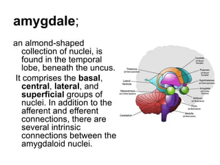
Download to read offline

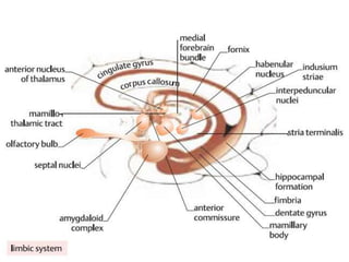
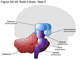
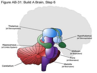
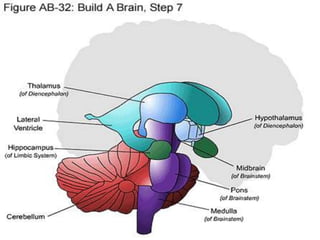
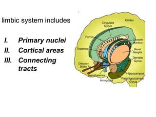
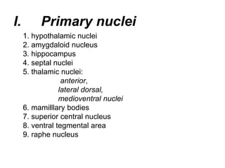
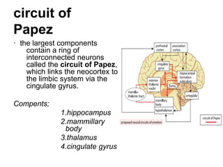
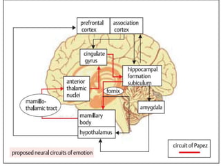
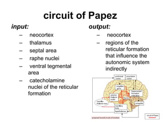
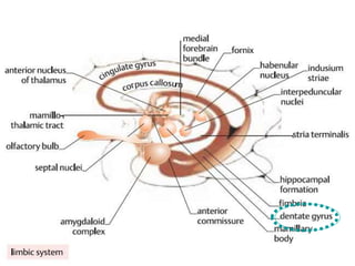
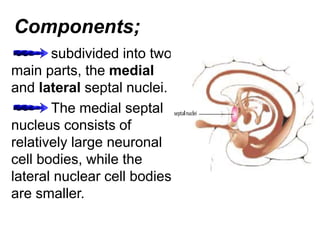
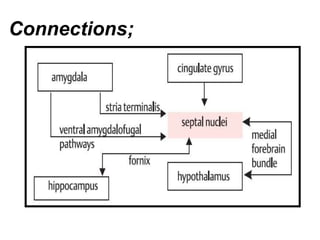
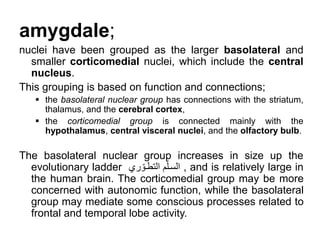
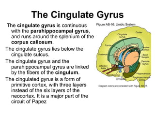


The limbic system includes structures involved in emotion processing and memory formation. It contains nuclei such as the hippocampus, amygdala, and septal nuclei, as well as connecting tracts. The hippocampus forms memories and is connected to the amygdala and septal nuclei. The amygdala processes emotions and regulates autonomic functions. Stimulation of the septal nuclei produces pleasurable sensations. Together, these structures form circuits like the circuit of Papez that are important for emotional processing and memory.


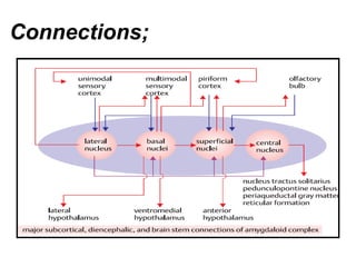
![ONFH[AVN HIP] -TRIPLE REGIME -A NOVAL SURGICAL CONCEPT .pptx](https://cdn.slidesharecdn.com/ss_thumbnails/onfhavnhip2026koaconcalicutdrgokuldevdrmashraf-260210064517-213ec005-thumbnail.jpg?width=640&height=640&fit=bounds)


