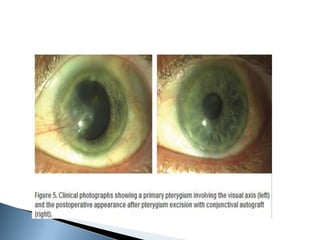
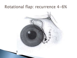
MODERATOR: DR SURESH.H.H
PRESENTER:DR ANJALI
 Derived from Greek word ‘pterygion’ means
wing.
 It is a non malignant slow growing
proliferation of wing shaped fibrovascular
tissue.
 Arises from subconjunctival tissue.
 May extend over the cornea thus disturbing
the vision.
 World wide distribution.
 More common in warm and dry climates.
 Prevalence : 22% equatorial areas.
<2% in latitudes between
28-38degree.
 Direct relation with amount of UV exposure.
 Sex: male : female= 2:1
 Age:>40years high prevalence
20-40years high incidence.
 In India prevalence is 9.5%.
 Morbidity: causes significant alteration in
visual function in advanced cases.
 Strong association between UV light exposure
and formation of pterygium.
 More common- in patients who worked
outdoors.
 In welders than other factory workers.
 Also associated with basal cell
carcinoma, porphyria cutanea
tarda, polymorphous light
eruptions, xeroderma pigmentosa.
 ANGIOGENESIS FACTOR:Prolonged UV
exposure causes biological changes in the
bowmans membrane.
 Altered protein so formed could act as
angiogenic/ pterygiogenic factor.
 UV Exposure: May induce hyperplasia in limbal
cells. These altered cells invade the cornea and
limbus which moves centripetally with them. This
explains wing shape of the pterygium.
 UV radiation causes depletion of langerhan cells
at limbus.(stocker’s line).
 Exposure to UVB+altered tear film
 Injury and susceptibility
 Loss of collagenase and dehydration
 Accumulation of Extracellular matrix
 Antigenic,type1 HS Pinguecula
 Fibroblastic reaction
Inflammation
 PTERYGIUM PTERYGIUM
 Light entering the temporal limbus at
90degree is concentrated at medial limbus.
 Related to corneal curvature.
 This explains the predominance of medial
pterygium.
 Dry and dusty environment.
 Drying of the tear film by wind devitalizes
tissue of medial 3rd of the palpebral aperture.
 This allows the actinic radiation to damage
the conjunctival, corneal epithelium and
bowmans membrane.
 MICROTRAUMA: mechanical irritation by dust
particles, enhanced by tear flow from lateral
to medial.
 IMMUNOLOGY: Cell bound IgE irritant
complexes initiate the release of
inflammatory mediators from mast cells.
Release of stimulatory factors.
Development of pterygium.
 Expression of vimentin.
 P53 mutation leads to decreased apoptosis
and increased TGF-b which leads to increased
growth.
 RECURRENT PTERYGIUM- stem cells are more
scattered and expression pattern is more
denser.
 HYPOXIA: increase in non perfusion areas and
attenuated vessels in nasal limbus during
early stage of pterygium causes recruitment
of progenitor cells.
 Viral markers: infection with HPV and herpes
virus is considered as risk factor(rare).
 Elastotic degeneration of collagen.(Not a true
elastic tissue)
 Fibro vascular proliferation with an overlying
covering of epithelium-characterized by
Cellular proliferation.
Tissue remodeling.
Neovascularisation.
 Subepithelial tissue shows basophilic
degeneration.
 Destruction of bowman’s membrane in the
cornea.
 So there is residual corneal scarring when
these growth are removed.
 Epithelium shows secondary changes like
orthokeratosis,acanthosis,dyskeratosis.
 Mast cells occur in increased number.
Normal conjunctiva Pterygium
CLINICAL STAGING PATHOLOGICAL
STAGING
Stage I Exposure
conjunctivitis
Size and number of
Conjunctival vessels
Mild – moderate congestion
S/S of dryness
No formed lesions
Altered tear film
Mild vascular
response
Stage II Pinguecula
and pterygium
Distinct raised lesion on
bulbar conjunctiva
With or w/o abnormal
vascularization and
inflammation
Cell injury
Inflammatory
response
Stage III Limbal pterygium Head is on or across
the limbus with or w/o
an iron line at the
conjunctival corneal
interface
Vascularization and
fibrous proliferation
Symptoms more
pronounced
Lesion
organization
Mixed
proliferation
and
degeneration
Stage IV Corneal
pterygium
Lesion 2mm or more
into cornea
Invasion of granulation
tissue
Zone of dellen
Stocker’s line
Infiltration of corneal
nerves- pain
Lesion b/w
epithelium and
bowman
Mixed
proliferative
and
degeneration
Stage V Compound
pterygium
Induced astigmatism
Symptoms more
frequent and severe
Lesion extended
into stroma
Mixed
proliferative and
degeneration
Proliferation- Small lymphocytes and plasma cells
Degeneration- Swirls of type I collagen
 Fuch’s patches.
 Stocker’s line.
 Hood.
 Head.
 Body.
 Base.
 Superior edge.
 Inferior edge.
 Progressive: thick
fleshy
marked vascularity.
It has opaque infitrative spot known as cap.
Stocker’s line.
 Atrophic/stationary: thin
attenuated
poor vascularity
no cap.
Progressive pterygium Atrophic pterygium
 Primary double pterygium.
 Recurrent pterygium.
 Pseudopterygium.
 Malignant pterygium(rare):recurrent
pterygium with restriction of ocular
movements.
 Asymptomatic
 Foreign body sensation
 Discomfort
 Congestion(redness)
 Irritation and grittiness-interference with
precorneal tear film.
 Interference with vision-obscuring visual axis
-inducing astigmatism
 Cosmesis.
 Type 1: extends <2mm on the cornea.
 Type 2: 4mm of cornea is involved.
 Type 3: encroaches onto >4mm of cornea
and involves visual axis.
Pseudopterygiu
m
Most often hx of
previous infective,
chemical, thermal, or
traumatic injury to the
cornea.
May occur at multiple
locations and is not
restricted to the 3 and 9
o'clock (interpalpebral)
positions.
-Slit-lamp examination:
reveals lesion to be
adhesion of a fold of
conjunctiva, which has
occurred as a response to
a previous peripheral
corneal
ulcer/inflammation.
-Lesion typically only fixed
at its apex to the cornea
so that a probe may be
passed underneath its
body at the limbus, while a
true pterygium adheres to
the underlying cornea
throughout its length.
Thinning of the underlying
cornea may be seen at its
head.
Condition Signs and symptoms Tests
Pinguecula Does not encroach on
the cornea.
Slit-lamp examination:
reveals exact extent and
nature of lesion. A
pingueculum is limited to
limbus and conjunctiva
and does not encroach
onto the cornea.
Marginal keratitis Associated with
blepharitis. Infiltrate on
corneal surface is
separated by a clear
zone from the limbus.
Occur at 2, 4, 8, and
10 o'clock position.
Does not have typical
pterygium shape. Often
superior and inferior.
Corneal swab/scraping:
microscopy and culture
positive for infecting
organism, but infecting
organisms are often not
detected, as many cases
are due to an
inflammatory reaction to
staphylococcal proteins
Corneal micropannus Hx of trachoma or lack
of corneal oxygenation
due to excessive
contact lens wear.
Slit-lamp examination:
reveals encroachment
of fine blood vessels
onto corneal surface.
Conjunctival
carcinoma in situ/
bowens epithlioma.
Rare. Does not have
typical pterygium
shape. Not restricted
to the 3 and 9 o'clock
(interpalpebral)
positions and can
occur at any position
on the cornea.
Slit-lamp examination:
gelatinous-appearing
mass.
Biopsy: cytological
features of a
squamous cell
carcinoma, but the
basal membrane of
the epithelium
remains intact.
Squamous cell
carcinoma
Rare. Does not have typical
pterygium shape. Not
restricted to the 3 and 9
o'clock (interpalpebral)
positions and can occur at
any position on the cornea.
May arise from a pterygium,
carcinoma in situ, or de
novo.
Slit-lamp examination:
surface may appear
keratinised and
friable.
Biopsy: well-
differentiated
squamous cell
carcinoma with
invasion of the basal
membrane.
Limbal dermoid Benign choriostomatous
tissue. MC site:inferior
temporal quadrant.
Histology contains
abberant tissue like
epidermal
appendages,connectiv
e tissue,skin,fatmuscle
teeth.
 Symptomatic patients- Tear substitutes
 Inflammation- Topical steroids
 Sunglasses- to reduce UV exposure and
decrease growth stimulus
1. Extension to the visual axis and induced
astigmatism.
2. Recurrent irritation.
3. Cosmetic- patient should be explained
there is fairly high risk of recurrence, which
may be more unsightly.
 Free conjunctival autograft for primary and
recurrent pterygium.
 Pre op evaluation:
1. Evaluation of pterygium.
2. Evaluation of superior bulbar conjunctiva.
3. Pre op preparation.
4. Anaesthesia and sedation.
Preparation and drape.
Place anaesthetic drops or topical
vasoconstrictor.
Ask patient to look opposite side of pterygium.
 Goal: Achieve a normal, topographically
smooth ocular surface
 Dissect a smooth plane toward the
limbus
 Preferable to dissect down to bare sclera
at limbus
 Bare sclera = remove loose Tenon’s layer
and leave episcleral vessels intact
 Mechanism of action: it acts forming a fibrin
clot between graft and host tissue.
 Advantages : decreases the post op pain.
reduces the surgical time as well
as recurrence rate.
Disadvantage : not FDA approved.
graft dehiscence.
infection, discomfort.
Recurrence rate: less as compared to suture.
 Avoid exposure to sunlight.
 Use of dark sun glasses.
 Topical steroid antibiotic drops, topical
NSAIDS, artificial tears.
 POD3/5 graft acquires redness.
 Complete healing expected between 6-
8weeks.
 Topical medications should be tapered.
Lubricants should remain for 3months.
 Instruction to patient: avoid exposure to
sunlight.
 Graft failure.
 Granuloma formation.
 Conjunctival infection.
 Suture detachment.
 Delayed healing.
 Recurrence.
Bare sclera technique:
-recurrence:
5-68%
(primary)
35-82%
(recurrent)
 Subconjunctival scarring limitation of
movements diplopia.
 Disinsertion of medial rectus muscle.
 Scleral perforation.
 Corneal irregularity due to deep stromal
excision.
 Growth of fibrovascular tissue across the limbus
onto cornea after initial removal.
 Excludes persistence of deeper corneal vessels
and scarring which may remain even after
adequate removal.
 Bunching of conjunctiva and formation of
parallel loops of vessels, which aim almost like
an arrowhead at the limbus, usually denotes a
conjunctival recurrence.
 Grade 1 – normal appearing
operative site.
 Grade 2 – fine episcleral
vessels in the site extending
to the limbus.
 Grade 3 – additional fibrous
tissues in site.
 Grade 4 – actual corneal
recurrence.
 AIM: To reduce recurrence.
1. Corticosteroids- post operative use of
topical steroids can reduce inflammatory
reaction and revascularization at the
operative site.
2. No significant role in prevention of
recurrence.
 Antibiotic and antineoplastic properties.
 Blocks the DNA synthesis.
 Concentration: 0.02%
 Use: intraoperative to the area of resection
with sponge for 2min followed by irrigating
with balanced salt solution.
 Side effects: pyogenic granuloma
dellen of sclera.
perforation of eye.
glaucoma.
cataract.
corneal edema.
 Recurrence: 3-25% (intraoperative)
5-54%(postoperative)
 Post operative
period LCAG MMC
3 month 1 -
6 month 1 2
12 month - 1
18 month - -
Total 02 (4%) 03 (6%)
 Nitrogen mustard alkylating agent.
antimitotic property.
 Radiomimetic- obliterates vascular
endothelial cells.
 Dose:1:2000 every 3 hours for 6 weeks.
 Used in bare sclera method.
 Complication: scleral thinning.
 Recurrence:10-16%
 Antiproliferative
 Inhibits thymidylate synthetase, thus inhibits
DNA.
 Only cells in the synthesis phase are
affected, allowing the remaining cells to
continue to proliferate after exposure to 5-
FU.
 Immunosuppresant drug.
 Dose: 0.05% topical for 3 months following
pterygium excision.
 Safe and effective.
 Low recurrence rate(3.4%)
 Inhibit neovascularisation.
 Stop the progression or prevent the
recurrence.
 Case reported by Wu and co workers.
Topical bevacizumab eye drops 25mg/ml
4times for 3weeks.
 No recurrence in 1year follow up period.
 Reduces mitosis in rapidly dividing vascular
endothelial cells.
 Dose : 15Gy units in single or divided doses.
 Recurrence: 4.3%-35% with bare sclera or
simple conjunctival closure.
 Complications: scleral necrosis
endophthalmitis
cataract formation.
conjunctival telangiectasia.
 The area of bare scleral was covered with
amniotic membrane, which was oriented with
the basement membrane side up.
 The amniotic membrane was sutured
through the episcleral tissue to the edge of
the conjunctiva along the bare sclera border
with 7-8 interrupted 8-0 Vicryl sutures.
 The eye was patched.
 Useful in:
 very large conjunctival defects.
 To preserve superior conjunctiva for future
glaucoma surgery.
 Advantages: faster healing rate
less discomfort.
lower recurrence rate(2% in
1year follow up)
 ANECORTANE ACETATE:
Angiostatic steroid: Inhibits the blood vessel’s.
Topical 1% have inhibitory effect on pterygium
regrowth following recurrenr pterygium
excision.
 Surgical and medical management of
pterygium-Ashok garg.
 Pterygium-a practical guide to
management,L. Alfred andeze.
 Kanski clinical ophthalmology.
pterygiumanditsmanagement-130115081420-phpapp02.pdf

pterygiumanditsmanagement-130115081420-phpapp02.pdf

  • 1.
  • 4.
     Derived fromGreek word ‘pterygion’ means wing.  It is a non malignant slow growing proliferation of wing shaped fibrovascular tissue.  Arises from subconjunctival tissue.  May extend over the cornea thus disturbing the vision.
  • 5.
     World widedistribution.  More common in warm and dry climates.  Prevalence : 22% equatorial areas. <2% in latitudes between 28-38degree.  Direct relation with amount of UV exposure.
  • 6.
     Sex: male: female= 2:1  Age:>40years high prevalence 20-40years high incidence.  In India prevalence is 9.5%.  Morbidity: causes significant alteration in visual function in advanced cases.
  • 7.
     Strong associationbetween UV light exposure and formation of pterygium.  More common- in patients who worked outdoors.  In welders than other factory workers.  Also associated with basal cell carcinoma, porphyria cutanea tarda, polymorphous light eruptions, xeroderma pigmentosa.
  • 8.
     ANGIOGENESIS FACTOR:ProlongedUV exposure causes biological changes in the bowmans membrane.  Altered protein so formed could act as angiogenic/ pterygiogenic factor.
  • 9.
     UV Exposure:May induce hyperplasia in limbal cells. These altered cells invade the cornea and limbus which moves centripetally with them. This explains wing shape of the pterygium.  UV radiation causes depletion of langerhan cells at limbus.(stocker’s line).
  • 10.
     Exposure toUVB+altered tear film  Injury and susceptibility  Loss of collagenase and dehydration  Accumulation of Extracellular matrix  Antigenic,type1 HS Pinguecula  Fibroblastic reaction Inflammation  PTERYGIUM PTERYGIUM
  • 11.
     Light enteringthe temporal limbus at 90degree is concentrated at medial limbus.  Related to corneal curvature.  This explains the predominance of medial pterygium.
  • 12.
     Dry anddusty environment.  Drying of the tear film by wind devitalizes tissue of medial 3rd of the palpebral aperture.  This allows the actinic radiation to damage the conjunctival, corneal epithelium and bowmans membrane.
  • 13.
     MICROTRAUMA: mechanicalirritation by dust particles, enhanced by tear flow from lateral to medial.  IMMUNOLOGY: Cell bound IgE irritant complexes initiate the release of inflammatory mediators from mast cells. Release of stimulatory factors. Development of pterygium.
  • 14.
     Expression ofvimentin.  P53 mutation leads to decreased apoptosis and increased TGF-b which leads to increased growth.  RECURRENT PTERYGIUM- stem cells are more scattered and expression pattern is more denser.
  • 15.
     HYPOXIA: increasein non perfusion areas and attenuated vessels in nasal limbus during early stage of pterygium causes recruitment of progenitor cells.  Viral markers: infection with HPV and herpes virus is considered as risk factor(rare).
  • 16.
     Elastotic degenerationof collagen.(Not a true elastic tissue)  Fibro vascular proliferation with an overlying covering of epithelium-characterized by Cellular proliferation. Tissue remodeling. Neovascularisation.  Subepithelial tissue shows basophilic degeneration.
  • 17.
     Destruction ofbowman’s membrane in the cornea.  So there is residual corneal scarring when these growth are removed.  Epithelium shows secondary changes like orthokeratosis,acanthosis,dyskeratosis.  Mast cells occur in increased number.
  • 18.
  • 20.
    CLINICAL STAGING PATHOLOGICAL STAGING StageI Exposure conjunctivitis Size and number of Conjunctival vessels Mild – moderate congestion S/S of dryness No formed lesions Altered tear film Mild vascular response Stage II Pinguecula and pterygium Distinct raised lesion on bulbar conjunctiva With or w/o abnormal vascularization and inflammation Cell injury Inflammatory response
  • 21.
    Stage III Limbalpterygium Head is on or across the limbus with or w/o an iron line at the conjunctival corneal interface Vascularization and fibrous proliferation Symptoms more pronounced Lesion organization Mixed proliferation and degeneration Stage IV Corneal pterygium Lesion 2mm or more into cornea Invasion of granulation tissue Zone of dellen Stocker’s line Infiltration of corneal nerves- pain Lesion b/w epithelium and bowman Mixed proliferative and degeneration
  • 22.
    Stage V Compound pterygium Inducedastigmatism Symptoms more frequent and severe Lesion extended into stroma Mixed proliferative and degeneration Proliferation- Small lymphocytes and plasma cells Degeneration- Swirls of type I collagen
  • 23.
     Fuch’s patches. Stocker’s line.  Hood.  Head.  Body.  Base.  Superior edge.  Inferior edge.
  • 25.
     Progressive: thick fleshy markedvascularity. It has opaque infitrative spot known as cap. Stocker’s line.  Atrophic/stationary: thin attenuated poor vascularity no cap.
  • 26.
  • 28.
     Primary doublepterygium.  Recurrent pterygium.  Pseudopterygium.  Malignant pterygium(rare):recurrent pterygium with restriction of ocular movements.
  • 30.
     Asymptomatic  Foreignbody sensation  Discomfort  Congestion(redness)  Irritation and grittiness-interference with precorneal tear film.  Interference with vision-obscuring visual axis -inducing astigmatism  Cosmesis.
  • 31.
     Type 1:extends <2mm on the cornea.  Type 2: 4mm of cornea is involved.  Type 3: encroaches onto >4mm of cornea and involves visual axis.
  • 32.
    Pseudopterygiu m Most often hxof previous infective, chemical, thermal, or traumatic injury to the cornea. May occur at multiple locations and is not restricted to the 3 and 9 o'clock (interpalpebral) positions. -Slit-lamp examination: reveals lesion to be adhesion of a fold of conjunctiva, which has occurred as a response to a previous peripheral corneal ulcer/inflammation. -Lesion typically only fixed at its apex to the cornea so that a probe may be passed underneath its body at the limbus, while a true pterygium adheres to the underlying cornea throughout its length. Thinning of the underlying cornea may be seen at its head. Condition Signs and symptoms Tests
  • 33.
    Pinguecula Does notencroach on the cornea. Slit-lamp examination: reveals exact extent and nature of lesion. A pingueculum is limited to limbus and conjunctiva and does not encroach onto the cornea. Marginal keratitis Associated with blepharitis. Infiltrate on corneal surface is separated by a clear zone from the limbus. Occur at 2, 4, 8, and 10 o'clock position. Does not have typical pterygium shape. Often superior and inferior. Corneal swab/scraping: microscopy and culture positive for infecting organism, but infecting organisms are often not detected, as many cases are due to an inflammatory reaction to staphylococcal proteins
  • 34.
    Corneal micropannus Hxof trachoma or lack of corneal oxygenation due to excessive contact lens wear. Slit-lamp examination: reveals encroachment of fine blood vessels onto corneal surface. Conjunctival carcinoma in situ/ bowens epithlioma. Rare. Does not have typical pterygium shape. Not restricted to the 3 and 9 o'clock (interpalpebral) positions and can occur at any position on the cornea. Slit-lamp examination: gelatinous-appearing mass. Biopsy: cytological features of a squamous cell carcinoma, but the basal membrane of the epithelium remains intact.
  • 35.
    Squamous cell carcinoma Rare. Doesnot have typical pterygium shape. Not restricted to the 3 and 9 o'clock (interpalpebral) positions and can occur at any position on the cornea. May arise from a pterygium, carcinoma in situ, or de novo. Slit-lamp examination: surface may appear keratinised and friable. Biopsy: well- differentiated squamous cell carcinoma with invasion of the basal membrane. Limbal dermoid Benign choriostomatous tissue. MC site:inferior temporal quadrant. Histology contains abberant tissue like epidermal appendages,connectiv e tissue,skin,fatmuscle teeth.
  • 36.
     Symptomatic patients-Tear substitutes  Inflammation- Topical steroids  Sunglasses- to reduce UV exposure and decrease growth stimulus
  • 37.
    1. Extension tothe visual axis and induced astigmatism. 2. Recurrent irritation. 3. Cosmetic- patient should be explained there is fairly high risk of recurrence, which may be more unsightly.
  • 38.
     Free conjunctivalautograft for primary and recurrent pterygium.  Pre op evaluation: 1. Evaluation of pterygium. 2. Evaluation of superior bulbar conjunctiva. 3. Pre op preparation. 4. Anaesthesia and sedation.
  • 39.
    Preparation and drape. Placeanaesthetic drops or topical vasoconstrictor. Ask patient to look opposite side of pterygium.
  • 40.
     Goal: Achievea normal, topographically smooth ocular surface  Dissect a smooth plane toward the limbus  Preferable to dissect down to bare sclera at limbus  Bare sclera = remove loose Tenon’s layer and leave episcleral vessels intact
  • 43.
     Mechanism ofaction: it acts forming a fibrin clot between graft and host tissue.  Advantages : decreases the post op pain. reduces the surgical time as well as recurrence rate. Disadvantage : not FDA approved. graft dehiscence. infection, discomfort. Recurrence rate: less as compared to suture.
  • 44.
     Avoid exposureto sunlight.  Use of dark sun glasses.  Topical steroid antibiotic drops, topical NSAIDS, artificial tears.  POD3/5 graft acquires redness.
  • 45.
     Complete healingexpected between 6- 8weeks.  Topical medications should be tapered. Lubricants should remain for 3months.  Instruction to patient: avoid exposure to sunlight.
  • 46.
     Graft failure. Granuloma formation.  Conjunctival infection.  Suture detachment.  Delayed healing.  Recurrence.
  • 47.
  • 51.
     Subconjunctival scarringlimitation of movements diplopia.  Disinsertion of medial rectus muscle.  Scleral perforation.  Corneal irregularity due to deep stromal excision.
  • 52.
     Growth offibrovascular tissue across the limbus onto cornea after initial removal.  Excludes persistence of deeper corneal vessels and scarring which may remain even after adequate removal.  Bunching of conjunctiva and formation of parallel loops of vessels, which aim almost like an arrowhead at the limbus, usually denotes a conjunctival recurrence.
  • 53.
     Grade 1– normal appearing operative site.  Grade 2 – fine episcleral vessels in the site extending to the limbus.  Grade 3 – additional fibrous tissues in site.  Grade 4 – actual corneal recurrence.
  • 54.
     AIM: Toreduce recurrence. 1. Corticosteroids- post operative use of topical steroids can reduce inflammatory reaction and revascularization at the operative site. 2. No significant role in prevention of recurrence.
  • 55.
     Antibiotic andantineoplastic properties.  Blocks the DNA synthesis.  Concentration: 0.02%  Use: intraoperative to the area of resection with sponge for 2min followed by irrigating with balanced salt solution.
  • 56.
     Side effects:pyogenic granuloma dellen of sclera. perforation of eye. glaucoma. cataract. corneal edema.  Recurrence: 3-25% (intraoperative) 5-54%(postoperative)
  • 57.
     Post operative periodLCAG MMC 3 month 1 - 6 month 1 2 12 month - 1 18 month - - Total 02 (4%) 03 (6%)
  • 58.
     Nitrogen mustardalkylating agent. antimitotic property.  Radiomimetic- obliterates vascular endothelial cells.  Dose:1:2000 every 3 hours for 6 weeks.  Used in bare sclera method.  Complication: scleral thinning.  Recurrence:10-16%
  • 59.
     Antiproliferative  Inhibitsthymidylate synthetase, thus inhibits DNA.  Only cells in the synthesis phase are affected, allowing the remaining cells to continue to proliferate after exposure to 5- FU.
  • 60.
     Immunosuppresant drug. Dose: 0.05% topical for 3 months following pterygium excision.  Safe and effective.  Low recurrence rate(3.4%)
  • 61.
     Inhibit neovascularisation. Stop the progression or prevent the recurrence.  Case reported by Wu and co workers. Topical bevacizumab eye drops 25mg/ml 4times for 3weeks.  No recurrence in 1year follow up period.
  • 62.
     Reduces mitosisin rapidly dividing vascular endothelial cells.  Dose : 15Gy units in single or divided doses.  Recurrence: 4.3%-35% with bare sclera or simple conjunctival closure.  Complications: scleral necrosis endophthalmitis cataract formation. conjunctival telangiectasia.
  • 63.
     The areaof bare scleral was covered with amniotic membrane, which was oriented with the basement membrane side up.  The amniotic membrane was sutured through the episcleral tissue to the edge of the conjunctiva along the bare sclera border with 7-8 interrupted 8-0 Vicryl sutures.  The eye was patched.
  • 65.
     Useful in: very large conjunctival defects.  To preserve superior conjunctiva for future glaucoma surgery.  Advantages: faster healing rate less discomfort. lower recurrence rate(2% in 1year follow up)
  • 66.
     ANECORTANE ACETATE: Angiostaticsteroid: Inhibits the blood vessel’s. Topical 1% have inhibitory effect on pterygium regrowth following recurrenr pterygium excision.
  • 67.
     Surgical andmedical management of pterygium-Ashok garg.  Pterygium-a practical guide to management,L. Alfred andeze.  Kanski clinical ophthalmology.